Original Article
Computed tomography-guided sacroiliac joint intra-articular injections: Success rate and technical issues
2023 Volume 14 Issue 6 Pages 878-883
Details
2023 Volume 14 Issue 6 Pages 878-883
Introduction: Periarticular sacroiliac joint (SIJ) injection should be first performed to diagnose SIJ pain. However, SIJ intra-articular injections are occasionally required in patients with severe and chronic SIJ pain. Radiation exposure of the operator's fingers is a major problem while performing intra-articular SIJ injections under fluoroscopy guidance. We attempted to perform this type of injection under CT guidance based on previous reports and report on the success rate and issues surrounding the injection techniques.
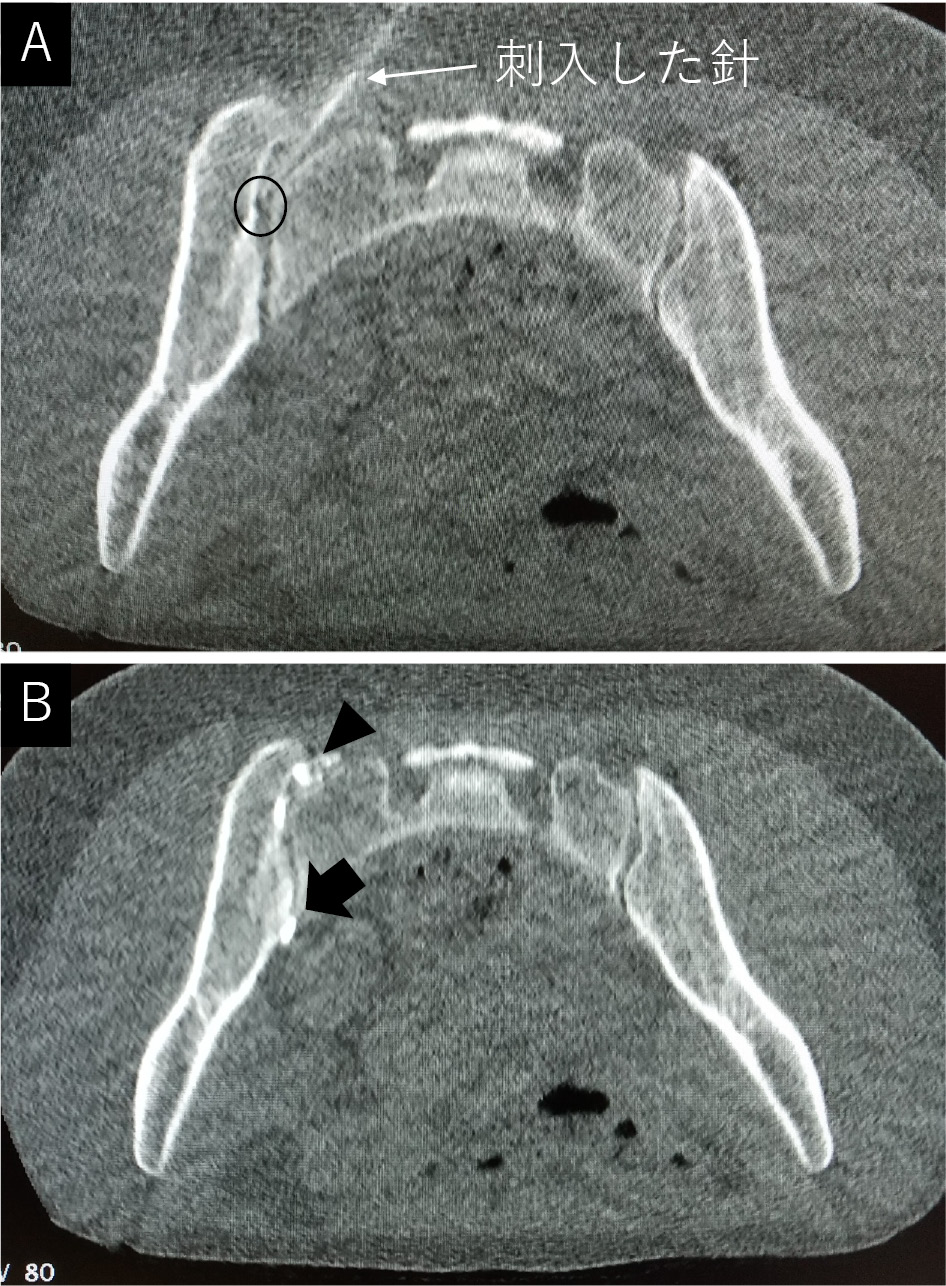
Methods: Fifty hospitalized patients (13 men, 37 women, mean 44±18 years) with 59 joints underwent CT-guided intra-articular block from June 2021 to October 2022. CT imaging was performed under minimum necessary conditions (80 kV, 10 mA, 1.0 s). The patient was placed in a prone position on a table, and the CT gantry was tilted 15° toward the head. A positioning laser was projected onto the patient's buttock to mark the entry point after setting the needle entry path on the CT image and measuring the depth and distance from the center. An angle meter was used to indicate the needle entry angle, and a 23 G needle (70 mm) was inserted along this angle. Once the operator felt that the needle tip had reached the cartilaginous surface of the iliac side, the needle position was confirmed by CT. The imaging was repeated until the optimal needle position was achieved after adjusting the direction. After confirming that the intra-articular was contrasted, a mixture of 2 ml of 2% lidocaine and 1.9 mg dexamethasone sodium phosphate was injected. The success rate of contrast of the joint, number of CT scans per procedure, time taken for a series of procedures (from needle insertion to the end of an intra-articular injection), and total radiation exposure dose (DLP) were investigated.
Results: Intra-articular contrast was identified in 34/59 joints (57.6%). The average number of CT scans was 4.9±1.0, total procedure time was 6 min 49 s (409±145 s), and total radiation exposure dose (DLP) was 9.06±2.91 [mGy・cm].
Conclusions: The success rate was low because of the inability to accurately reproduce the angles of measurement on the images at the time of needle insertion. The needle tip position could not be adjusted as frequently as in fluoroscopy. The advantage of this method is that the total exposure to the patient is low, and no exposure to the operator's fingers was made.