Journal of Spine Research
Online ISSN : 2435-1563
Print ISSN : 1884-7137
Volume 14, Issue 6
Displaying 1-25 of 25 articles from this issue
Editorial
Review Article
  • Yoichi Sato, Norio Yamamoto, Naoya Inagaki, Yusuke Iesaki, Takamune As ...
    2023 Volume 14 Issue 6 Pages 818-823
    Published: June 20, 2023
    Released on J-STAGE: June 20, 2023
    JOURNAL FREE ACCESS

    In Japan, the number of patients with osteoporosis is increasing as the population ages, but the therapeutic intervention rate is not high. For efficient and effective interventions, we hypothesized that the evaluation of osteoporosis can be performed (1) without special equipment and (2) without changing the conventional flow of medical care, thereby helping to solve this social problem. Focusing on chest X-rays, which are common and frequently taken, two AI algorithms to evaluate osteoporosis from chest X-rays. The first AI algorithm was developed to predict vertebral fractures from chest X-rays to detect vertebral fractures. A total pf 5,791 chest X-rays were used, and the AI algorithm predicted the presence of vertebral fractures in chest X-rays with Area Under the Curve = 0.98. The second AI algorithm was developed to predict bone mineral density from chest X-rays to evaluate bone mass. A total of 17,899 chest X-rays and bone mineral density measurements were used, and the AI algorithm predicted bone mineral density with a correlation coefficient (R) of 0.75. The AI algorithms developed in this study were able to detect vertebral fractures and evaluate bone mineral density with high accuracy from chest X-rays. Accurate detection and evaluation of these factors are important aspects of planning appropriate interventions for osteoporosis.

    Download PDF (1162K)
Original Article
  • Tomohito Mukaihata, Satoshi Maki, Yawara Eguchi, Seiji Ohtori
    2023 Volume 14 Issue 6 Pages 824-830
    Published: June 20, 2023
    Released on J-STAGE: June 20, 2023
    JOURNAL FREE ACCESS

    Study Design: Retrospective analysis of magnetic resonance imaging (MRI).

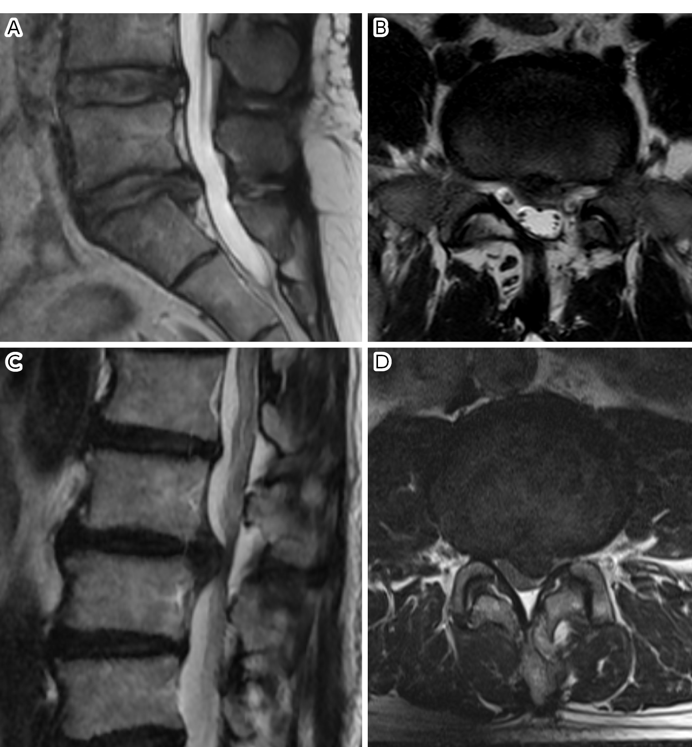
    Introduction: Discriminating between spinal pyogenic spondylitis and Modic change is crucial. A deep-learning approach based on convolutional neural networks (CNNs) is attracting attention in the medical imaging field. The aim of this study was to evaluate the performance of our CNN in differentiating between spinal Pyogenic spondylitis and Modic change on MRI. We compared the performance of the CNN and that of four specialists.

    Materials: Data from patients with spinal pyogenic spondylitis and Modic change who had undergone MRI. There were 50 patients with Modic change and 50 patients with pyogenic spondylitis. Sagittal T1-weighted MRI (T1WI) and sagittal T2-weighted MRI (T2WI) and short TI inversion recovery (STIR) were used for the CNN training and validation. The deep learning framework Tensorflow was used to construct the CNN architecture. To evaluate the performance of the CNN, the receiver operating characteristic curve (ROC) was plotted, and the area under the curve (AUC) was caluculated. We calculated and compared the accuracy, sensitivity, and specificity of the diagnosis by the CNN and a radiologist and a spine surgeon, and two orthopedic surgeons were compared.

    Results: The CNN-based AUCs of the ROC from the T1WI, T2WI, and STIR were 0.95, 0.94, and 0.95, respectively. The accuracy of the CNN was significantly better than that of four specialists on T1WI and STIR (p<.05), and better than a radiologist and one orthopedic surgeon on the T2WI (p<.05). The sensitivity was significantly better than that of four specialists on T1WI and STIR (p<.05), and better than a radiologist and one orthopedic surgeon on the T2WI (p<.05). The specificity was significantly better than one orthopedic surgeon on T1WI and T2WI (p<.05) and better than both orthopedic surgeons on STIR (p<.05).

    Conclusions: Spinal pyogenic spondylitis and Modic change were successfully differentiated using the CNN with high diagnostic accuracy comparable to that of four specialists.

    Download PDF (1627K)
  • Kazuma Murata, Kenji Endo, Takato Aihara, Hidekazu Suzuki, Yasunobu Sa ...
    2023 Volume 14 Issue 6 Pages 831-837
    Published: June 20, 2023
    Released on J-STAGE: June 20, 2023
    JOURNAL FREE ACCESS

    Plain radiography of the spine is an essential method for the evaluation of spinal diseases. However, the accuracy of radiography for assessing spinal diseases is not optimal when image diagnosis of radiography for spinal diseases is highly specialized and often difficult for primary physicians. Skilled spine surgeons are not always available in a primary care setting, which is where most patients with spinal diseases are initially examined. Therefore, minimizing the diagnostic errors of spinal diseases by radiography is crucial. Image identification based on artificial intelligence (AI) has been recognized as being potentially effective as a diagnostic strategy for orthopedic diseases; however, the accuracy of detecting vertebral fractures (VF) and ossification of posterior longitudinal ligaments (OPLL) using AI has remained uncertain. In this study, the mean accuracies of AI for rhe detection of VF and OPLL were 86.0% and 98.9%, respectively. Thus, AI should be considered in the assistance of clinicians for the image diagnosis of spinal diseases.

    Download PDF (1427K)
Review Article
  • Kazuta Yamashita, Fumitake Tezuka, Kosuke Sugiura, Daiki Nagajima, Yos ...
    2023 Volume 14 Issue 6 Pages 838-847
    Published: June 20, 2023
    Released on J-STAGE: June 20, 2023
    JOURNAL FREE ACCESS

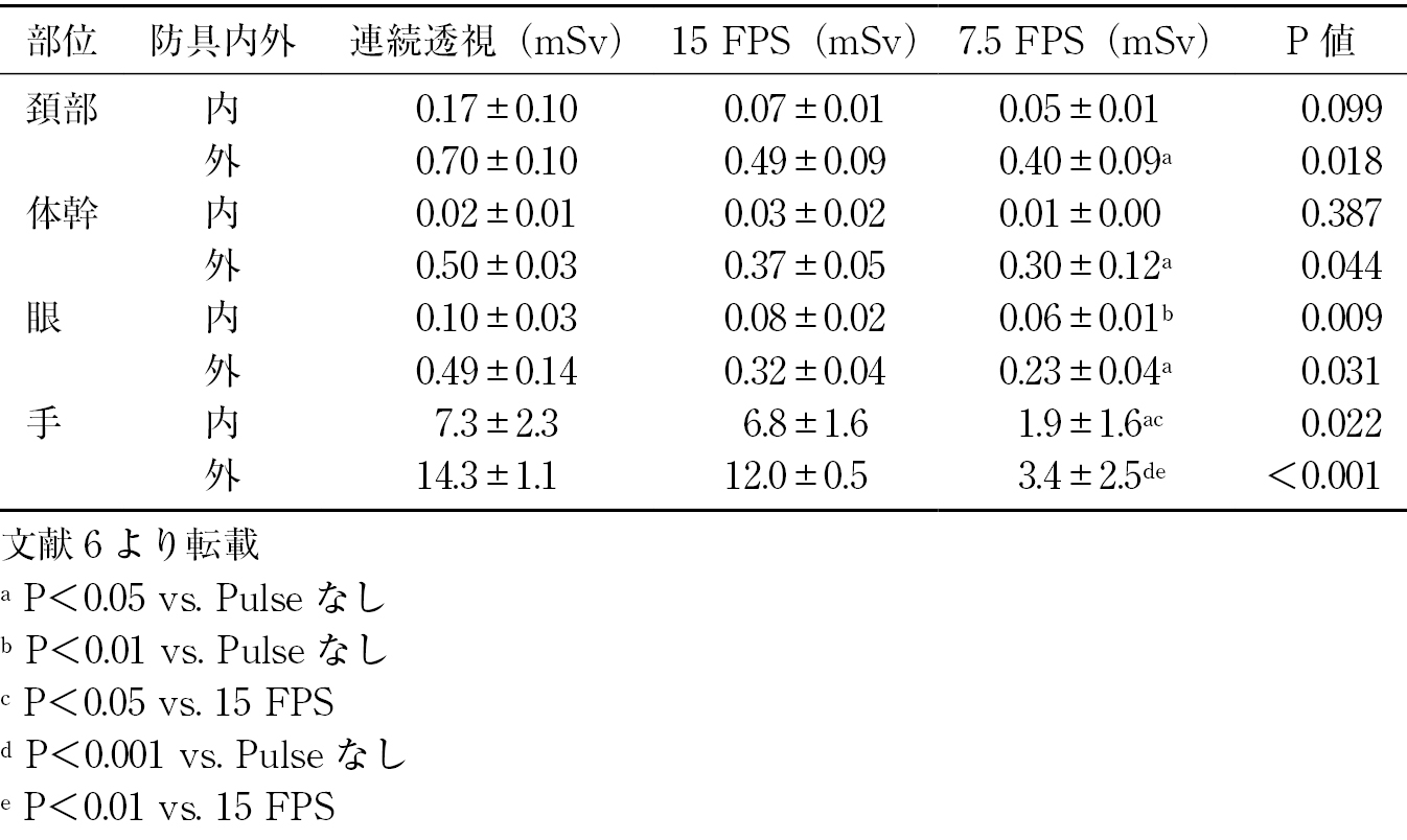
    Awareness of the harmful effects of long-term low-dose radiation is increasing. It seems to be able to guess the radiation exposure dose to each body site by visualizing them in our research. There are significant differences in the radiation exposure dose under different conditions and for different irradiated areas. The risk of occupational radiation exposure was the greatest with the lateral position. Medical staff, especially spine surgeons, should consider how to reduce radiation exposure and protect themselves by using appropriate shielding and techniques.

    Download PDF (1697K)
  • Kazuko Ohno, Kanichiro Wada
    2023 Volume 14 Issue 6 Pages 848-852
    Published: June 20, 2023
    Released on J-STAGE: June 20, 2023
    JOURNAL FREE ACCESS

    Fluoroscopic and radiographic-guided treatment has rapidly developed as a less invasive treatment for patients. However, during the past few years, several cases of radiation injury to the operators have been reported. For fluoroscopic guided treatment to continue to develop and be beneficial to patients, operators need to always be aware of radiation exposure in their daily practice. Four important considerations for operators are to: wear protective clothing and dosimeters; recognize that the reduction in radiation exposure to the medical staff is almost synonymous with the reduction in radiation exposure to the patient; narrow down the image; and simulate procedures in which the operator's hands are visible and try to reduce image quality.

    Download PDF (1129K)
Original Article
  • Kentaro Yamane, Shinichiro Takao, Kazuhiro Takeuchi
    2023 Volume 14 Issue 6 Pages 853-857
    Published: June 20, 2023
    Released on J-STAGE: June 20, 2023
    JOURNAL FREE ACCESS

    Long-term exposure to radiation can lead to gene mutations and increase the risk of malignant neoplasms. Spine surgeons have to perform various radiographic procedures. Selective nerve root block is the essential radiographic procedure used by spine surgeons to diagnose the specific source of nerve root pain, and provide therapeutic relief of leg pain. It is impossible for the spine surgeon to overlook the radiation exposure throughout his entire career. However, there has been no detailed report on the actual radiation exposure doses of spine surgeons. Spine surgeons should make efforts to reduce radiation exposure during radiographic procedures.

    Download PDF (1030K)
Review Article
  • Ko Matsudaira, Satoshi Kasahara, Mie Sakai, Shinsuke Inoue, Tomoko Tet ...
    2023 Volume 14 Issue 6 Pages 858-868
    Published: June 20, 2023
    Released on J-STAGE: June 20, 2023
    JOURNAL FREE ACCESS

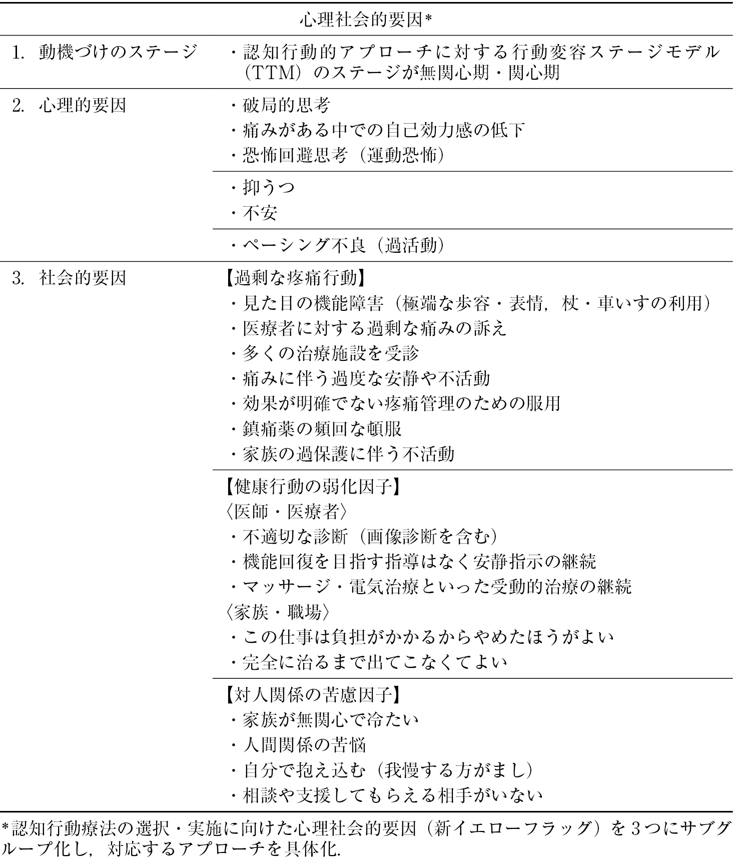
    Psychosocial factors are often involved in chronic non-specific low back pain patients, in which case an accurate assessment of these factors and a corresponding cognitive-behavioral approach are required. Multidisciplinary treatment based on a biopsychosocial model is essential in the treatment of chronic pain such as musculoskeletal pain. Multidisciplinary treatment for chronic low back pain is rated 1 (strong) A (high) in Clinical Practice Guidelines for the Management of Chronic Pain in Japan. We introduced an original psychosocial flags system for Japanese workers that was developed by members of the Ministry of Health, Labour and Welfare's Policy Research Project on chronic pain. The "psychosocial flags framework" originated in the U.K. for the multidisciplinary treatment of musculoskeletal pain, which was recommended in the neck and back pain guidelines in Europe. We focused on the yellow flags (i.e., psychosocial factors used to screen for cognitive-behavioral therapy) that will be an appropriate operational system for motivated therapists.

    Download PDF (2259K)
  • Junzo Fujitani, Koichi Sairyo
    2023 Volume 14 Issue 6 Pages 869-877
    Published: June 20, 2023
    Released on J-STAGE: June 20, 2023
    JOURNAL FREE ACCESS

    Exercise therapy is an important treatment option for low back pain, as indicated in the Guidelines for the Treatment of Low Back Pain 2019 (Revision 2). In recent years, the effectiveness of Pilates, based on the motor control concept, has received increasing attention. Based on Joint by Joint Theory, the concept of Pilates is to reduce and disperse mechanical stress on the spine by improving mobility in the low-mobility thoracic spine and hip joint and stability in the hypermobile cervical spine and lumbar spine.

    Pilates exercise therapy is based on a constant awareness of the draw-in and bracing of the trunk musculature with breathing, axial elongation of the spine and limbs, spine articulation, isolated movement of the limbs, and whole-body integration. In addition, the approach differs depending on the disease. For example, in the case of lumbar spine kyphosis, the mobility of the thoracic spine is improved and the stability of the lumbar spine and pelvis (core) is worked on. On the other hand, in the case of postoperative lumbar disc herniation, the first step is to stabilize the core to prevent recurrence.

    Currently, we are promoting Pilates exercise therapy based at our hospital in collaboration with related hospitals. In the future, we would like to continue to study Pilates therapy to make it the gold standard of exercise therapy for patients with low back pain.

    Download PDF (2130K)
Original Article
  • Daisuke Kurosawa, Eiichi Murakami, Shuhei Nishidate, Kazushi Sato, Yuk ...
    2023 Volume 14 Issue 6 Pages 878-883
    Published: June 20, 2023
    Released on J-STAGE: June 20, 2023
    JOURNAL FREE ACCESS

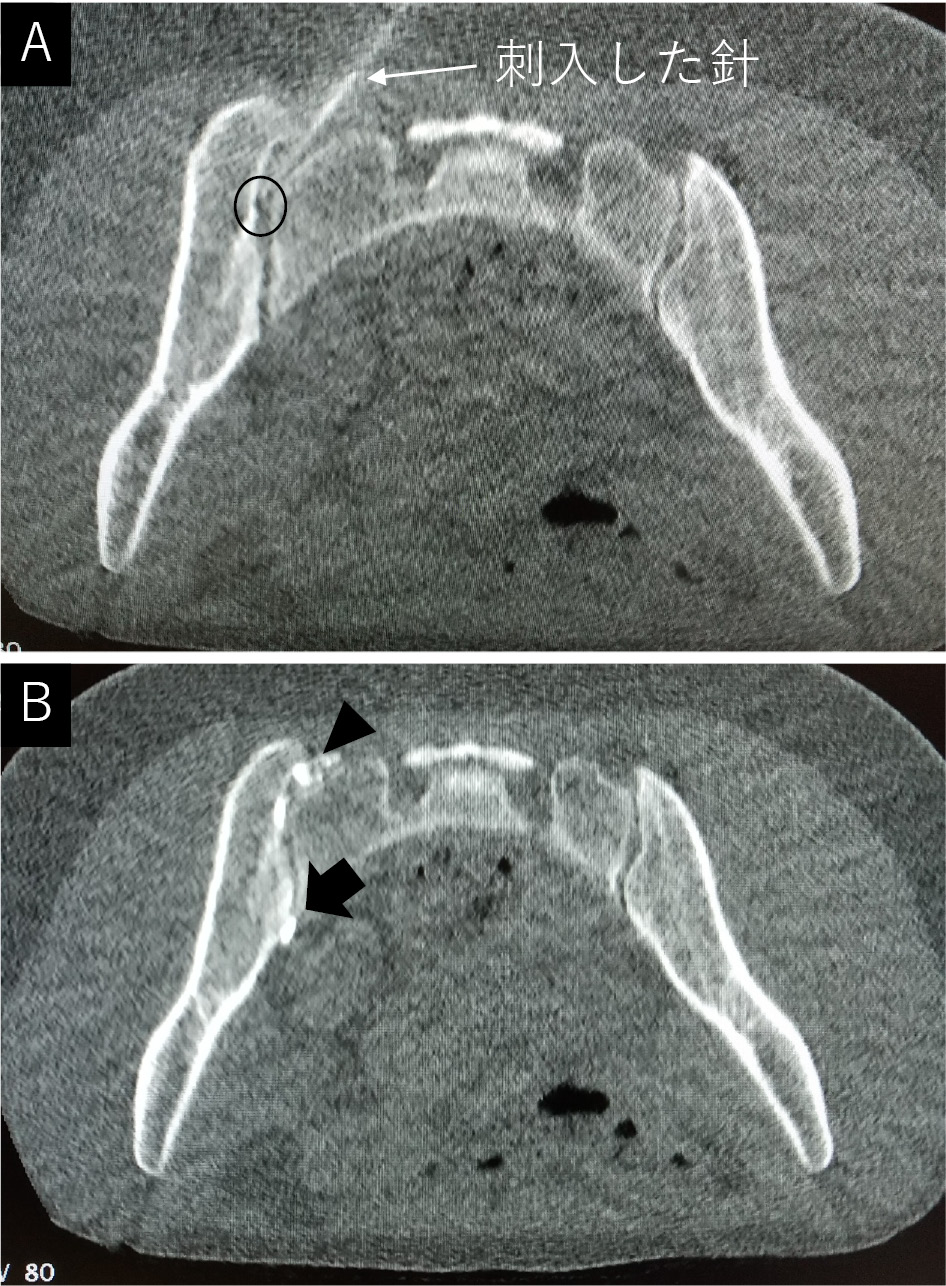
    Introduction: Periarticular sacroiliac joint (SIJ) injection should be first performed to diagnose SIJ pain. However, SIJ intra-articular injections are occasionally required in patients with severe and chronic SIJ pain. Radiation exposure of the operator's fingers is a major problem while performing intra-articular SIJ injections under fluoroscopy guidance. We attempted to perform this type of injection under CT guidance based on previous reports and report on the success rate and issues surrounding the injection techniques.

    Methods: Fifty hospitalized patients (13 men, 37 women, mean 44±18 years) with 59 joints underwent CT-guided intra-articular block from June 2021 to October 2022. CT imaging was performed under minimum necessary conditions (80 kV, 10 mA, 1.0 s). The patient was placed in a prone position on a table, and the CT gantry was tilted 15° toward the head. A positioning laser was projected onto the patient's buttock to mark the entry point after setting the needle entry path on the CT image and measuring the depth and distance from the center. An angle meter was used to indicate the needle entry angle, and a 23 G needle (70 mm) was inserted along this angle. Once the operator felt that the needle tip had reached the cartilaginous surface of the iliac side, the needle position was confirmed by CT. The imaging was repeated until the optimal needle position was achieved after adjusting the direction. After confirming that the intra-articular was contrasted, a mixture of 2 ml of 2% lidocaine and 1.9 mg dexamethasone sodium phosphate was injected. The success rate of contrast of the joint, number of CT scans per procedure, time taken for a series of procedures (from needle insertion to the end of an intra-articular injection), and total radiation exposure dose (DLP) were investigated.

    Results: Intra-articular contrast was identified in 34/59 joints (57.6%). The average number of CT scans was 4.9±1.0, total procedure time was 6 min 49 s (409±145 s), and total radiation exposure dose (DLP) was 9.06±2.91 [mGy・cm].

    Conclusions: The success rate was low because of the inability to accurately reproduce the angles of measurement on the images at the time of needle insertion. The needle tip position could not be adjusted as frequently as in fluoroscopy. The advantage of this method is that the total exposure to the patient is low, and no exposure to the operator's fingers was made.

    Download PDF (1645K)
  • Yoshihito Sakai, Norimitsu Wakao, Hiroki Matsui, Naoaki Osada, Tsuyosh ...
    2023 Volume 14 Issue 6 Pages 884-890
    Published: June 20, 2023
    Released on J-STAGE: June 20, 2023
    JOURNAL FREE ACCESS

    Introduction: There is a lack of medical evidence regarding the prognosis and course of chronic low back pain in elderly individuals. In this study, longitudinal data on chronic low back pain from a project aimed at elucidating the pathology of chronic pain in elderly people and developing treatment methods were used, and factors that contribute to intractability in patients with nonspecific chronic pain were analyzed.

    Methods: A one-year longitudinal evaluation was performed involving 341 patients aged 65 years or older with chronic low back pain that persisted for 3 months or longer (mean age 78.5 years; 133 males, 208 females). After 1 year follow-up including the therapeutic exercise and/or pharmacotherapy, those who improved to 3 cm or less on the visual analogue scale (VAS) were classified as the improvement group, and multivariate analysis was performed to evaluate the factors related to improvement of low back pain.

    Results: There were 141 patients (41.3%) in the improvement group with VAS < 3 cm within 1 year. The non-improvement group had a significantly longer disease duration, a higher pretreatment VAS, and a significantly lower limb skeletal muscle mass, and the erythrocyte volume distribution width (RDW) was significantly higher. Logistic regression analysis with age and sex added as explanatory variables to the factors with p<0.1 in the comparison between the two groups showed that VAS, RDW, and L4/5 high trunk muscle cross-sectional area were significant factors.

    Conclusions: In chronic low back pain, maintaining trunk muscles can be expected to improve low back pain. On the other hand, the fact that the RDW, which is reported to reflect aging, was high in the non-improved group suggests an irreversible aspect of chronic low back pain in elderly people.

    Download PDF (1133K)
  • Takashi Hirai, Kyohei Sakaki, Tsuyoshi Kato, Shoji Tomizawa, Masato Yu ...
    2023 Volume 14 Issue 6 Pages 891-896
    Published: June 20, 2023
    Released on J-STAGE: June 20, 2023
    JOURNAL FREE ACCESS

    Chemonucleolysis with condoliase has been anticipated as a novel therapy for lumbar intervertebral disc herniation. A total of 124 cases treated with intradiscal injection with condoliase and followed for at least 6 months after the injection in 5 institutions were reviewed. Ninety-seven patients had sufficient relief of the lower extremity pain caused by radiculopathy, although 6 cased underwent herniotomy even after the injection therapy within 6 months. Approximately 80% of patients in whom limb pain was reduced to 50% compared to that before the injection obtained pain relief within 3 months after intradiscal injection.

    Download PDF (1280K)
  • Tetsuto Yamaura, Keishi Maruo, Fumiaki Okada, Fumihiro Arizumi, Ryo Iw ...
    2023 Volume 14 Issue 6 Pages 897-902
    Published: June 20, 2023
    Released on J-STAGE: June 20, 2023
    JOURNAL FREE ACCESS

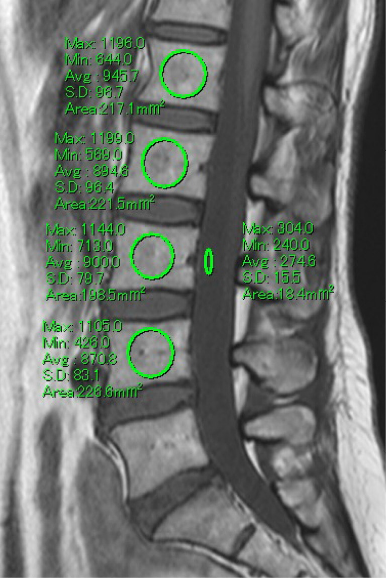
    Introduction: The vertebral bone quality score (VBQ) was recently reported as a tool for bone quality evaluation based on magnetic resonance imaging (MRI). The purpose of this study was to investigate the correlation between the VBQ score and dual-energy X-ray absorptiometry (DEXA), as well as the association of osteoporotic vertebral fractures (OVFs) with the VBQ score.

    Methods: Of 257 patients who underwent DEXA at our hospital between January 2021 and December 2022, 99 patients who underwent lumbar MRI within 3 months were enrolled. The VBQ score was based on the lumbar MRI T1-weighted sagittal midline slice and calculated by dividing the median signal intensity of the L1-4 vertebral bodies by the signal intensity of cerebrospinal fluid (CSF) at the level of L3. The patients were divided into two groups based on MRI diagnosis: the OVF group including acute OVF and prevalent OVF and the non-OVF group. Multiple logistic regression analysis was performed to identify associated factors.

    Results: The mean VBQ score was 3.7±0.6. The VBQ score was negatively correlated with the lowest T-score, lumbar bone mineral density (BMD), and proximal femur BMD. The OVF group was significantly older and had a significantly higher VBQ score and lower BMD and T-score than the non-OVF group. Multiple logistic regression analysis showed that the VBQ score was an independent factor associated with OVF.

    Conclusion: This study suggested that the VBQ score may be a useful predictor of OVF.

    Download PDF (1108K)
  • Yusuke Oshita, Haruka Emori, Ichiro Okano, Kazuyuki Segami, Yoshifumi ...
    2023 Volume 14 Issue 6 Pages 903-908
    Published: June 20, 2023
    Released on J-STAGE: June 20, 2023
    JOURNAL FREE ACCESS

    Introduction: A high controlling nutritional status (CONUT) score represents a poor nutritional status and is associated with infection, but its association with pyogenic spondylitis (PS) has not been well examined. The aim was to investigate nutritional status using the modified CONUT score in patients with PS.

    Methods: Using the data from one university hospital between January 2020 and May 2022, 26 patients (15 males, 11 females) were included. Their average age was 71 (23-89) years. Baseline nutritional status was calculated using the modified CONUT tool (serum albumin, lymphocytes, hemoglobin). Baseline MRI was used to evaluate the spread of infections.

    Results: No patient had a normal nutritional status; 6, 9, and 11 patients had light, moderate, and severe malnutrition, respectively, with no significant difference in age among the three groups (average age 65, 71, and 73 years, respectively). In the light malnutrition group, 4 patients had vertebral discitis (VD), and 2 had VD and spinal epidural abscess (SEA). In the moderate malnutrition group, 5 patients had VD, 1 had VD and SEA, 2 had VD and iliopsoas abscess (IA), and 1 had VD, SEA, and IA. In the severe malnutrition group, 5 patients had VD, 1 had VD and SEA, 2 had VD and IA, and 3 had VD, SEA, and IA. Participants with a high modified CONUT score had more infection sites. The length of hospital stay was 45, 34, and 62 days, and the discharge to home rate was 66.7%, 55.6%, and 36.4% in the light, moderate, and severe malnutrition groups, respectively.

    Conclusions: Poor nutritional status was found to be associated with more abscesses and a low discharge rate in patients with PS. Nutritional assessment with the modified CONUT score suggests that the score may predict clinical outcomes.

    Download PDF (1468K)
  • Satoru Tanaka, Tetsuhiro Iguchi, Keisuke Kinoshita, Takashi Sadamitsu, ...
    2023 Volume 14 Issue 6 Pages 909-914
    Published: June 20, 2023
    Released on J-STAGE: June 20, 2023
    JOURNAL FREE ACCESS

    The relationship between postpartum back pain and breastfeeding was investigated from the perspective of low bone mineral density. A total of 297 women who delivered at our hospital were included in this study, and a questionnaire created by our hospital was sent to women 4 months after delivery; the 215 patients who responded were included. The survey items were age, pre-pregnancy BMI, number of births, ultrasonic bone density, presence or absence and severity of back pain, breastfeeding rate and breastfeeding posture. Back pain was evaluated by VAS in each month after delivery. The severity was calculated based on the average value for each month; women with a score ≥4 were defined as having back pain. Bone mineral density was measured by ultrasound after delivery, and breastfeeding rates were categorized as in 100%, 75%, 50%, 25%, and 0%. It was found that 134 women (62.3%) had low back pain after delivery. In relation to breastfeeding, the 100% breastfeeding group had higher frequency and severity of back pain. In relation to bone mineral density, there was no difference in the rate and severity of back pain between the two groups, low bone mineral density and high mineral density, in the 100% breastfeeding women. The rate of back pain was significantly higher in those with postural abnormalities during lactation (p< 0.05). In recent years, some reports have implicated low bone density as the cause of postpartum back pain. In the present study, postural abnormalities were more involved than breastfeeding and low bone mineral density.

    Download PDF (912K)
  • Tomohiro Nakatani, Masakazu Minetama, Masatoshi Teraguchi, Yoshio Enyo ...
    2023 Volume 14 Issue 6 Pages 915-922
    Published: June 20, 2023
    Released on J-STAGE: June 20, 2023
    JOURNAL FREE ACCESS

    Introduction: Preventing subsequent vertebral fractures (SVFs) is a common challenge in surgical and conservative treatment for osteoporotic vertebral fractures.

    This study examined whether continuation of multiple types of exercise after balloon kyphoplasty (BKP) can prevent SVF and progression of kyphosis.

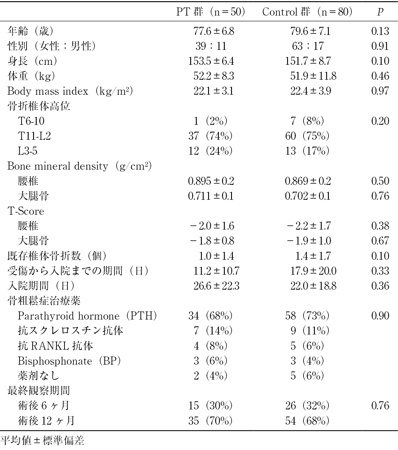
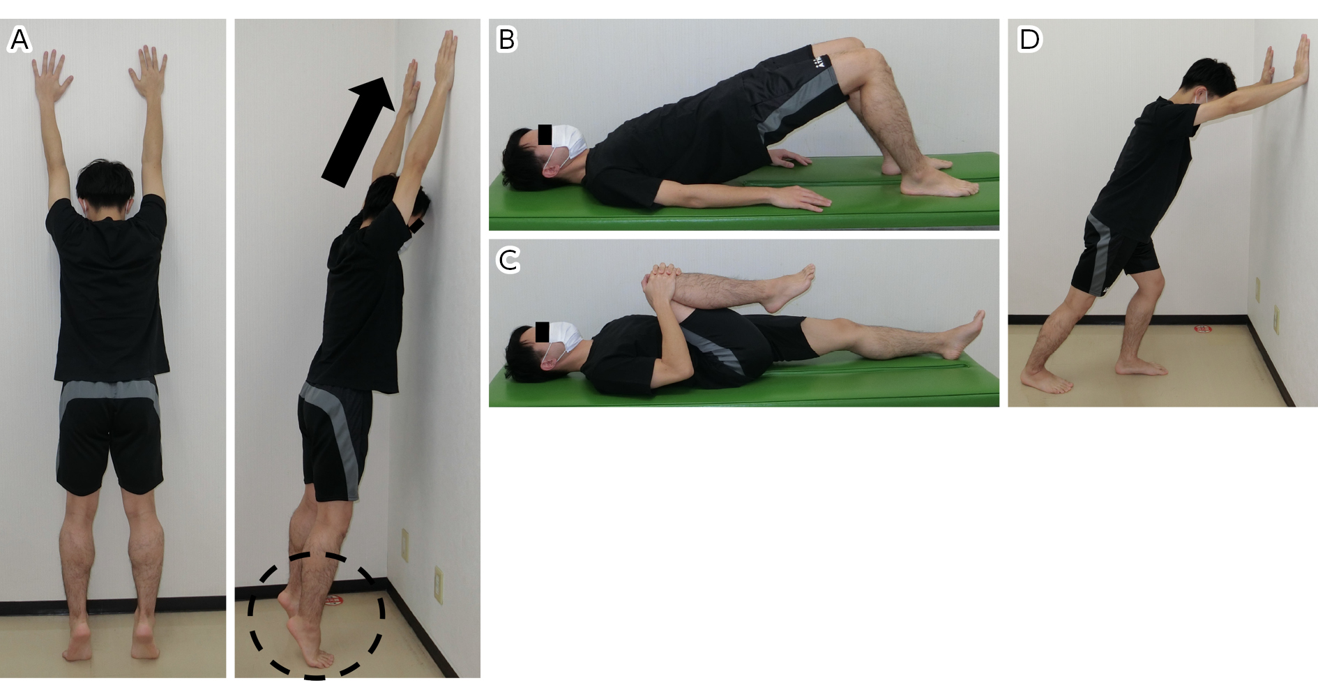
    Methods: Included were 130 patients who received a first BKP and had a 6- or 12-month follow-up assessment. All patients received postoperative treatment, including pharmacotherapy for osteoporosis, use of a thoracolumbar corset for ≥3 months, and had postoperative inpatient rehabilitation. Outpatient physical therapy consisted of strengthening, aerobic, and balance exercises, and was offered once or twice a week for 3 months to patients willing to continue rehabilitation (PT or control group). The primary outcome was the incidence of SVFs. Secondary outcomes were the incidence of falls, radiographic spinopelvic alignment, physical function (muscle strength and gait performance), and patient-reported outcomes on a numerical rating scale, Oswestry disability index, MOS 36-item short-form health survey, and EuroQol 5 dimension 5 level at baseline and 3, 6, and 12 months after BKP.

    Results: Fifty and 80 patients were included in the PT and control groups, respectively. At baseline, only thoracic kyphosis differed between the groups (PT: 33.5° vs. control: 42.1°). The incidence of SVFs was lower in the PT group than in the control group at 3 months (2% vs. 14%) and at the final follow-up (4% vs. 19%). Trunk extensor muscle strength (4.1 kg vs. 0.4 kg) and the EuroQol 5 dimension 5 level at 3 months (0.38 vs. 0.23) and the Oswestry disability index at the final follow-up (−34.1 vs. −22.5) improved significantly from the baseline in the PT group compared with the control group.

    Conclusions: Continued exercise, including back muscle-strengthening, aerobic, and balance exercises, seems to be effective for preventing SVF after BKP. The best means of keeping patients exercising after BKP is a challenge for future study.

    Download PDF (1140K)
  • Eriko Mochizuki, Hiromi Ataka, Natsumi Shida, Izumi Nobe, Takaaki Tann ...
    2023 Volume 14 Issue 6 Pages 923-930
    Published: June 20, 2023
    Released on J-STAGE: June 20, 2023
    JOURNAL FREE ACCESS

    Introduction: Age-related degenerative thoracolumbar kyphosis negatively affects health including chronic low back pain, physical mobility, and quality of life (QOL). Exercise interventions are potentially effective treatments for affected patients. The aims of the present study were to determine the effects of physical therapy interventions for 3 months on flexed posture, walking ability and health-related QOL in patients with degenerative thoracolumbar kyphosis and chronic low back pain.

    Methods: The subjects were 111 patients with degenerative thoracolumbar kyphosis and chronic low back pain. All patients received physical therapy that targeted spinal mobility and postural alignment provided by physiotherapists once per week for 3 months. The exercise program consisted of standardized posture training, individual muscle strengthening exercises, and stretch-altered flexibility according to the patient's physical ability. The patients were divided into two groups: patients with a flexible kyphosis [group F] (n = 72) and those with a rigid kyphosis [group R] (n = 39). Outcome measures included gait speed, patient-reported QOL scores (Japanese Orthopaedic Association Back Pain Evaluation Questionnaire [JOABPEQ]), back pain (visual analogue scale [VAS]), and spinal sagittal alignment (the flexicurve-measured kyphosis index or radiographic sagittal parameters). All measurements were evaluated before and 3 months after the intervention.

    Results: Gait speed showed significant increases (p<0.01) in both groups. Preoperative JOABPEQ category scores for low back pain were significantly lower in group R than in group F. However, both groups achieved good functional outcomes, with similar scores at 3- month follow-up in the category of low back pain. There were no significant differences between the groups in the therapeutic effectiveness for the four categories, with the exception of lumbar function. Forty percent of the patients in group F and 50% in group R showed an improvement of VAS greater than 20 mm. Spinal sagittal alignment showed no significant improvement after the intervention in both groups.

    Conclusion: Although the intervention did not improve clinical measures of kyphosis, gait speed was significantly improved in both groups, and back pain was reduced in 40-50% of the patients after 3-month exercise interventions. Thus, regardless of spinal mobility, a kyphosis-specific exercise program could have beneficial effects on physical function in patients with thoracolumbar kyphosis and chronic low back pain.

    Download PDF (1456K)
  • Hidehisa Torikai, Keigo Enomoto, Masatosi Inoue
    2023 Volume 14 Issue 6 Pages 931-937
    Published: June 20, 2023
    Released on J-STAGE: June 20, 2023
    JOURNAL FREE ACCESS

    Introduction: The effects of antineuropathic pain drugs on lumbar spinal stenosis (LSS) in the midterm are unknown. Thus, the effects of antineuropathic pain drugs on LSS were retrospectively investigated.

    Methods: Patients who could be followed for more than 6 months in the outpatient clinic and 533 matched patients were investigated. The patients were divided into two groups. One group (Group A, n=264) was treated before pregabalin, mirogabalin or opioids were started. The other group (Group P, n=269) was treated after they were started. Outcomes were classified into 5 categories: good, no change, deteriorated, surgical treatment, and unknown. The types and rates of each medicine or other treatments, and the outcomes of each group were obtained from the medical records.

    Results: Pregabalin was used in 45.1%, mirogabalin in 19.5%, opiods in 20.5% and duloxetine in 8.5% in group P. Good results were obtained in 54.2%, no change was seen in 28.8%, deterioration was seen in 2.8%, surgical treatment was conducted in 12.9%, and the outcome was unknown in 1.5% in group A. Good results were obtained in 46.1%, no change was seen in 23.2%, deterioration was seen in 3.3%, surgical treatment was conducted in 26.2%, and the outcome was unknown in 1.1% in group P. There was no significant difference in the rate of good cases between them.

    The rate of surgical treatment was higher in Group P than in Group A. However, the number of surgical treatments by one year was not different between them. Furthermore, group P was subdivided into two subgroups. One was the group treated by pregabalin or mirogabalin and the other was the group without them. There was no significant difference between them in the rate of good results and surgical treatment. The effect of antineuropathic pain drugs seemed better in mild or moderate cases. On the other hand, the rate of surgical treatment did not decrease after the start of antineuropathic pain drugs.

    Conclusion: The midterm effect of antineuropathic pain drugs in the treatment of lumbar spinal stenosis was investigated.

    Surgical treatment still needs to be considered for severe cases.

    Download PDF (1036K)
  • Koki Tohgei, Tetsuhiro Iguchi, Hiroo Souda, Keisuke Kinoshita, Takashi ...
    2023 Volume 14 Issue 6 Pages 938-944
    Published: June 20, 2023
    Released on J-STAGE: June 20, 2023
    JOURNAL FREE ACCESS

    Introduction: Spinal fracture of pregnancy and lactation-associated osteoporosis (PLO) is rare, and its incidence is unknown. Experiences and awareness of the spinal fractures with PLO were studied among orthopedic surgeons in Hyogo prefecture.

    Methods: A total of 436 members and colleges of the Hyogo Clinical Orthopaedic Association (HCOA) and the chief orthopedic surgeons of 38 hospitals performing delivery in Hyogo prefecture during 2021-22 were interviewed using a questionnaire about their experiences and awareness of spinal fractures with PLO by e-mail, mail, and face-to-face survey. The questionnaire was composed of the same items published elsewhere (Obori et al. JJOS, 2018). The final response rate to the questionnaire was 73% in HCOA members. Studies considering their age and working status (clinic, hospital, academic, and others) were also performed.

    Results: The average age of the orthopedic surgeons was 60.1 (37- 85) years. There were 177 clinic doctors with an average age of 60.1 years, 117 hospital doctors with an average age of 59.9 years, 11 academic doctors with an average age of 52.5 years, and 12 doctors working in other fields with an average age of 69.9 years. A total of 32 surgeons (10.1%) saw 49 spinal PLO fractures. Previous experience and the number of cases in different working conditions were 5 cases within a year (clinic: 3 cases, hospital: 2 cases), 11 cases in 2-5 years (clinic: 3, hospital: 8), 18 cases in 6-10 years (clinic: 8, hospital: 7, others: 3), 12 cases in 11-20 years (clinic: 9, hospital: 3), and 3 cases over 21 years (clinic: 1, others: 2). The percentage of the doctors who did not know about PLO disease were 57.6% in the clinic, 59.0% in the hospitals, 36.4% in the academic field, and 33.3% in the others; more than half of the orthopedic surgeons were unaware of the disease.

    Conclusions: More than 10% of the orthopedic surgeons had seen spinal fractures with PLO, and it was thought not to be rare. Because 5 cases were found within a year, its incidence is estimated to be 0.014%/year in Hyogo prefecture. This was a survey performed of a limited numbers of orthopedic surgeons; therefore, the expected incidence of PLO spinal fractures is thought to be higher.

    Download PDF (1218K)
  • Masaru Kanda, Takuya Kitamura, Isamu Konishi, Kei Watanabe, Naritoshi ...
    2023 Volume 14 Issue 6 Pages 945-952
    Published: June 20, 2023
    Released on J-STAGE: June 20, 2023
    JOURNAL FREE ACCESS

    Low back pain (LBP), a common symptom experienced by all age groups, has been reported to be the primary cause of disability worldwide. The aim of this study was to investigate changes in relative circulation over time in the lumbar multifidus in different trunk extension exercises between subjects with and without LBP.

    Twelve subjects (mean age, 21.2±0.4 years) with LBP (LBP group) for the past three months and 18 healthy subjects (mean age, 21.1±0.3 years) without LBP (non-LBP group) were recruited. They received a full explanation of the study, and all agreed to participate. Near-infrared spectroscopy (NIRS) was used to non-invasively measure oxygenated hemoglobin (Oxy-Hb), deoxygenated hemoglobin (Deoxy-Hb), and total hemoglobin (Total-Hb) of the lumbar multifidus at the L5-S1 segment. All measurements were obtained in the neutral position, at trunk extension of 20 degrees, and during trunk extending exercise while standing. The subjects were asked to move into either the trunk extended position or extending exercise movement from the starting (neutral) position in 3 seconds, timed by a metronome, and to maintain the position for 10 seconds. The angles of the extended positions were measured with a goniometer, and self-made devices were used for extending exercise movement to properly maintain these positions. All participants received education and practice time to be able to perform the proper body movements and positions prior to assessment. The measurements were obtained at −3 seconds (neutral position), 0, and 10 seconds in each extended and extending exercise position while standing and compared between the LBP and non-LBP groups.

    In extension, there were no significant interactions in both the LBP and non-LBP groups. In both groups, significant increases were noted in Deoxy-Hb and Total-Hb.

    In extending exercise, there were no significant interactions in both the LBP and non-LBP groups in Oxy-Hb and Total-Hb. In the non-LBP group, significant increases were noted in Deoxy-Hb.

    The results of this study showed that the intramuscular circulation of lumbar multifidus increased with trunk extension, but the circulation increased more with extending exercise in both the LBP and non-LBP groups.

    Download PDF (1532K)
  • Hiroshi Kobayashi, Takuya Nikaido, Kazuyuki Watanabe, Kinshi Kato, Yos ...
    2023 Volume 14 Issue 6 Pages 953-958
    Published: June 20, 2023
    Released on J-STAGE: June 20, 2023
    JOURNAL FREE ACCESS

    Objective: This study aimed to investigate and characterize falls of inpatients admitted to our department, focusing on spinal disorders and nocturnal falls.

    Methods: Study 1: Patients who had fallen while admitted to the department between April 2019 and November 2021 were included. The number of falls, age, sex, time of occurrence, disease site, presence or absence of surgery, sleeping medication, and BMI were investigated. Study 2: A nocturnal fall group and a non-fall group of patients who were hospitalized at the same time and matched for sex and age were established, and the site of disease, medications taken, and BMI were compared between the groups.

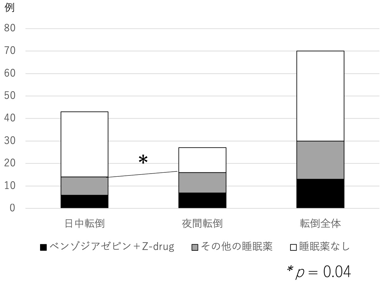
    Results: Study 1: Of the 2,372 cases, falls were observed in 71 cases (3.0%, mean age 68.6 years; 32 males and 39 females). Nocturnal falls occurred in 39.4%, 49.3% had a spinal disease, and 69.0% had postoperative falls. Study 2: Spinal disease was present in 53.6% of nocturnal fall cases. The percentage of patients taking sleeping pills was significantly higher in the nocturnal fall group (57.1%) than in the non-fall group (14.3%).

    Discussion: Falls occurred in 3.0% of hospitalized patients with spinal disease, with about half of fall cases having spinal disease, and about 40% of nocturnal. Nocturnal falls were more common in spinal disease patients, and the percentage of patients taking sleeping pills was higher. A fall prevention approach based on the results of this study may be practical.

    Download PDF (1003K)
  • Atsushi Terakado, Toshifumi Nakamura, Ayano Mitsuhashi, Koshiro Okachi ...
    2023 Volume 14 Issue 6 Pages 959-965
    Published: June 20, 2023
    Released on J-STAGE: June 20, 2023
    JOURNAL FREE ACCESS

    The rate of bone fusion in lumbar spondylolysis during the growth period changes depending on the disease stage (morphology) of the separation, and the corset used. In this study, the subjects were limited to junior high school and high school students with unilateral lumbar spondylolysis, all of whom used a semi-rigid corset, and the rate of bone fusion according to disease stage (morphology) classification was investigated.

    A total of 127 subjects/134 sites, with unilateral lumbar spondylolysis and who underwent treatment with a semi-rigid corset were included from among the junior and senior high school students who visited our hospital between June 2015 and November 2019. Bone fusion was determined by CT scan after the patients' back pain on extension disappeared.

    The fusion rate was 100% (type Ia−) in the very early stage, 95.2% (type Ia) and 87.5% (type Ib) in the early stage, 40% (type II) in the advanced stage, and 0% (type III) in the terminal stage.

    It is possible to obtain sufficient bone fusion with a semi-rigid corset for lumbar spondylolysis in the growth period in junior high school and high school students, unilateral cases, and very early-to-early stages.

    Download PDF (1353K)
  • Shinji Kotaka, Yasushi Fujiwara, Yoriko Toten, Ryo Ota, Kazutoshi Iwas ...
    2023 Volume 14 Issue 6 Pages 966-972
    Published: June 20, 2023
    Released on J-STAGE: June 20, 2023
    JOURNAL FREE ACCESS

    Introduction: In this study, changes in low back pain after posterior decompression surgery without spinal fusion for lumbar spinal canal stenosis in patients who presented primarily with low back pain were investigated.

    Methods: The study included 51 patients who presented primarily for evaluation of low back pain and underwent microscopic posterior decompression surgery without spinal fusion for lumbar spinal canal stenosis. Preoperative and postoperative low back pain, leg pain, lower extremity numbness, Japanese Orthopaedic Association back pain evaluation questionnaire (JOABPEQ) scores, lumbar kyphosis angle and range of motion, intervertebral joint edema, vertebral end plate Modic changes, spinous process kissing, and vertebral body slippage were recorded. Low back pain visual analog scale (VAS) scores were compared between patients who improved (improvement group) and those without improvement (non-improvement group).

    Results: Low back pain improved in 72.5% of the patients. The preoperative low back pain VAS and postoperative JOABPEQ severity scores associated with pain-induced disability, gait dysfunction, social disability, psychological disability, and preoperative lumbar spine range of motion were significantly greater and Modic changes were significantly fewer in the improvement group.

    Conclusions: The present suggest that patients with high levels of preoperative low back pain and a low degree of lumbar vertebral body endplate injury show improvement in low back pain after the aforementioned surgery. In contrast, patients with vertebral endplate lesions may experience residual low back pain, and additional treatment such as spinal fusion should be considered in such cases.

    Download PDF (1164K)
  • Yusuke Masumoto, Masaki Tomori, Yoshiyasu Arai, Kenichiro Sakai, Ichir ...
    2023 Volume 14 Issue 6 Pages 973-977
    Published: June 20, 2023
    Released on J-STAGE: June 20, 2023
    JOURNAL FREE ACCESS

    Introduction: Lumbar disc herniation (LDH) is a common disease in daily clinical practice. However, there are conflicting reports on the effectiveness of early surgery for drop foot due to LDH, a typical symptom of radiculopathy, and no consensus has been reached. The purpose of this study was to investigate the effectiveness of early surgery within 7 days for drop foot due to LDH.

    Methods: Patients who underwent surgical decompression for drop foot (tibialis anterior [TA] Medical Research Council grade ≤2) due to LDH from September 1, 2017 to November 31, 2021 were categorized into the early surgery group (within 7 days, 15 patients) and the late surgery group (after 8 days, 16 patients), and the postoperative TA MRC grades were compared between the two groups.

    Results: Six months after surgery, 11 patients (73.3%) in the early surgery group and 10 patients (62.5%) in the late surgery group had TA MRC grade ≥3 (P=0.704). One year after surgery, 12 patients (80.0%) and 12 patients (75.0%) in each group had TA MRC grade ≥3 (P>0.999). The mean TA MRC grade improvements 6 months after surgery were 2.07 in the early surgery group and 1.56 in the late surgery group (P=0.317). Improvements 1 year after surgery were 2.47 and 1.88 in the two groups, respectively (P=0.107). There was no significant difference between the two groups in either analysis.

    Conclusions: This study did not demonstrate the effectiveness of early surgery within 7 days for drop foot due to LDH.

    Download PDF (781K)
Case Report
feedback
Top