Abstract
Introduction: The aim of this study was to examine the association between vertebral collapse and magnetic resonance imaging (MRI) signal changes and risk factors in osteoporotic vertebral fractures (OVF).
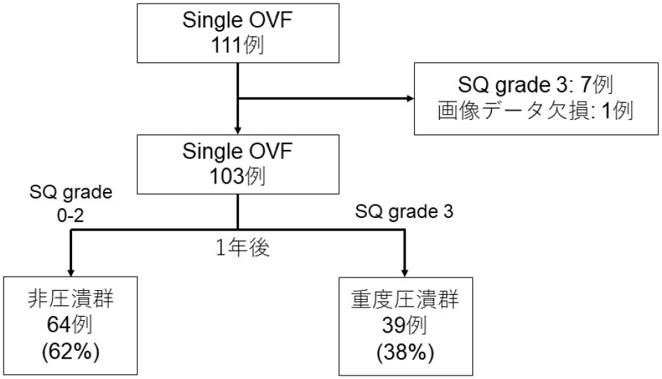
Methods: A total of 103 patients met the inclusion criteria (age ≥ 60 years, single OVF, semi-quantitative [SQ] grade of 0-2 at baseline, and minimum follow-up ≥1 year) with a mean age of 79.3±7.1 years. The severe collapse group was defined as SQ grade 3 at 1-year follow-up. Patient characteristics and clinical outcomes (JOABPEQ, ODI and VAS) were compared between the severe collapse and noncollapse groups. Additionally, we evaluated bone marrow edema (BME) using MRI-STIR images at baseline, 3 months, and 1 year, categorizing it into four levels (none: 0%, minor: 1%-24%, moderate: 25%-74%, severe: 75%-100%) and comparing the two groups.
Results: Severe collapse was observed in 39 cases (38%). There were no significant differences in patient characteristics between the two groups. Clinical outcomes showed significant improvement in VAS scores at 3 months and 1 year in both groups. No significant differences were found between the two groups in terms of JOABPEQ and ODI at baseline, 3 months and 1 year. The severe collapse group had a higher prevalence of confined high signal and diffuse low signal on T2-weighted images at baseline. Regarding BME, there was a significant difference at 3 months, with the severe collapse group having a 71% rate of severe BME compared with 34% in the noncollapse group. This difference was also significant at 1 year, with the severe collapse group at 5% for "none" compared with 50% in the noncollapse group.
Conclusions: Severe vertebral collapse was observed in 38% of cases, but clinical outcomes significantly improved in both groups. In cases of noncollapse, half of them showed resolution of bone marrow edema on MRI, suggesting bone healing.