Journal of Spine Research
Online ISSN : 2435-1563
Print ISSN : 1884-7137
Volume 16, Issue 2
Displaying 1-6 of 6 articles from this issue
Editorial
Original Article
  • Kazuma Nagao, Keishi Maruo, Tomoyuki Kusukawa, Tetsuto Yamaura, Masaru ...
    2025 Volume 16 Issue 2 Pages 50-57
    Published: February 20, 2025
    Released on J-STAGE: February 20, 2025
    JOURNAL FREE ACCESS

    Introduction: The aim of this study was to examine the association between vertebral collapse and magnetic resonance imaging (MRI) signal changes and risk factors in osteoporotic vertebral fractures (OVF).

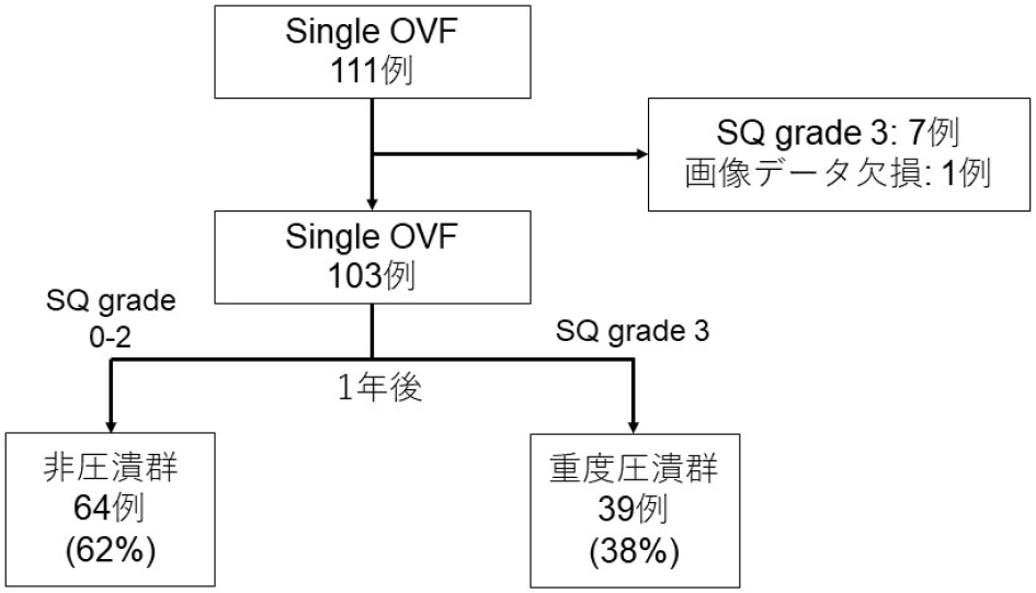
    Methods: A total of 103 patients met the inclusion criteria (age ≥ 60 years, single OVF, semi-quantitative [SQ] grade of 0-2 at baseline, and minimum follow-up ≥1 year) with a mean age of 79.3±7.1 years. The severe collapse group was defined as SQ grade 3 at 1-year follow-up. Patient characteristics and clinical outcomes (JOABPEQ, ODI and VAS) were compared between the severe collapse and noncollapse groups. Additionally, we evaluated bone marrow edema (BME) using MRI-STIR images at baseline, 3 months, and 1 year, categorizing it into four levels (none: 0%, minor: 1%-24%, moderate: 25%-74%, severe: 75%-100%) and comparing the two groups.

    Results: Severe collapse was observed in 39 cases (38%). There were no significant differences in patient characteristics between the two groups. Clinical outcomes showed significant improvement in VAS scores at 3 months and 1 year in both groups. No significant differences were found between the two groups in terms of JOABPEQ and ODI at baseline, 3 months and 1 year. The severe collapse group had a higher prevalence of confined high signal and diffuse low signal on T2-weighted images at baseline. Regarding BME, there was a significant difference at 3 months, with the severe collapse group having a 71% rate of severe BME compared with 34% in the noncollapse group. This difference was also significant at 1 year, with the severe collapse group at 5% for "none" compared with 50% in the noncollapse group.

    Conclusions: Severe vertebral collapse was observed in 38% of cases, but clinical outcomes significantly improved in both groups. In cases of noncollapse, half of them showed resolution of bone marrow edema on MRI, suggesting bone healing.

    Download PDF (1420K)
  • Tetsuto Yamaura, Keishi Maruo, Tomoyuki Kusukawa, Masaru Hatano, Masak ...
    2025 Volume 16 Issue 2 Pages 58-64
    Published: February 20, 2025
    Released on J-STAGE: February 20, 2025
    JOURNAL FREE ACCESS

    Introduction: This study aimed to evaluate the natural course and risk factors associated with severe residual disability in patients with osteoporotic vertebral fractures (OVFs) undergoing conservative treatment using the Oswestry disability index (ODI).

    Methods: This prospective multicenter study was conducted at eight hospitals from July 2020 to April 2022. We evaluated 125 patients (mean age 79 years) with acute OVF who had been treated conservatively and were followed up for 1 year. Clinical outcomes were evaluated using the ODI and JOA back pain evaluation questionnaire (JOABPEQ) at baseline and 3 and 12 months. The ODI > 40% at 12 months was defined as residual disability. Clinical course, demographic factors, conservative treatment, and radiological factors in both groups were compared in the univariate analysis. Multivariate analysis was performed using variables with a p-value < 0.1 were used as covariate to detect risk factors for residual disability.

    Results: At 12 months, 34 patients (27%) had residual disability. All clinical outcomes in both groups improved up to 3 months; however, the clinical outcomes thereafter in the no residual disability group improved and those in the residual disability group worsened. The residual disability group was significantly older and had worse JOABPEQ walking disability, social life function, mental health, and ODI at baseline. There were no significant differences in demographic data and radiographic factors. Multivariate logistic regression analysis identified age and baseline ODI as independent risk factors.

    Conclusions: Residual disability was observed in 27% of patients with acute OVFs at 12 months. Clinical outcomes improved at 3 months but thereafter worsened. Pseudoarthrosis, severe vertebral body collapse, and subsequent OVFs were not significantly associated with residual disability.

    Download PDF (1179K)
  • Ryosuke Nishi, Nodoka Manabe, Daisuke Tsunoda, Sho Isiwata, Kazuro Tsu ...
    2025 Volume 16 Issue 2 Pages 65-71
    Published: February 20, 2025
    Released on J-STAGE: February 20, 2025
    JOURNAL FREE ACCESS

    Introduction: The purpose of this study was to investigate the following: (a) basic patient information, (b) preoperative physical function, and (c) preoperative patient-based outcomes to identify factors that delay gait acquisition after lumbar fusion surgery.

    Methods: A total of 129 patients who had undergone lumbar fusion surgery within 3 years from April 2020 were included. The evaluation included (a) age, sex, BMI, duration of disease, number of fixed intervertebral spaces, and number of days since surgery in which the patient acquired a walking style at discharge (hereafter referred to as "days of gait acquisition" ). (b) SLR, NRS, time to stand on one leg, and 6-min walk test. (c) JOABPEQ, PSEQ, CSI, HADS, PCS, and TSK scores were investigated. For statistical analysis, the Mann-Whitney U test or chi-square test was used to compare two groups: those who acquired gait within 14 days after surgery (normal group) and those who acquired gait more than 14 days after surgery (delayed group), and the significance level was 5%.

    Results: The results showed significant differences in age, BMI, NRS, time to stand on one leg, 6-min walk test, JOABPEQ gait function, and PSEQ score (p < 0.05).

    Conclusions: We observed that not only basic attributes such as age, preoperative balance function, and walking ability, but also low preoperative self-efficacy influenced gait acquisition after lumbar fusion surgery.

    Download PDF (1034K)
  • Teruaki Miyake, Sanshiro Saito, Takaki Yoshimizu, Tetsutaro Mizuno, Us ...
    2025 Volume 16 Issue 2 Pages 72-77
    Published: February 20, 2025
    Released on J-STAGE: February 20, 2025
    JOURNAL FREE ACCESS

    Introduction: The minimally invasive cervical laminoplasty performed at our hospital is an evolution of Hirabayashi laminoplasty and can expand the spinal canal in the same manner as conventional methods. In this study, we examined the characteristics of minimally invasive laminoplasty.

    Methods: A total of 177 cervical laminoplasty cases (average age, 65.7 years; 44 men and 133 women) performed at our hospital from January 1, 2016 to December 31, 2020 were included to evaluate surgical outcomes and Japanese Orthopaedic Association Cervical Myelopathy Evaluation Questionnaire (JOACMEQ) scores preoperatively and at 1 and 3 years postoperatively.

    Results: The results for the included groups were a skin incision of 3.8±0.6 cm, operative time of 73.4±25.1 min, blood loss 49.8±87.1 g, and hospital stay of 11.8±5.8 days. JOACMEQ efficacy rates for each domain (1 and 3 years after surgery) compared with the preoperative values were cervical function (48% and 49%); upper-extremity function (51% and 47%); lower-extremity function (36% and 34%); urinary function (24% and 26%); and quality of life (26% and 29%). Posterior neck pain and upper-extremity numbness on the visual analog scale of the JOACMEQ questionnaire showed significant improvement in pain at 1 and 3 years postoperatively compared with preoperative pain.

    Conclusions: The results of the JOACMEQ for minimally invasive cervical laminoplasty were as good as those for conventional methods.

    Download PDF (1161K)
Secondary Publication
  • Toshiki Endo, Tomoo Inoue, Masaki Mizuno, Ryu Kurokawa, Kiyoshi Ito, S ...
    2025 Volume 16 Issue 2 Pages 78-86
    Published: February 20, 2025
    Released on J-STAGE: February 20, 2025
    JOURNAL FREE ACCESS

    Objective: We performed a retrospective observational study to demonstrate the surgical risks and long-term prognoses of intramedullary tumors in Japan, using a multicenter registry authorized by the Neurospinal Society of Japan.

    Methods: Data from 1,033 consecutive patients with intramedullary tumors, treated between 2009 and 2020, were collected from 58 centers. Patients with spinal lipomas or myxopapillary ependymomas were excluded from the study. Patient characteristics, clinical presentations, imaging characteristics, treatments, and outcomes were analyzed. The modified McCormick scale was used to classify the functional status. Survival was described using Kaplan-Meier curves, and multivariable logistic regression analyses were performed.

    Results: The mean age of the patients was 48.4 years. Data of 361 ependymomas, 196 hemangioblastomas, 168 astrocytic tumors, 160 cavernous malformations, and the remaining 126 cases including subependymomas, metastases, schwannomas, capillary hemangiomas, and intravascular B-cell lymphomas were analyzed. Twenty-two patients remained undiagnosed. The mean follow-up duration was 46.1±38.5 months. Gross total tumor removal was achieved for 672 tumors (65.1%). On the modified McCormick scale, 234 patients (22.7%) had worse postoperative grades at discharge. However, the patient's neurological status gradually improved. At 6 months postoperatively, 251 (27.5%), 500 (54.9%), and 160 patients (17.6%) had improved, unchanged, and worsened grades, respectively. Preoperative functional status, gross total tumor removal, and histopathological type were significantly associated with mortality and functional outcome.

    Conclusion: Our findings demonstrated better postoperative functional outcomes in patients with fewer preoperative neurological deficits. The degree of resection, postoperative treatment, and prognosis are closely related to the histology of intramedullary tumors.

    Download PDF (1394K)
feedback
Top