Abstract
Giant congenital melanocytic nevi of the scalp are rare congenital melanocytic lesions associated with cranial bone defects. Cranioplasty can be technically challenging, particularly in the absence of normal subcutaneous tissues and dissection planes. However, few surgical reports have focused on these technical difficulties.
A girl (age: 6 years and 5 months) with a congenital pigmented scalp lesion, slightly to the right of the midline, underwent postnatal cranial computed tomography scanning that revealed a large cranial bone defect extending from the frontal to parietal bones. Histopathological examination of a biopsy specimen revealed a blue nevus. Considering the patient's normal neurodevelopment and the non-progression of the bone defect during follow-up, elective cranioplasty was planned before school admission. Cranioplasty using a custom-made artificial bone was performed in collaboration with plastic surgeons. Intraoperative findings revealed the absence of normal subcutaneous tissue and the subgaleal dissection plane. The dermis directly adhered to the cranial bone and dura mater, and partially to arachnoid-like tissue. Sharp dissection under careful tension was undertaken with minimal dural injury to preserve scalp viability. Postoperatively, subcutaneous fluid collection and minor scalp necrosis occurred and were successfully managed with local wound care and continuous swimming-cap compression.
Congenital melanocytic scalp lesions may be associated with cranial bone defects and loss of normal tissue planes, with severe adhesions between the scalp and intracranial structures. Surgeons should consider these anatomical abnormalities when planning cranioplasty and customize surgical strategies to preserve scalp viability while minimizing intracranial complications.
Introduction
Giant congenital melanocytic nevi (GCMN) are rare congenital melanocytic lesions frequently discussed in dermatology and plastic surgery from the perspectives of cosmetic concerns and risk of malignant transformation. In most cases, the lesion is primarily cutaneous; however, it may extend into the deep dermis and subcutaneous tissue. In cases of GCMN occurring on the scalp, sporadic reports of associated cranial bone defects or thinning in the same region as the skin lesions have been reported.1,2) However, very few reports are available on cranioplasty requiring neurosurgical techniques in cases accompanied by loss of subcutaneous tissue and severe adhesions between the scalp and meningeal tissues. Detailed reports focusing on surgical techniques in cases in which surgical manipulation is difficult, owing to the disappearance of normal dissection planes, are limited.
Herein, we report a case of GCMN with an associated cranial bone defect for which cranioplasty was performed. Dissection was challenging owing to the loss of subcutaneous tissue and severe adhesion between the scalp and the bone, dura mater, and arachnoid. In this report, we discuss the surgical strategy and techniques, as well as the underlying pathophysiological background and perioperative considerations.
Technical Notes
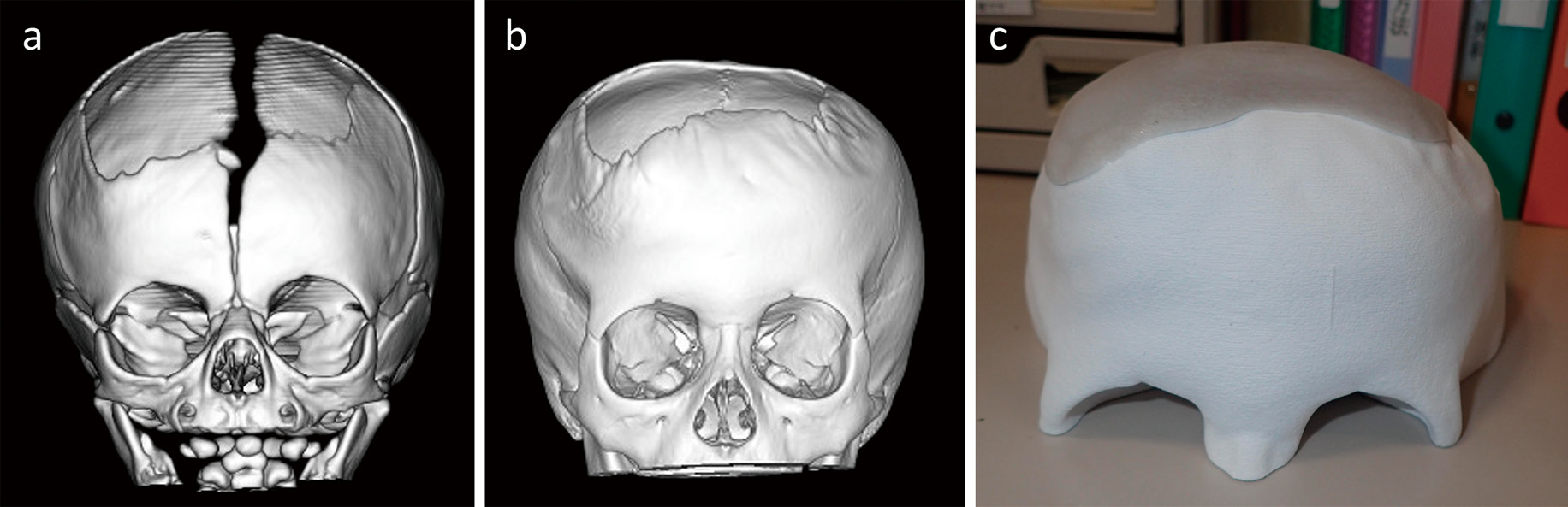
A girl aged 6 years and 5 months presented with a congenital blue nevus located slightly to the right of the midline on the scalp (Figure 1). Cranial computed tomography (CT) performed on day 2 of life revealed a cranial bone defect, and the patient was subsequently referred to our department (Figure 2a). The congenital skin lesion was extensive and was clinically managed as a scalp GCMN. However, histopathological examination of a skin biopsy at 11 months of age revealed a blue nevus. Periodic skull radiographs confirmed neither enlargement nor reduction of the cranial bone defect, and no neurological abnormalities or developmental delays were observed. On palpation of the lesion corresponding to the cranial bone defect, the findings were similar to those observed after decompressive craniectomy. The margin of the bone defect was palpable as a ridge. No specific characteristic findings were noted in the skin lesion itself, and palpation did not provide additional information for surgical planning. Therefore, we planned single-stage cranioplasty after allowing further growth of the patient. Psychomotor development was normal, and at 5 years and 10 months of age, the patient's score on the Tanaka-Binet Intelligence Scale V revealed an Intelligence Quotient of 100. Elective cranioplasty was performed at 6 years and 5 months, before school enrollment.

Preoperative cranial CT (Figure 2b) revealed a large defect extending from the frontal to the parietal bones. Under general anesthesia, surgery was performed in collaboration with plastic surgeons. Based on preoperative CT data, a custom-made cranial implant (SKULPIO®; Kyocera Medical Corporation, Kyoto, Japan) was fabricated to completely reconstruct the bone defect (Figure 2c).
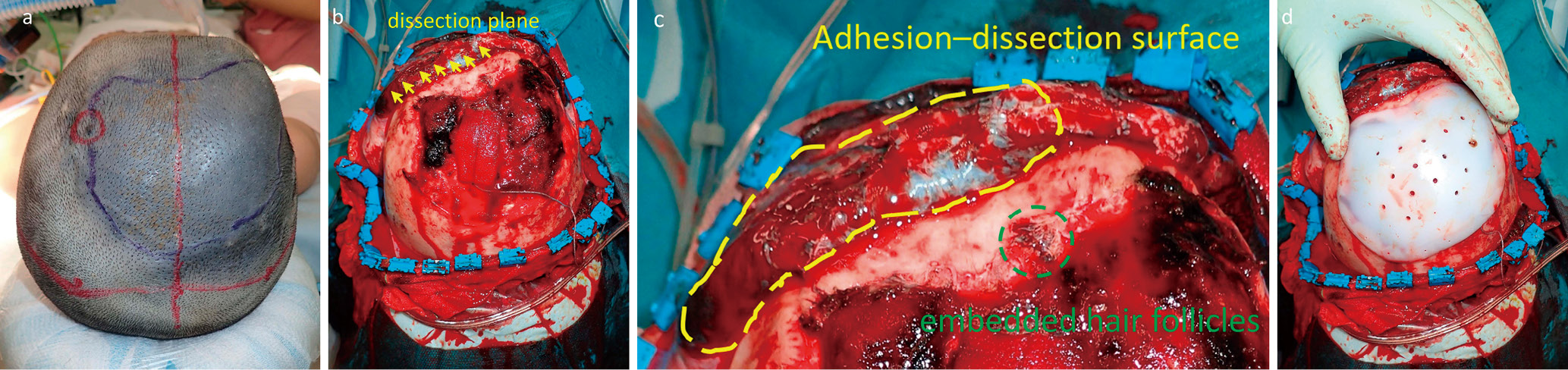
The head was fixed on a horseshoe headrest, and a coronal skin incision was made posterior to the nevus to avoid the lesion (Figure 3a). Although the incision was made down to the periosteum, separation of the periosteum integrated with the incomplete subcutaneous tissue from the dura carried a high risk of tissue loss; therefore, the periosteum was elevated en bloc with the scalp flap. The bone-defect area was sharply dissected using a scalpel while applying appropriate tension with skin hooks to avoid thinning of both the scalp flap and dura. Subcutaneous fat and galea were absent in the region of the nevus, and the dermis strongly adhered to the bone and dura mater (Figure 3b and c). Partial dural defects occurred, and dissection from arachnoid-like tissue proved challenging. The partial dural and arachnoid injuries were sealed using fibrin glue. Oxidized cellulose was used to ensure hemostasis for bleeding from the dissected surface. The implant was fixed with six 4-mm and four 5-mm screws (Figure 3d). The subcutaneous tissue was closed with 3-0 absorbable polyglycolic acid sutures, and the normal scalp was closed with staples. The nevus area was closed using interrupted 4-0 nylon sutures, including closure of a partial skin defect. The intraoperative blood loss was 597 mL, and the operative time was 3 hours 56 min. The patient's body weight was 20.8 kg; thus, the estimated blood loss corresponded to approximately 28.7 mL/kg. Although this volume was relatively high for a pediatric case, it was considered acceptable under controlled transfusion management, and hemodynamic stability was maintained throughout the procedure. The main source of bleeding was venous bleeding from the adhesion-dissection surface, and coagulation was minimized to preserve flap perfusion. Perioperatively, blood transfusions were administered, in collaboration with anesthesiologists and intensive care specialists, to maintain hemodynamic stability. The transfusion volume consisted of 240 mL red blood cells and 240 mL of fresh frozen plasma.
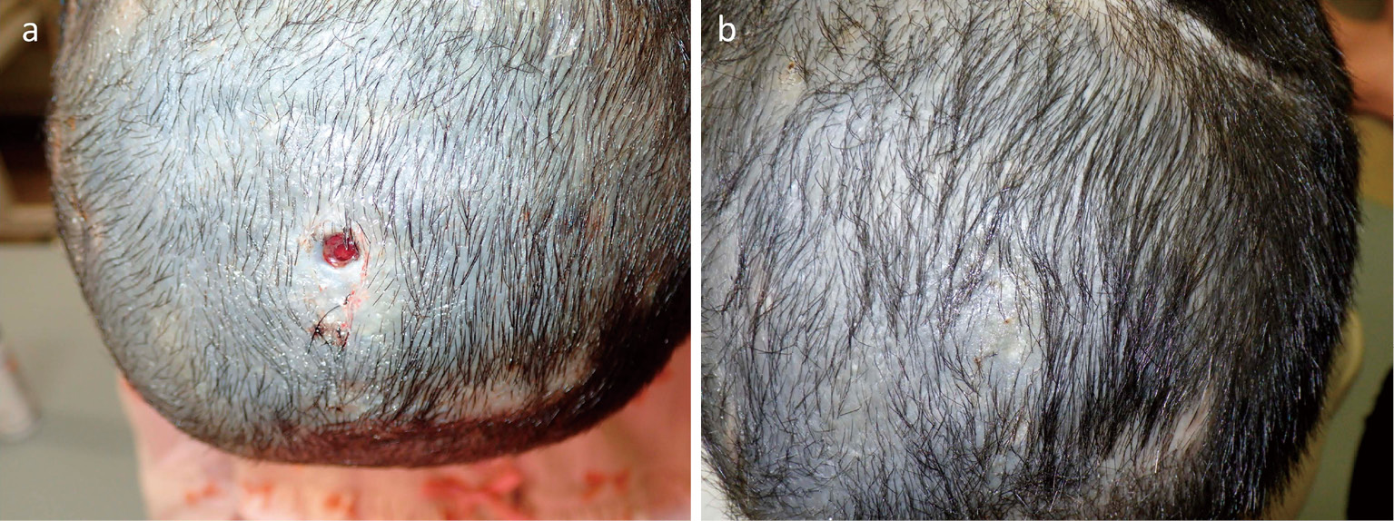
On postoperative day (POD) 6, a subcutaneous fluid collection was observed; therefore, compression bandaging of the head was performed. The color of the sutured skin defect area was good. On POD 9, subcutaneous fluid leakage from the suture site was observed, and as much fluid as possible was drained, followed by re-suturing of the wound using 5-0 nylon. As improvement was limited, continuous compression using a swimming cap was initiated on POD 14. On the same day, a 7-8 mm area of skin necrosis and ulceration around the suture site of the skin defect was observed (Figure 4a). However, no implant exposure was noted macroscopically; subsequently, application of a prostaglandin ointment was initiated. The necrotic area initially showed pulsation; however, as epithelialization progressed, the pulsation disappeared. Epithelialization was achieved 20 days after the initiation of the prostaglandin ointment treatment (Figure 4b), and the patient was discharged on POD 37.
Discussion
Relationship between pigmented nevus and cranial bone defect
GCMN is a congenital melanocytic lesion that forms during embryogenesis and is not limited to the superficial skin; instead, GCMNs extend into deep dermal and subcutaneous structures.3) Some reports have described cranial bone defects or thinning in the same region as skin lesions, suggesting that skin lesions and cranial bone formation abnormalities potentially represent a continuous pathological process.1,2) Furthermore, scalp GCMNs have been reported to extend beyond the skin and galea to involve the full thickness of the skull, dura mater, and venous sinuses, whereby scalp lesions may disrupt normal tissue boundaries.2) Even in cases histopathologically diagnosed as blue nevi, invasion of the skull and dura have been reported, demonstrating that pigmented nevi may not be confined to the superficial skin layer.4) In this case, the cranial bone defect was present at birth and showed neither enlargement nor reduction during follow-up, suggesting an origin from a congenital developmental abnormality, rather than trauma or secondary factors. Although the lesion was histopathologically diagnosed as a blue nevus, its extensive congenital presentation and clinical behavior were consistent with GCMN. Congenital melanocytic lesions are heterogeneous and may show overlapping clinical and pathological features.3) Therefore, in this case, the lesion was clinically managed within the spectrum of GCMN.
Pathophysiological background of subcutaneous tissue loss and adhesion
The normal scalp structure consists of 5 layers: (1) epidermis, (2) subcutaneous fat and loose connective tissue, (3) galea aponeurotica, (4) loose connective tissue (subgaleal layer), and (5) periosteum5). The subcutaneous connective tissue and subgaleal layers function as important dissection planes during craniotomy. In GCMN, the presence of nevus cells may extend from the deep dermis into the subcutaneous tissue, and potentially replace the subcutaneous fat and disrupt the formation of the normal skin layer.3) Reports of scalp GCMN extending into the galea, skull, and dura2) suggest that, in our patient, a normal layer was not formed and that the loss or fibrosis of subcutaneous tissue led to the direct adhesion of the scalp to the bone, dura, and arachnoid.
Surgical strategy
The timing and method of cranioplasty for cranial bone defects should be determined individually, considering the patient's age, skull growth, defect progression, and scalp condition; however, no unified consensus has been established.6,7) In this case, management was initially guided by the progression of the defect. As no progression was observed, cranioplasty was planned after skull growth had stabilized and was undertaken at an age suitable for single-stage reconstruction, prior to school enrollment. Moreover, the child's ability to understand and cooperate was considered with regard to safety during postoperative management.
Given the poor scalp condition, single-stage cranioplasty using a custom-made implant requiring minimal intraoperative adjustment was considered appropriate. Intraoperative curvature adjustment of the implant margins was performed owing to the thin scalp flap, with particular attention being paid to the absence of screw prominence. As the patient was of school age and at future risk of head trauma, an ultra-high-molecular-weight polyethylene implant was selected for its capacity for impact resistance.
Various materials have been used for pediatric cranioplasty, including autologous bone, titanium, hydroxyapatite, and polyethylene. Autologous bone offers excellent biocompatibility and potential for growth; however, it carries a risk of resorption, particularly in younger patients. Titanium provides high mechanical strength but may be associated with poor contour adaptation and potential limitations in growing skulls. Hydroxyapatite has favorable osteoconductive properties but is relatively brittle. In contrast, ultra-high-molecular-weight polyethylene provides flexibility and impact resistance, which may be advantageous for school-aged children at increased risk of head trauma. Therefore, material selection should be individualized based on the patient's age, defect size, and scalp condition.
The timing of surgical intervention should also be carefully considered. In cases without progression of the defect, delayed reconstruction after a certain degree of cranial growth may be reasonable, whereas earlier intervention may be required for patients with large defects at risk of trauma or neurological complications.
Surgical technique
Owing to the loss of subcutaneous tissue and cranial bone, the normal subgaleal dissection plane used in craniotomy was absent, which necessitated the creation of a dissection plane. As thinning of the subcutaneous tissue in the nevus area was anticipated, dissection was initiated from the normal subcutaneous tissue. After entering beneath the periosteum and proceeding toward the nevus region, blunt dissection was difficult because of the absence of a clear boundary with the meningeal tissue; therefore, sharp dissection with a scalpel was required. Appropriate tension on the scalp was essential. The dissection plane was defined between the dermis and the meningeal adhesion surface. Punctate venous bleeding frequently occurred on adhesion surfaces and served as a reference for identifying the dissection layer. During dissection, a balance was maintained between the risk of flap necrosis secondary to impaired blood flow and the risk of cerebrospinal fluid leakage. Preservation of the scalp flap was prioritized, while accepting a certain degree of arachnoid-side injury. Ultimately, a minor flap injury occurred; however, the overall dissection was successfully completed. Oxidized cellulose and fibrin glue are useful for preventing cerebrospinal fluid leakage. Although the sharp dissection resulted in continuous venous bleeding requiring substantial transfusion, flap perfusion was preserved. Coagulation was minimized to avoid further compromise of flap blood flow.
Postoperative management
Postoperatively, serous subcutaneous fluid collection occurred, but did not worsen, suggesting that cerebrospinal fluid leakage was not the primary cause of the collection. Flap dehiscence occurred during the course of treatment and required additional suturing; moreover, localized necrosis at the suture site was observed several days later. The necrotic area was small and healed with prostaglandin ointment treatment. Wound management was performed in collaboration with plastic surgeons. Management by neurosurgeons alone should be avoided to reduce complications. Necrosis may be caused when multiple sutures are placed in close proximity to each other with excessive suture tension. In cases of suspected poor blood flow, the use of 5-0 or finer sutures and reduced suture tension may be beneficial. Compression using a swimming cap is highly effective for managing subcutaneous fluid collection. The purpose of compression is to reduce the subcutaneous dead space and suppress fluid accumulation, thereby contributing to wound stabilization. After the initiation of swimming cap compression, the fluid collection gradually decreased, and the wound stabilized without further drainage. Bandage compression frequently loosens; in contrast, swimming caps are available in various sizes and may be more useful if appropriately fitted.
Limitation
This report has several limitations. First, it describes a single case, and the generalizability of the surgical strategy remains uncertain. Second, long-term follow-up is necessary to evaluate implant durability, cranial growth, and potential complications related to growth mismatch. Third, although the lesion was histopathologically diagnosed as a blue nevus, its clinical presentation as a giant congenital melanocytic nevus suggests heterogeneity within congenital melanocytic lesions. Further accumulation of similar cases is required to clarify the optimal management of scalp GCMN associated with cranial bone defects.
Scalp GCMN may be associated with cranial bone defects and loss of normal tissue planes, with severe adhesions between the scalp and intracranial structures. In such cases, surgeons should consider the non-feasibility of conventional dissection techniques and adopt flexible surgical strategies that minimize cerebrospinal fluid leakage and meningeal injury while preserving scalp perfusion.
Acknowledgement
We would like to express our gratitude to Editage (www.editage.jp) for their assistance with English language editing.
Conflicts of Interest Disclosure
All authors have no conflict of interest.
Statement of Ethics
Written informed consent for publication of this case and accompanying images was obtained from the patient's parents.
This study was approved by the Ethics Committee of Aichi Children's Health and Medical Center (Approval No. 2025166).
References
- 1) Kwon YS, Park JM, Kim HJ, et al. Congenital melanocytic nevus of the scalp with underlying skull defect. Int J Dermatology. 2010;49 (8):963-5. doi: 10.1111/j.1365-4632.2009.04311.x
- 2) Madaree A, Ramdial PK, Du Trevou M. Giant congenital naevus of the scalp and cranium: case report and review of the literature. Br J Plast Surg. 1997;50 (1):20-5. doi: 10.1016/S0007-1226 (97) 91278-9
- 3) Viana ACL, Gontijo B, Bittencourt FV. Giant congenital melanocytic nevus. An Bras Dermatol. 2013;88 (6):863-78. doi: 10.1590/abd1806-4841.20132233
- 4) Spetzler RF, Marano SR, Brooks RA, et al. Giant congenital cellular blue nevus of the scalp of a newborn with an underlying skull defect and invasion of the dura mater. Neurosurgery. 1986;18 (1):85-9. doi: 10.1227/00006123-198601000-00015
- 5) Standring S, ed. Gray's anatomy: the anatomical basis of clinical practice. 41st ed. London: Elsevier; 2016. 1584 p.
- 6) Fearon JA. Cranioplasty for large skull defects in children. Plast Reconstr Surg. 2005;115 (6):1700-5.
- 7) Honeybul S, Morrison DA, Ho KM, et al. A randomized controlled trial comparing autologous cranioplasty with custom-made titanium cranioplasty. J Neurosurg. 2017;126 (1):81-90. doi: 10.3171/2015.12.JNS152004