Case Reports
Double-lumen Carotid Plaque Associated with Severe Stenosis Treated with Staged Angioplasty: A Case Report
2021 年 8 巻 1 号 p. 359-365
詳細
2021 年 8 巻 1 号 p. 359-365
Double-lumen carotid plaque is a rare pathological condition, and only few reports about this condition have been recorded in the literature. However, no study has used endovascular therapy (EVT) for the treatment of double-lumen carotid plaque. Herein, we present a unique case of double-lumen carotid plaque associated with severe stenosis that was successfully treated with staged angioplasty (SAP). Moreover, a literature review of its pathology and other treatment options has been conducted. SAP is a two-stage carotid artery stenting (CAS) that can prevent hyperperfusion syndrome after revascularization. In this study, a 62-year-old man developed walking disturbance and left hemiparesis. Magnetic resonance imaging (MRI) revealed ischemic lesions in the watershed area of the right hemisphere and an irregular plaque in the right cervical internal carotid artery (ICA). Ultrasonography showed 84% stenosis in the area and a double lumen distal to the stenosis in the right ICA. Digital subtraction angiography (DSA) revealed a double-lumen plaque with 70% stenosis based on the North American Symptomatic Carotid Endarterectomy Trial criteria. SAP was performed after medication therapy and rehabilitation, and the surgery was uneventful. A double-lumen carotid plaque associated with severe stenosis is a rare condition with a high risk of emboli and stroke. In an unstable lesion, carotid endarterectomy is the first option. However, since the patient in this case was at high risk for general anesthesia, SAP was performed. Hence, if an appropriate device is used, EVT can be a safe treatment strategy for unstable and atypical plaques as in this case.
Internal carotid artery (ICA) with a double-lumen structure is a rare pathological condition, and only a few reports about this condition are recorded in the literature. Furthermore, the outcome of ICA stenosis with a double-lumen structure treated by endovascular therapy (EVT) is unknown.
Herein, we present a unique case of double-lumen carotid plaque associated with severe stenosis treated with staged angioplasty (SAP), a two-stage carotid artery stenting (CAS). Moreover, a literature review of its pathology and other treatment options was performed.
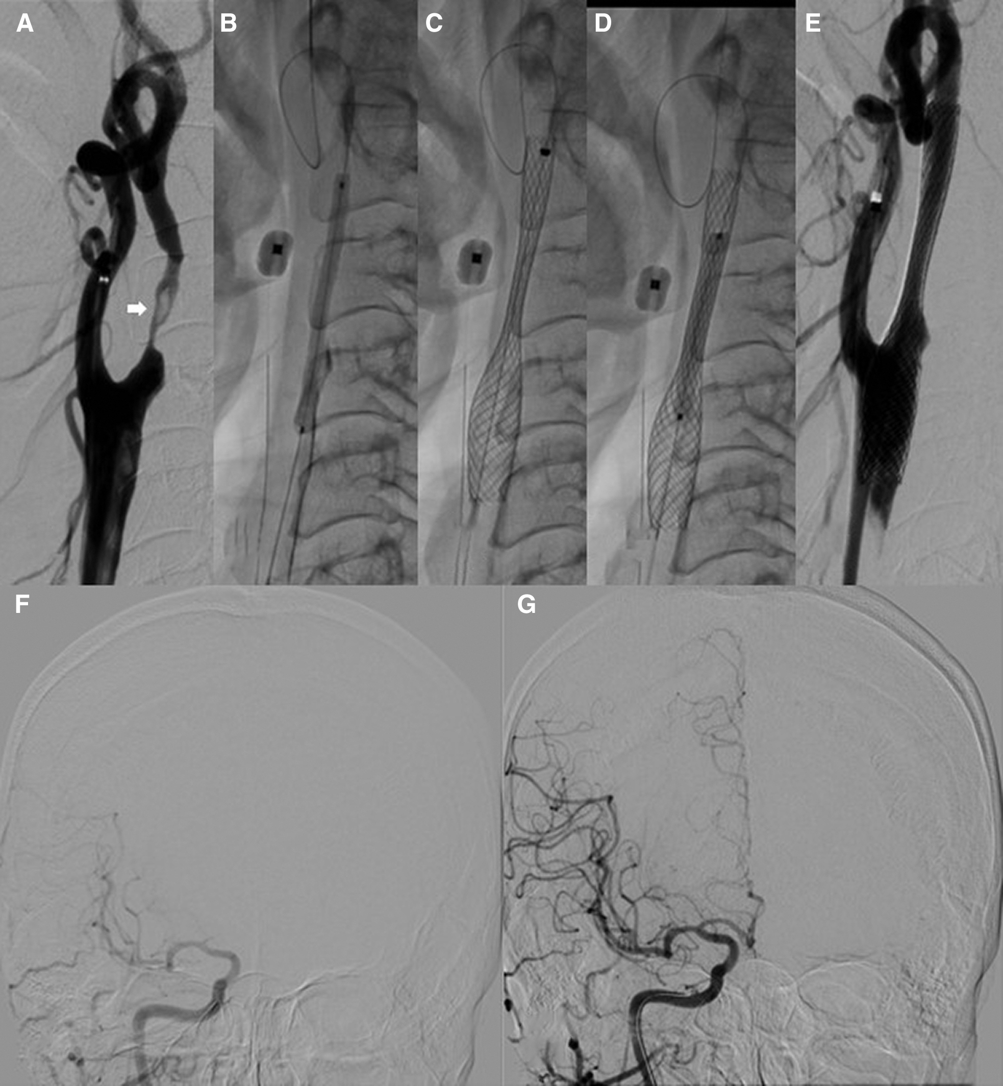
A 62-year-old man developed walking disturbance and left hemiparesis. He was a smoker and had a previous history of lung cancer, rheumatoid arthritis, chronic obstructive pulmonary disease, hypertension, and lipid disorder. He was immediately transported to our hospital after the onset. Upon arrival, he was awake and alert but presented with left hemiparesis, with a manual muscle test score of 4. Magnetic resonance imaging (MRI) revealed an ischemic lesion in the watershed area of the right hemisphere (Fig. 1A). Magnetic resonance angiography showed severe stenosis and an irregular plaque in the right cervical ICA (Fig. 1B). The plaque was hyperintense on T1- and T2-weighted imaging (Figs. 1C and 1D). Carotid angiography revealed 70% stenosis based on the North American Symptomatic Carotid Endarterectomy Trial criteria and a double lumen distal to the right ICA (Figs. 1E and 1F). Ultrasonography revealed a heterogeneous plaque (mixed hyperechoic, hypoechoic, and isoechoic) with 84% area stenosis and a double lumen distal to the stenosis in the right ICA (Fig. 1G). The peak systolic velocity (PSV) of the ICA was 535 cm/s. N-isopropyl-p-[123I]-iodoamphetamine (IMP) single-photon emission computed tomography (SPECT) showed moderate decreased cerebral blood flow in the frontal lobe and parietal lobe at rest. Thus, medication therapy (aspirin 100 mg, clopidogrel 75 mg, and pravastatin 10 mg) was initiated. The patient’s neurological symptoms improved after rehabilitation, with a Modified Rankin Scale score of 2; percutaneous coronary intervention was performed for 90% stenosis in the right coronary artery. For the secondary prevention of ischemic stroke, SAP was then considered for the management of severe ICA stenosis.

In the first procedure, lumen dilatation was reduced to prevent hyperperfusion. After systemic heparinization, the Mo.Ma Ultra device (Medtronic, Minneapolis, MN, USA) was inserted into the right external carotid artery (ECA) (Fig. 2A). Total proximal carotid flow arrest was induced with balloon occlusion of the common carotid artery (CCA) and proximal ECA. SHIDEN PTA balloon measuring 2.0 × 40 mm (Kaneka Medical Products, Osaka, Japan) was passed through the anterior part of the double lumen and percutaneous transluminal angioplasty (PTA) was performed (Figs. 2B and 2C). Then, the patient’s cerebral blood flow improved. The patient did not present with new neurological symptoms after the procedure. MRI conducted 1 day after PTA did not show any ischemic infractions, and IMP SPECT performed 2 days after PTA revealed no signs of hyperperfusion. The PSV of the ICA was 336 cm/s 11 days after PTA.

Next, CAS was performed 13 days after PTA. After systemic heparinization, the Mo.Ma Ultra device (Medtronic) was inserted into the right ECA (Fig. 3A). After balloon occlusion of the CCA and proximal ECA, pre-dilation was conducted using the SIDEN PTA balloon measuring 3.0 × 40 mm (Kaneka Medical Products) through the anterior part of the double lumen (Fig. 3B). Then the Carotid WALLSTENT measuring 10 × 31 mm (Boston Scientific, St Albans, UK) was deployed, and post-dilatation was performed using Rx-Genity measuring 3.5 × 30 mm (Kaneka Medical Products) (Figs. 3C–3E). Subsequently, intracranial digital subtraction angiography (DSA) revealed a higher volume of cerebral blood flow (Figs. 3F and 3G). MRI and IMP SPECT conducted 1 day after CAS revealed no signs of ischemic infarction and hyperperfusion (Figs. 4A and 4B). The patient, with a Modified Rankin Scale score of 1, was then discharged 10 days after surgery.

To date, the mechanism underlying the double-lumen structure has not been fully elucidated. However, several hypotheses have been proposed, and two types of double lumen, namely, true-double lumen (two true lumens) and a pseudo-double lumen (true lumen with a false lumen), are described.
The most common cases of a true-double lumen of the ICA involve fenestration and duplication as an anatomical variation.1–7) Killien et al.8) have shown the duplication results from the fusion of the primitive carotid plexus into two channels and described the embryological abnormalities as a pathology.8)
Gailloud et al.9) hypothesized that the pseudo-double lumen of the cervical ICA can be an anatomic variant predisposing to arterial dissection.9) In 2002, Yu et al. reported another possible mechanism of pseudo-double lumen of the ICA, which is the plaque anatomy, a channel dissecting through the atherosclerotic plaque.10) In this study, 320 plaque specimens were analyzed with high-resolution MRI, and five cases of double lumen were found, with an incidence rate of 1.6%. The rate of neurologic symptoms in these cases was significantly higher, indicating that the irregular surface of the false lumen may cause embolization. This carotid plaque morphologic abnormality may help identify a lesion with high risk for associated emboli and stroke.
In our case, based on the cerebral angiography and MRI findings, the double lumen was considered a pseudo-double lumen due to arteriosclerosis, as Yu et al.10) have proposed, rather than the embryological true-double lumen. The patient was old and had a high risk of atherosclerotic carotid artery stenosis due to a history of smoking, hypertension, and lipid disorder.11–13) Moreover, the patient had right coronary artery stenosis, which is typical in atherosclerotic carotid artery stenosis.14–16) However, he did not have a history of neck pain, and there were no signs of dissection in the computed tomography scan and MRI images.17,18) These findings also support the diagnosis.
Several reports showed that CAS and SAP could be considered a treatment option for severe ICA stenosis.19–22) Patients with unstable plaque are considered at high risk of distal embolism during EVT.23,24) To prevent stroke in an unstable region as in this case, carotid endarterectomy may be the first option.10) However, in this case, we chose CAS because the patient had a history of lung cancer and chronic obstructive pulmonary disease, suggesting high risk of general anesthesia.
IMP SPECT showed moderate decreased cerebral blood flow. Reduced collateral circulation was also observed on DSA. Both observations are considered as a risk factor of hyperperfusion syndrome.25,26) Furthermore, it was difficult to predict the true risk of hyperperfusion after CAS in a unique irregular plaque like this. SAP is reported as an effective method to prevent hyperperfusion. Therefore, we selected SAP.
In this case, the carotid plaque was heterogeneous and suspected unstable.27) The passage of the device across an unstable lesion is an inherent risk of embolization; a proximal balloon protection device could be a better option in this type of plaque.28) Total proximal carotid flow arrest was induced using the Mo.Ma Ultra device (Medtronic), and the procedure was safely performed.
There are several reports comparing closed-cell and open-cell stents for the treatment of ICA stenosis. A large-scale clinical study showed that the 30-day incidence of ipsilateral stroke was significantly lower for closed-cell stents than for open-cell stents.29) However, there are reports showing that the embolic complications do not differ according to stent types.30,31) At present, there is no consensus on the appropriate stent that must be used in CAS. In this case, a closed-cell stent was utilized because its strut is thinner than that of an open-cell stent, and its use is correlated to a lower plaque protrusion. Thus, the procedure was safely performed.
We could not distinguish the true lumen with the false lumen in this case. Therefore, we are not sure which lumen the CAS was performed. We selected the anterior part of the double lumen because the blood flow was natural and the devices could be easily inserted, so it is unknown whether CAS could be accomplished through the false lumen. On the other hand, in the cardiovascular field, PTA and stenting against the false lumen are performed.32) However, it is uncertain whether this could be adapted in the carotid artery. Optical coherence tomography and intravascular ultrasound could be an option to detect the intima and identify the true lumen.
We presented a case of double-lumen carotid plaque associated with severe ICA stenosis. SAP was performed to prevent hyperperfusion, and the patient had a good outcome. If an appropriate device is used, EVT can be a safe treatment strategy for unstable plaques with an as in this case.
Y. Sato, S. Osawa, and N. Narita contributed to the data acquisition. Y. Sato and S. Osawa wrote the manuscript. T. Tominaga conceived the study. All authors have read and approved the final manuscript.
This study did not receive any financial support or grants. None of the authors have a personal or institutional financial interest in the drugs, materials, or devices used in this study.