抄録
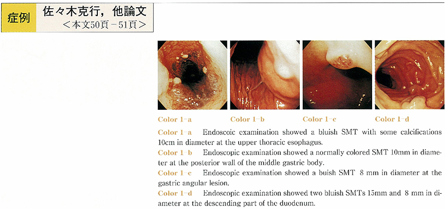
The patient was a 40-year-old woman. She visited our hospital because of hoarseness and difficulty of breathing. A computed tomographic (CT) scan of the chest revealed isodensity mass of esophagus with some calcifications. Endoscopic examination showed a bluish submucosal tumor (SMT) at the upper thoracic esophagus with some calcifications. It also showed two SMTs in stomach and two SMTs at the duodenum. These were similar color to the esophgeal lesion. A magnetic resonance imaging (MRI) of the chest showed the esophageal tumor that was very high intensity on T2-weighted. We suspected these SMTs hemangiomas. In other studys there was one hemangioma of the liver.
Many cases of bleeding of the gastrointestinal hemangioma are reported, therefore it is necessary for this patient screening of small and large intestin. But it is not done yet because of her rejection.
Gastrointestinal hemangioma is a benign tumor comparatively rare. We report a very rare case which had been susupected multiple hemangiomas at the upper gastrointestinaltruct.