抄録
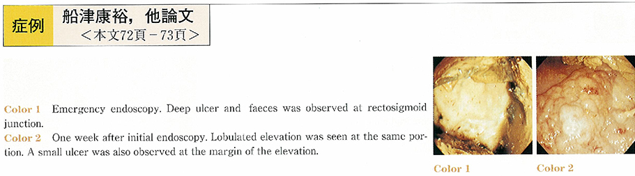
A 74-year-female was admitted to our hospital complaining of lower abdominal pain and hematochezia. A large and deep ulcer was observed at rectosigmoid junction by emergency colonoscopy. After one week TPN therapy, follow up study was performed. The ulcer was altered into elevated lesion with lobulation like laterally spreading tumor granular type (LST-G) . A small ulcer was also detected at the margin of the elevation. The endoscopic diagnosis was confused whether neoplastic tumor or stercoraceous ulcer. The initial endoscopy was performed in poor preparation. Large faeces interrupted observation and only a part of the ulcer was seen. However, there was no pit on the surface of the elevated lesion in magnification. The biopsy revealed the elevation consisting of inflammatory granulation tissue. The elevation was disappeared completely and only ulcer scar was observed after 4 month later. The inflammatory Granulomatous elevation appears in gastric ulcer healing processes, especially treated with PPI. There was no case report of stercoraceous ulcer making inflammatory Granulomatous elevation in Japanese literature. And it is also very rare colonic ulcer making inflammatory Granulomatous elevation. This case is very rare.