抄録
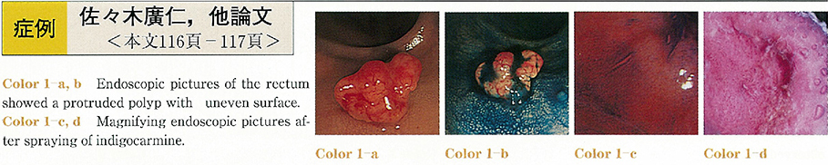
A 58-year-old woman was admitted to our hospital because of positive fecal occult blood which was pointed out by regular medical check-up. The patient underwent colonoscopic examination which revealed a protruded polyp with uneven surface, located in the rectum and measuring about 10mm in diameter. In magnifying colonoscopic view after dye spraying, most of the surface area was shown to be non-structural (Kudo's type VN pit pattern) , but only the periphery of the lesion was covered with the normal mucosa (type I pit pattern) . The lesion was excised with endoscopic mucosal resection (EMRtechnique) . The pathological diagnosis was a carcinoid tumor.
The additional surgery including low anterior resection of the rectum and lymph node dissection was performed because the lesion showed rather large size, high grade atypia, 2.3% of Ki 67 index and uncertain cut margin. The case was peculiar in gross appearance, which made the diagnosis of carcinoid difficult.