抄録
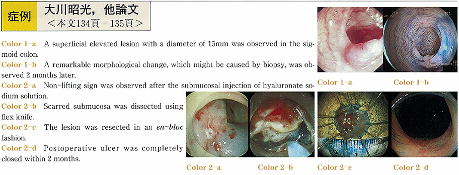
A 69-year-old woman visited her local medical doctor with a chief complaint of diarrhea. As the facal occult blood test showed a positive result, she undertook a colonoscopy, which revealed 0-IIa typed tumor (superficial elevated lesion) with a diameter of 15mm. A histological examination of biopsy specimens from the lesion showed Group IV. In order to undertake an endoscopic resection of the tumor, she was introduced to this hospital. The colonoscopy at our outpatients clinic demonstrated remarkable converging folds. The tumor showed the non-lifting sign when we injected saline into the submucosal layer around the lesion. Endoscopic Ultrasound demonstrated a remarkable thickness of submucosal layer. Although we understood that a surgical operation should be one of the best options for its treatment, we performed an endoscopic submucosal dissection with informed consent because the histological diagnosis was Group IV, not Group V. En-bloc resection was achieved using of our newly devised flex knife and hyaluronate sodium solution without any complication. The size of resected specimen was 12mm in the greatest diameter. Histopathological examination revealed that the tumor was tubular adenoma with moderate to severe atypia, and the resected margin was negative. Colonoscopy two-month after the procedure showed a smooth ulcer scar with slight redness. She has been making good progress at the present time. We have learned a valuable lesson from this case. We demonstrated that a biopsy have to avoid before EMRs.