抄録
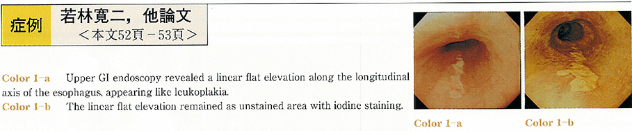
A 71-year-old woman was detected to have an ulcer-like lesion in the gastric corpus during a health examination and was referred to the Tokyo Electrical Power Company hospital. Upper GI endoscopy revealed no ulcer-like lesions in the stomach, but a linear flat elevation along the longitudinal axis of the esophagus, appearing like leukoplakia. The lesion stained positively with iodine staining. It was diagnosed histopathologically as squamous cell carcinoma of the esophagus in situ, and could be resected completely via endoscopy and use of an EEMR tube. After EMR, a repeat upper GI endoscopy revealed an ulcer scar, but no stenosis of the esophagus. Esophageal carcinoma in situ developing in this shape only along the longitudinal axis of the esophagus is very rare. The patient was not a smoker and was not habituated to alcohol. There were no other risk factors, either, and the cause of formation of an esophageal carcinoma lesion of this shape is unknown. An association with reflex esophagitis may be possible.