抄録
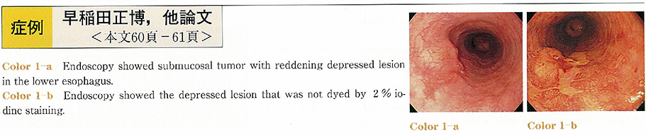
A 71-year-old man who was diagnosed a cancer of the esophagus was admitted to our hospital. Endoscopic examination revealed an elevated lesion covered with normal mucosa with a depressed lesion of the esophagus. The depressed lesion was not dyed by 2% iodine staining and pathological findings showed squamous cell carcinoma. And the elevated lesion was soft and gradual. Therefore, we diagnosed the lesion as a submucosal tumor (SMT) with early cancer of the esophagus. Then we performed endoscopic mucosal resection (EMR) .
The pathological findings of the specimen showed the lesion size was 20×15mm and it was non-small cell type undifferentiated carcinoma with early moderately differentiated squamous cell carcinoma. The tumor was located in submucosal layer covered normal mucosa of esophagus. And the surgical margin was negative.
After EMR, no additional therapy was done. He has no recurrence for 16 months.