抄録
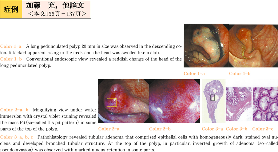
A 60-year-old female underwent total colonoscopy in February 2010 for a positive fecal occult blood test. A long pedunculated polyp 20 mm in size was observed in the descending colon. It lacked apparent rising in the neck and the head was reddish and swollen like a club. Normal mucosa extended beyond the neck and magnified views after crystal violet staining revealed the mass Pit (so-calledIIIs pit pattern) in some parts of the top of the mass. Endoscopic ultrasonography demonstrated a low-echoic cystic mass with inner high-echoic spots from the head to the neck. Pedunculated mucosal polyp was suspected by endoscopy and was resected endoscopically from the base because pseudoinvasion of adenoma or cancer invasion to the mucosal lake was differentially diagnosed by magnified endoscopy and ultrasonography. Pathohistology revealed tubular adenoma that comprised epithelial cells with homogeneously dark-stained oval nucleus and developed branched tubular structure. At the top of the polyp, in particular, inverted growth of adenoma (so-called pseudoinvasion) was observed with marked mucus retention in some parts. At the neck, on the other hand, relatively large blood vessels accompanying wall thickening and hyalinization were present.
We here report a case of adenoma with pseudoinvasion which was difficult to differentiate from a pedunculated mucosal polyp.