Progress of Digestive Endoscopy
Online ISSN : 2187-4999
Print ISSN : 1348-9844
ISSN-L : 1348-9844
症例
内視鏡的切除を行った十二指腸Peutz-Jeghers型ポリープの1例
遠藤 佑香藤原 崇林 星舟小泉 理美千葉 和朗岩崎 将來間 佐和子桑田 剛江頭 秀人小泉 浩一神澤 輝実田畑 拓久藤原 純子荒川 丈夫門馬 久美子堀口 慎一郎
著者情報
キーワード: Peutz-Jeghers型ポリープ
ジャーナル フリー

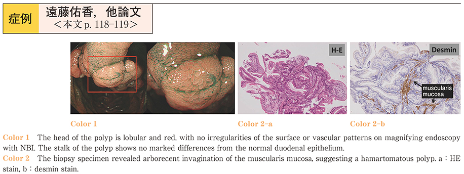
2014 年 84 巻 1 号 p. 118-119

詳細
抄録

The patient was a 67-year-old woman in whom endoscopic retrograde cholangiopancreatography (ERCP) performed to treat choledocholithiasis revealed a 1.5-cm pedunculated polyp in the duodenum in October 2010. Pathological examination of a biopsy specimen showed no evidence of malignancy. In July 2012, the polyp had grown to 3.5 cm in diameter. The head of the polyp was lobular and red, and revealed no irregularity of the surface or vascular pattern on magnifying endoscopy with narrow-band imaging (NBI) . The stalk of the polyp showed no marked differences from the normal duodenal epithelium. Another biopsy specimen revealed arborecent hyperplasia of the muscularis mucosa, and a hamartomatous polyp was suspected. The patient was admitted to the hospital for endoscopic treatment in view of the possibility of malignancy. She underwent polypectomy; her subsequent clinical course was uneventful, and she was discharged from the hospital 8 days after treatment. The resected specimen showed a lobular polyp measuring 34×18×14 mm, and pathological examination confirmed the diagnosis of a hamartomatous polyp. She did not have any family history of Peutz-Jeghers (P-J) syndrome and showed no pigmented spots; therefore, P-J-type polyp was diagnosed. No recurrence or evidence of other polyps was seen on gastrointestinal endoscopy or colonoscopy performed 1 year after the treatment. P-J-type polyps are hamartomatous polyps with the same pathological findings as the polyps associated with P-J syndrome without the family history or pigmented spots. P-J-type polyps can cause bleeding and obstruction, and show potential for malignant transformation; therefore, treatment such as surgery or endoscopic removal is warranted. In addition, other areas of the gastrointestinal tract, including the small intestine, need to be examined to rule out multiple lesions.

Fullsize Image
著者関連情報
© 2014 一般社団法人 日本消化器内視鏡学会 関東支部
前の記事 次の記事
feedback
Top