Abstract
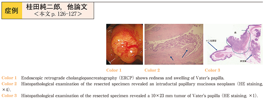
A 70-year-old man was admitted to our hospital with jaundice. Abdominal ultrasonography (US) and computed tomography (CT) showed a multilobular cystic lesion in the head of the pancreas. Abdominal contrast-enhanced CT suggested a tumor in the papilla of Vater. Endoscopic retrograde cholangiopancreatography (ERCP) showed redness and swelling of the papilla of Vater, a dilated common bile duct and a multilobular cystic lesion in the head of the pancreas. ERCP and endoscopic biliary drainage (EBD) were performed. Because the fever remained persistent even after the drainage, we performed a CT, which showed a liver abscess. Percutaneous transhepatic abscess drainage (PTAD) was therefore performed, and histopathological examination of a biopsy specimen from the papilla of Vater revealed adenocarcinoma. On the basis of these findings, pancreaticoduodenectomy (PD) was performed. Histopathological examination of the resected specimen revealed a 10×23 mm tumor of the papilla of Vater and an intraductal papillary mucinous neoplasm (IPMN) . The patient was discharged without complications.