A 74-year-old woman was admitted to our hospital complaining of a sense of abdominal fullness. Abdominal CT with a radiocontrast agent showed marked ascites, opacification of the mesenteric fat, and several enhancing lesions, including swollen lymph nodes in the right groin area, a small nodule in the pouch of Douglas, and nodules in the anorectal area. Ascitic cell block cytology showed tumor cells containing blackish brown granules. Based on these findings, we made a preliminary diagnosis of malignant melanoma. Colonoscopy revealed black elevated lesions in the anorectal area, and histology of biopsy specimens obtained from the lesions showed tumor cells containing blackish brown granules. Based on these findings, a final diagnosis of stage IV malignant anorectal melanoma was made.
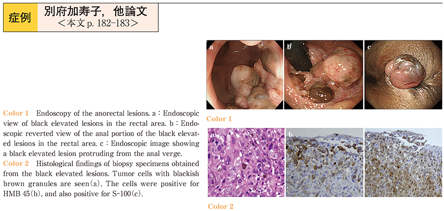
A definitive diagnosis of malignant anorectal melanoma can be challenging, because the colonoscopic findings of anal polyps, anal canal cancer and malignant lymphoma can be similar. Careful examination for detecting the colonic elevated lesions and obtaining biopsies from these lesions may allow early diagnosis of anorectal melanoma at a higher frequency.