Abstract
Esophageal anisakiasis is a rare disease, accounting for about 0.2% of all cases of anisakiasis.
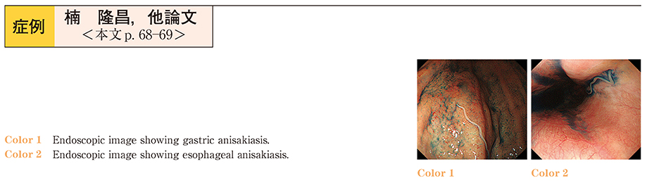
A 26-year-old woman visited our hospital because of severe epigastric pain and heart burn several hours after eating pickled mackerel, which progressively worsened, and subsequently presented with fever. Contrast CT showed marked hypertrophy of the gastric wall and a significant edematous change in the lower esophagus, in addition to inflammation of the mediastinum. Upper gastrointestinal endoscopy revealed an Anisakis worm in the gastric corpus and lower esophagus. After removing it by endoscopy, her condition immediately improved.
The present patient was complicated with mediastinitis. It was suggested that anisakiasis can become severe in an esophagus with a thin wall, as can be the case with the small intestine, where the worm may burrow in some cases. As in this case, we can achieve the marked amelioration of anisakiasis through the removal of worms. Thus we should perform endoscopy in the early stage. In addition, since some patients may ingest several worms of Anisakis, we should carefully check whether or not worms burrow in sites other than the stomach, where worms have already been identified.