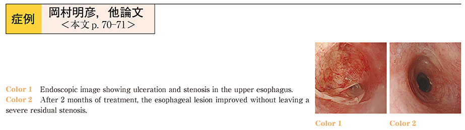
-
Akihiko Okamura
Department of Surgery, School of Medicine, Keio University
-
Tai Omori
Department of Surgery, School of Medicine, Keio University Center for Diagnostic and Therapeutic Endoscopy, Keio University Hospital
-
Kenjiro Ishii
Department of Surgery, School of Medicine, Keio University
-
Rieko Nakamura
Department of Surgery, School of Medicine, Keio University Center for Diagnostic and Therapeutic Endoscopy, Keio University Hospital
-
Tsunehiro Takahashi
Department of Surgery, School of Medicine, Keio University
-
Norihito Wada
Department of Surgery, School of Medicine, Keio University
-
Hirofumi Kawakubo
Department of Surgery, School of Medicine, Keio University
-
Yoshiro Saikawa
Department of Surgery, School of Medicine, Keio University
-
Hiroya Takeuchi
Department of Surgery, School of Medicine, Keio University
-
Jun Yamagami
Department of Dermatology, School of Medicine, Keio University
-
Masayuki Amagai
Department of Dermatology, School of Medicine, Keio University
-
Yuko Kitagawa
Department of Surgery, School of Medicine, Keio University