Abstract
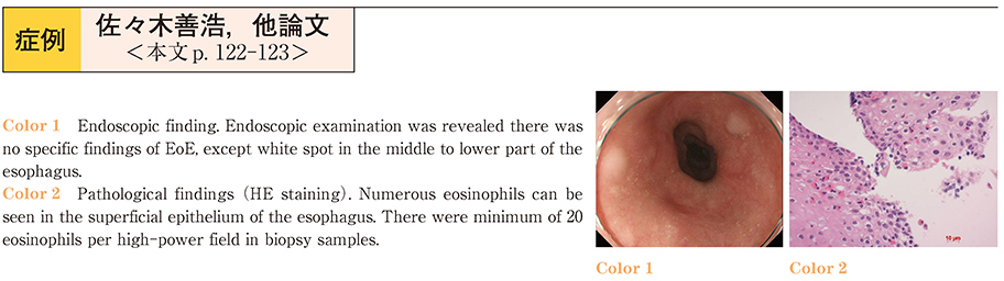
A 62 year-old man visited our hospital because of chest discomfort and chest pain. He had no abnormality by cardiovascular examination. Thoracic CT study revealed edematous wall thickening of middle to lower part of the esophagus, suggesting eosinophilic esophagitis (EoE) . But esophagogastrodupdenoscopy (EGD) revealed no specific findings of EoE, except white spot in the middle to lower part of the esophagus, and biopsy samples were non-specific.
His symptoms continued, and EGD was performed again one month later. EGD findings were the same, but the diagnosis of EoE was done by biopsy samples.
After he was administered predonisolone, his condition was improved.
EoE is said to caused by the chronic stimulation of the antigens such as meals.
The prevalence of EoE is estimated as 1/5,000 by some report, but the real one is uncertain. It is possible that more people are not properly diagnosed as EoE. Biopsy should be done for patients complaining chest discomfort, even if there is no specific findings of EoE on EGD.