抄録
A 55-year-old female was introduced to our hospital for a further evaluation of the duodenal lesion.
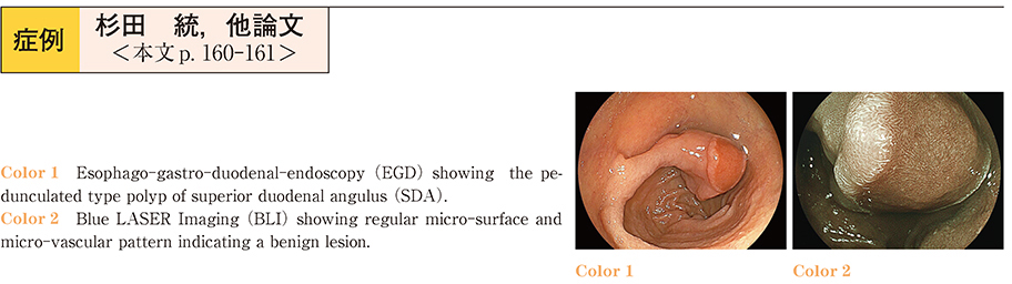
Esophago-gastro-duodenal-endoscopy (EGD) demonstrated the pedunculated polyp about 30mm in diameter at superior duodenal angulus (SDA) . The lesion had several red depressed areas, and image-enhanced magnified endoscopy by Blue LASER Imaging (BLI) showed regular micro-surface and micro-vascular pattern indicating a benign lesion. Biopsies was taken from the depressed area, but the definitive diagnosis was not provided.
Considering a malignant lesion, we resected the lesion by polypectomy. Histo-pathological findings showed Brunner’s gland hyperplasia, and microscopic examination demonstrated the surface of the depressed lesion was covered by regenerated epithelial cell with non-atypical glands hyperplasia.
We speculated this unique lesion was developed by a mechanical stimulation.