Abstract
A 55-year-old man visited our clinic with chief complaints of upper abdominal discomfort and tarry stools. An upper endoscopic examination showed a submucosal tumor on the upper intrathracic esophagus and a gastric ulcer stage A2. The tumor was elastic soft, thumbtip sized and located at the 9 o'clock direction of the esophagus at about 25cm distal from the incisors. It showed an equable low echoic lesion that existed at the second layer by an endoscopic ultrasonic examination.
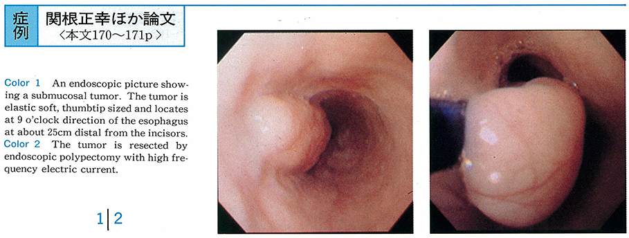
It was resected by endoscopic polypectomy with high frequency electric current. The resected specimen was 23×13×15mm in size, was resected completely, and the leiomyoma originated from muscularis mucosae by a histopathological examination. Four weeks after polypectomy on the esophagus, the ulcer healed and was covered with normal esophageal epithelium.
This case was dignosed as leiomyoma of the esophagus originating from muscularis mucosae due to endoscopic ultrasonic findings. Even though its size was over 20mm, it was resected safely and surely by endoscopic polypectomy, and the patient was cured with no complications.