Progress of Digestive Endoscopy(1972)
Online ISSN : 2189-0021
Print ISSN : 0389-9403
Volume 44
Displaying 1-47 of 47 articles from this issue
Technology and instrument
  • Minoru Hanashiro, Tohru Hagiwara, Taro Ishii, Chiaki Abe, Eisuke Katsu ...
    1994 Volume 44 Pages 39-42
    Published: June 06, 1994
    Released on J-STAGE: May 25, 2015
    JOURNAL FREE ACCESS
    We employ the laser Doppler method in the measurement of gastric mucosal blood flow, but anemia or like conditions render it difficult to evaluate data obtained by this method. To cope with this problem, gastric mucosal oxygen tension and blood flow were directly measured at the same time.
    An endoscope was introduced into the stomach of mongrel dogs in Nembutal anesthesia, and gastric mucosal tissue oxygen tension and blood flow were measured before and after the intravenous administration of butylscopolamine bromide, using a biological tissue oxygen tension continuous measuring apparatus (PO2100, Intermedical) and PeriFlux PF3, respectively.
    The measurement of gastric mucosal tissue oxygen tension seems to provide information not only on oxygenation of the local site of measurement but also on blood flow there.
    Download PDF (650K)
  • Yukihide Takahashi, Masahiro Igarashi, Tomoe Katsumata, Yoshitaka Nait ...
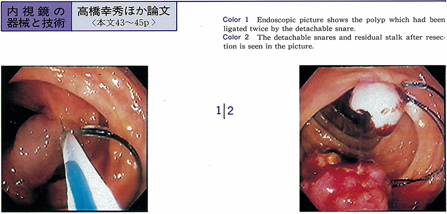
    1994 Volume 44 Pages 43-45
    Published: June 06, 1994
    Released on J-STAGE: May 25, 2015
    JOURNAL FREE ACCESS
    We studied about the effect of the detachable snare for patients with large pedunculated polyp of the colon. The materials were as follows : sixteen cases were applied for preventing hemorrhage, and the other for hemostasis after polypectomy on emergency. The size of the polyps were from 13mm to 30mm at the maximum diameter of their head (mean 19.8mm) . Seven cases were treated at out-patient ward and the others were treated in hospital. To strengthen the effect of the snare, we ligated the stalk of the polyp twice by the snare in one case, and in five cases, endoscopic hemo-clipping procedure was used in combination. No bleeding was found after polypectomy in every cases. In the case of emergent hemostasis, we could ligate the residual stalk easily by the snare and succeeded in arrest of bleeding.
    We concluded that the detachable snare is very useful and safe instrument for preventing hemorrhage or hemostasis after polypectomy of pedunculated polyps especially with large and wide stalk.
    Download PDF (275K)
  • Masahiro Igarashi, Tomoe Katsumata, Hideyuki Takahashi, Yoshitaka Nait ...
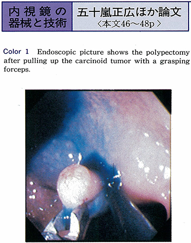
    1994 Volume 44 Pages 46-48
    Published: June 06, 1994
    Released on J-STAGE: May 25, 2015
    JOURNAL FREE ACCESS
    We had an opportunity to use a new 2-channels electronic videocolonoscope (XCF-2T200 : Olympus) and evaluated the value for diagnostic instrument and endoscopic polypectomy. The scope has two instrument channels (left channel with 3.2mm and right channel with 2.8mm in diameter respectively) and its calibre is 0.9mm, being larger than the ordinary scopes of Olympus made.
    This scope was applied in 112 patients, and 127 lesions were resected endoscopically in a period of 7 months between April to October in 1993. By using this instrument, total colonoscopy was successful in all 99.1%. Especially, the mucosal resection of flat polyps was easily manipulated by the combined use of a grasping forceps and a polypectomy snare.
    In conclusion this scope is considerably valuable as a diagnostic as well as treatment tool.
    Download PDF (451K)
Clinical study
  • Nobuhiko Nagamine, Norio Ueno, Ken Kimura
    1994 Volume 44 Pages 49-52
    Published: June 06, 1994
    Released on J-STAGE: May 25, 2015
    JOURNAL FREE ACCESS
    We evaluated the usefulness of an ultrasonic microprobe (UMP) which was able to scan the esophageal varices via biopsy channel on endoscopic variceal ligation (EVL) by Stiegmann's method.
    Eight patients with liver cirrhosis and 2 patients with idiopathic portal hypertension were studied from July through November 1993.
    As a result, we determined the following usefulness of UMP on EVL ; (1) Judgement of efficacy of the treatment with EVL by visualization of esophageal wall structure and variceal venous channel including perforating vein (the ideal site of ligation) which communicated varix and collateral vein, (2) Prediction of RC sign appearance in early stage after EVL, hyperechoic change of varices which we named“snow ball”sign in this study could be observed. According to our experiment, we speculated some acoustic change in the varices by EVL caused such hyperechoic view of the ligated varices.
    In conclusion, this non-invasive new method is considered to be useful for evaluation of variceal blood flow on EVL clinically.
    Download PDF (1137K)
  • Susumu Shibuya, Yasuhiro Takase, Shigehiro Kokubu, Shyun Yamamoto, Sho ...
    1994 Volume 44 Pages 53-56
    Published: June 06, 1994
    Released on J-STAGE: May 25, 2015
    JOURNAL FREE ACCESS
    It is well known fact that treatment of giant straight esophageal varix is almost difficult. Therefore, feeders of the giant straight varix is examined by endoscopic varicealography during injection sclerotherapy (EVIS) . From December 1977 to September 1992, 32 cases of the straight varix were treated with EVIS.
    Our technique is to embolize esophageal varices and their feeders by using oral side balloon attached to the tip of the endoscope that was inflated to stop the blood flow of the varices during treatment and intravariceally injecting 5% EO with iopamidol or meglumine amidotrizoate.
    It was reported that their feeders were classifed with left gastric vein (LGV) , short gastric vein (SGV) , fundic plexus (FP, LGV communicated with SGV) and unknown by EVIS. Also, it was reported that major shunts except esophageal varices were done with abdominal type (between systemic circulation and part of the feeders) and thoracic type (between systemic circulation and parts of esophageal varix) .
    Results : In 31 cases, feeders were LGV and 7 of them had shunt of thoracic type. Remaining one case had unknown feeders and two shunts of thoracic type. None had SGV, FP and shunts of abdominal type. In the cases of shunt of thoracic type, recurrence rates were greater and numbers of injection during sclerotherapy to eradicate the varices were larger than in the cases without shunt of thoracic type.
    As a result, the giant straight esophageal varix had direct effect of pressure from portal vein. It was thought to be especially difficult to treat the straight varices with shunt of thoracic type. However, all straight esophageal varices were thrombosed by sclerotherapy with the sclerosant-contrast medium mixture and the oral side balloon (EVIS technique) .
    Download PDF (1093K)
  • Tetsurou Arai, Toru Endo, Hiroyuki Uemura, Takashi Tazoe, Nobuyuki Sak ...
    1994 Volume 44 Pages 57-59
    Published: June 06, 1994
    Released on J-STAGE: May 25, 2015
    JOURNAL FREE ACCESS
    A clinical study on efficacy and complications of endoscopic variceal ligation (EVL) was carried out on 17 patients with esophageal varices (16) and gastric varices (1) . Those were devided by the stages of treatment into 3 groups ; the first group was 10 cases of prophylactic stage, the 2nd was 2 cases of elective stage and the 3rd was 5 cases of emergent stage.
    The effective rate was 94.1% inclusive of, 29.4% in eradicative rate. It was possible to control hemorrhage in all emergent cases by EVL. Complications occurred in 2 cases ; One was chest pain in esophageal varices and the other a small amount of bleeding in gastric varices.
    The endoscopic variceal ligation for management of esophago-gastric varices was thought to be effective and safe. This simple technique could promote a good hemostatic effect especially in emergent cases.
    Download PDF (337K)
  • Hisao Nakai, Masahito Ohida, Sayuri Yamagata, Keita Ishii, Hiroshi Ima ...
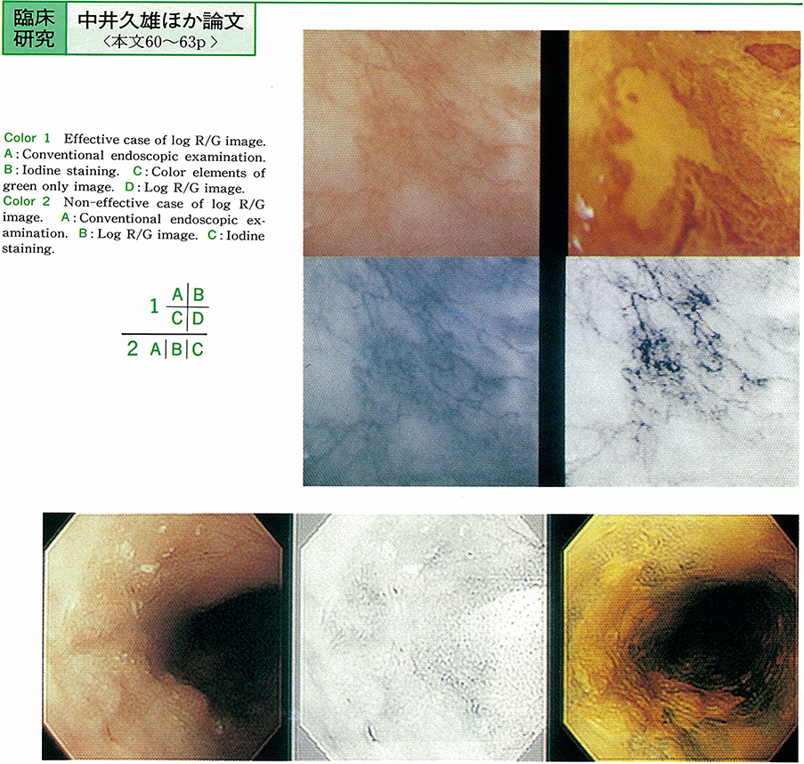
    1994 Volume 44 Pages 60-63
    Published: June 06, 1994
    Released on J-STAGE: May 25, 2015
    JOURNAL FREE ACCESS
    Log R/G images using an Olympus image input and processing system (IP system) were studied to determine the extent of superficial spread and the depth of invasion of resected six cases of superficial esophageal cancers at Kitasato University East Hospital.
    The capillary networks in lesions were more clearly defined in intraepithelial cancers compared with before processing. This image was useful to diagnosis for intraepithelial cancers, but was not of mucosal and more invasive ones. It seems this finding are useful for differential diagnosis between intraepithelial cancers and more invasive ones. However, this image was unable to exceed iodine staining method to diagnose the extent of infiltration.
    Download PDF (960K)
  • Hiroyuki Hisai, Hajime Yamaguchi, Kuniaki Shirao, Daizo Saito, Hitoshi ...
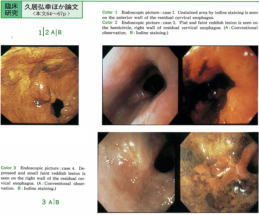
    1994 Volume 44 Pages 64-67
    Published: June 06, 1994
    Released on J-STAGE: May 25, 2015
    JOURNAL FREE ACCESS
    We report four cases of superficial esophageal cancer, arising from residual cervical esophagus after the surgical resection of thoracic esophageal cancers. In these cases the second lesions were detected endoscopically and macroscopic types were 0-IIc in three and 0-IIb in one. Surgical treatments were performed for two cases, irradiation therapy for one case and endoscopic laser treatment for one case.
    Out of 417 patients with esophageal squamous cell carcinoma who underwent esophagectomy without preoperative treatment during the period between January 1985 to June 1993 in the National Cancer Center Hospital, 85 patients (20.4%) had additional primary esophageal cancers.
    Such a high incidence of multicentricity of esophageal cancer and our experiense reported here suggested that a careful endoscopic examination using iodine staining method is important not only for the whole esophagus before treatment but also for the residual esophagus after treatment in patients with esophageal cancer.
    Download PDF (929K)
  • Tooru Honda, Kimiya Takeshita, Masao Tani, Fumio Kando, Naoya Saito, M ...
    1994 Volume 44 Pages 68-72
    Published: June 06, 1994
    Released on J-STAGE: May 25, 2015
    JOURNAL FREE ACCESS
    Magnifying electronic endoscopic examination using Olympus GIF-V10Z, XGIF-200Z and XGIF-200HM was done to assess the mucosal microstructure (gastric pits and finger like processus) of the gastric elevated lesions.“Band enhancement”was also used to get the clear image of the mucosal microstructure. These magnifying endoscopic images were compared with the dissecting microscopic views of the resected specimens and pathological examination was performed. The results obtained were as follows.
    1) Hyperplastic foveolar polyp had reddish and enlarged finger like processus which reflected congestion and edema of the interstitium.
    2) Fundic polyp had regularly distributed, round gastric pits.
    3) Gastric adenoma had whitish, regularly distributed gastric pits and finger like processus.
    4) Protruded early gastric cancer had irregularly shaped gastric pits and finger like processus.
    These results indicated that mucosal observation with the magnifying electronic endoscopy and“band enhancement”leaded to more accurate diagnosis of the gastric elevated lesions.
    Download PDF (1304K)
  • Masao Tani, Haruhiro Inoue, Toshihisa Ashikawa, Fumio Kando, Naoya Sai ...
    1994 Volume 44 Pages 73-76
    Published: June 06, 1994
    Released on J-STAGE: May 25, 2015
    JOURNAL FREE ACCESS
    We have already introduced an endoscopic mucosal resection using a cap-fitted panendoscope (EMRC) , and this time we developed a pre-looped cap which has a hook at the tip of the cap to simplify the snaring.
    EMRC-cap is fitted at the tip of the panendoscope. More than 10 ml of saline solution mixed with low-volume epinephrine is injected into the submucosal layer around the lesion. A fine snare is passed through the biopsy channel of the endoscope, and is pre-looped at the tip of the cap using the hook. Under full endoscopic suction, the lesion-involved mucosa is packed into the cap, and then snared tightly. And then mucosal resection is performed with high-frequency electrocautery.
    Eleven patients with 12 gastric lesions were treated with this EMRC procedure using a pre-looped cap. Twelve lesions consist of 7 early cancers (4 IIa types, 1 IIa+IIc type, 1 IIc type, 1 IIb type) , 4 adenomas, and 1 hyperplastic polyp. Almost all lesions were resected without any complication. Only one case had remnant cancer, and underwent laser therapy.
    We believe that EMRC using a pre-looped cap is a simple and useful method of treatment for tumorous mucosal lesions of the stomch.
    Download PDF (862K)
  • Yukio Watanabe, Hitoshi Kondo, Daizo Saito, Toshihiro Yokota, Kuniaki ...
    1994 Volume 44 Pages 77-81
    Published: June 06, 1994
    Released on J-STAGE: May 25, 2015
    JOURNAL FREE ACCESS
    Eight hundred and ninety adenomas of 836 cases were diagnosed during the period from June, 1962 to February, 1993 at National Cancer Center Hospital. We studied relationship between changes of tumor size and pathological findings in 77 lesions (70 cases) which are followed endoscopically and histologically for more than 3 years. Fourteen (18%) lesions increased in size (increased group) and 63 (82%) lesions almost unchanged (unchanged group) .
    The male was significantly dominant in the increased group, and lesions larger than 2cm or those with flat polypoid type were more frequent in the increased group. The follow up periods were various, until the increase in size was noticed.
    No relationship was identified between changes of size and grades of histological atypia. Histological atypia at the first examination was not correlated with changes in size. Histological diagnosis by the method of morphometrical analysis with image processing was done for 30 lesions, 23 unchanged and 7 changed cases. In increased group, aspect ratio of nuclei was decreased and area and maximum width of tubuli were increased in the final biopsy specimens. These changes mean morphometrically rounding of nuclei and widening of tubuli in pathological findings.
    At the first examination, coefficient variation of maximum width of tubuli (MWCV) was higher in the increased group (50.64) than in the unchanged group (36.95) . This finding shows that there are morphometrical difference between the two groups.
    Our study suggests that 4 factors of sex, size, shape and MWCV may be helpful for predicting increase in size of the adenoma.
    Download PDF (995K)
  • Michiru Yamada, Shoko Midorikawa, Tetsuya Sanji, Yutaka Handa, Shigefu ...
    1994 Volume 44 Pages 82-86
    Published: June 06, 1994
    Released on J-STAGE: May 25, 2015
    JOURNAL FREE ACCESS
    We investigated early gastric cancer according to the relationship between sex, age, depth, tumor size, location, macroscopic type of cancer, histological type of cancer and lymph node metastasis. One thousand and 137 patients (1,291 focuses) of early gastric cancer were analysed retrospectively.
    The lymph node metastatic rate was 7.1% among the total early gastric cancer. Metasatatic rate of the single focus of gastric cancer was 7.6%. Especially male, young (20・30's) and elderly ages (70・80's) showed a high rate. Among the single submucosal cancer, 14.9% were metastatized to the lymph node, significantly higher than that of mucosal cancer rate (2.5%) (P<0.05) .
    According to the tumor size, the lymph node metastasis were negative in cases of mucosal cancer less than 2.0cm and submucosal cancer less than 0.5cm. Macroscopically, combined type of mucosal cancer and elevated type of submucosal cancer showed high lymph node metastatic rate than the other type. Histologically, mucinous adenocarcinoma showed the highest rate of metastasis, and papillary adenocarcinoma did second. 7.3% and 8.1% metastasis were seen in differenciated and undifferenciated type of early gastric cancer respectively.
    Multifocal gastric cancer showed 3.1% lymph node metastasis. In particular female and middle ages (40・50・60's) showed a high rate metastasis. Concerning to the depth of cancer, no metastasis were seen in the mucosal cancer, and 6.2% in submucosal cancer. With respect to the tumor size, 13.0% were positive over 4.1cm, while 0.9% in less than 4.0cm tumor size.
    In conclusion ; early gastric cancer without lymph node metastasis were follows ; histologically differenciated type of mucosal cancer, macroscopically without ulcer, and tumor size less than 3.0cm. And futher IIb type of mucosal cancer in any size have showen no metastasis. From the point of view of lymph node metastasis, we consider these early gastric cancer are the indication of endoscopic mucosal resection.
    Download PDF (599K)
  • Kazuya Yoshimoto, Tadayoshi Kakemura, Syunichiro Ishitsuka, Kougo Kata ...
    1994 Volume 44 Pages 87-91
    Published: June 06, 1994
    Released on J-STAGE: May 25, 2015
    JOURNAL FREE ACCESS
    We studied the usefullness of the newly developed slim ultrasonic probe for the colorectal lesions comparison with the ultrasonic scope in the diagnosis of endoscopic ultrasonography (EUS) . From May 1993 to October 1993, 54 lesions (50 cases) were examined by EUS using slim ultrasonic probe[UM-2R (12MHz), UM-3R (20MHz) developed by Olympus Optical Co]. These result were compared with those of ultrasonic scope [CF-UM3 (12MHz), CF-UM20 (12MHz)]and ultrasonic probe〔UM-1W (7.5MHz)].
    These slim ultrasonic probe were easily inserted into the ordinary scope instrument channel diameter of 2.8mm or more because of slim 2.4mm diameter. These probe was easily contact to the purpose lesions. Only 1 case located in the cecum bottom were failed to insert the probe into the instrument channel because of over scope bending. But other lesions were approached to the lesion completely.
    Visualizing rate were almost well or fair, but 2 cases located in the cecum and transeverse colon were not well visualized because of the lesion was located on the intestinal fold. Concerning to the diagnostic ability, diagnostic accuracy of the depth of the cancer invasion was 94% (early cancer : 86%, advanced cancer : 100%) . These result was same as the data of UM-1W and better than those of ultrasonic scope. Concerning to the lymph node swelling detectability, only 1 case out of 6 cases was visualized because of echo dumping using slim ultrasonic probe.
    From the relation to the thickness of the cancer lesion and the EUS visualization, superficial lesions were obtained diagnosable image using high ultrasonic frequency probe (20MHz) . But thick lesions were obtained inadequate image because of echo dumping. So concerning to the thickness of the lesion, under 15mm lesions would be better to use slim ultrasonic probe and over 15mm lesions should be used ultrasonic scope except stenotic sections.
    In conclusion, these equipments provided advancement of operational and ultrasonic imaging capabilities and seemed to be much useful in the ultrasonic diagnosis of colorectal lesions because of greater sonographic definition.
    Download PDF (1453K)
  • Yasuhiko Ohno, Takeshi Terai, Osamu Kobayashi, Yasushi Imai, Toshio Mu ...
    1994 Volume 44 Pages 92-94
    Published: June 06, 1994
    Released on J-STAGE: May 25, 2015
    JOURNAL FREE ACCESS
    Nine lesions in 9 cases (8 males and 1 female ranging in age from 45 to 69 years) of colorectal tumors of IIa+I type were examined stereomicroscopically and histologically. Of the 9 lesions, 6 were located at the sigmoid colon and the remaining 3 at the rectum. All of the lesions ranging in size from 5 to 13 mm in diameter were removed by endoscopic mucosal resection (EMR) and submitted to stereomicroscopy. Histologically, 9 lesions included 3 adenomas with moderate atypia, 3 adenomas with severe atypia and 3 adenomas with focal carcinoma, indicating that local treatments including EMR for the IIa+I type of colorectal tumor must be cutious, because of their high malignant potential (3/9) .
    Download PDF (1022K)
  • Kaori Ohguri, Masato Murakami, Keiji Shirasaki, Masaki Satou, Akitaka ...
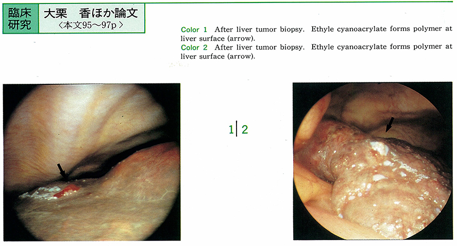
    1994 Volume 44 Pages 95-97
    Published: June 06, 1994
    Released on J-STAGE: May 25, 2015
    JOURNAL FREE ACCESS
    As to the hemostatic efficiency for laparoscopic liver biopsy, we studied the method of hemostasis using ethyl cyanoacrylate (ECA) . Hemostasis using ECA at laparoscopic liver biopsy was performed in 3 cases of metastatic liver carcinoma, 2 cases of cholangioma, one case of liver hemangioma, one case of liver cirrhosis, and 7 cases of liver cirrhosis with hepatocellular carcinoma.
    Twelve of the cases were hypervascular tumor on angiography, 2 cases were hypovascular tumor, but had liver cirrhosis. After the liver biopsy, all cases were hemostated instantly with ECA. No complications were observed after the biopsy. By using ECA, the laparoscopic tumor biopsy is performed safety even if the tumor is hypervascular.
    Download PDF (351K)
Case report
  • Mako Hino, Seirou Shiomi, Hideki Sakurai, Masashi Daibou, Yuuji Fujii, ...
    1994 Volume 44 Pages 99-101
    Published: June 06, 1994
    Released on J-STAGE: May 25, 2015
    JOURNAL FREE ACCESS
    A 31-year-old woman visited our institution with chief complaints of epigastralgia and hemoptysis on March 2, 1993. Endoscopic examination showed longitudinal mucosal redness in whole esophagus and irregular, geographical erosion coverd by a whitish exudate in the distal side. The brushing cytology were taken from erosive lesion. Multinuclear giant cells and acute inflammatory changes were observed in light microscopic examination. The serum antiherpes simplex Type 1 (HSV-1) antibody remarkably elevated. So we diagnosed that this esophagitis was caused by HSV-1 infection.
    The herpes esophagitis is observed almost in autopsy cases of immunosuppressive patients suffering from malignancy, after chemotherapy and radiation therapy. We report a rare case of herpes simplex esophagitis in a healthy adult.
    Download PDF (344K)
  • Izumi Yamazaki, Masato Murakami, Shigehiro Kokubu, Norie Ishigaki, Yas ...
    1994 Volume 44 Pages 102-105
    Published: June 06, 1994
    Released on J-STAGE: May 25, 2015
    JOURNAL FREE ACCESS
    A 47-year-old man with liver cirrhosis had had repeated endoscopic injection sclerotherapy for bleeding of esophageal varices since 1976. He had had also transcatheter arterial embolisation for hepatocelluler carcinoma in 1991. He was admitted to our hospital because of ascites, hepatic encephalopathy and melena in 1992.
    Endoscopic examination revealed red plaque in the esophageal varices. Endoscopic variceal ligation (EVL) was selected as a treatment of choice because he was in a grave condition with hyperbilirubinemia and hypoalbuminemia. Rebleeding of esophageal varices did not occur before he died of hepatic failure ten days after EVL. Autopsy revealed no dilatation of vein and bleeding at the site of the esophagus where EVL had been performed.
    It is suggested that effectiveness of EVL for a short period of time is promised even in the patients with esophageal varices in a grave condition.
    Download PDF (1036K)
  • Jun-ichi Fukushima, Tomoyuki Goya, Cyo-o Kaku, Noboru Suzuki, Shojiro ...
    1994 Volume 44 Pages 106-109
    Published: June 06, 1994
    Released on J-STAGE: May 25, 2015
    JOURNAL FREE ACCESS
    A case of blue rubber bleb nevus syndrome that was cured of esophageal hemangioma by endoscopic sclerotherapy is reported.
    The patient complained of discomfort in swallowing and was sent to our hospital. For hemangioma of esophagus, endoscopic sclerotherapy was performed using etanolamine olaete and cyanoacrylate. After this treatment, the hemangioma of the patient disappeared. The endoscopic sclerotherapy was an effective treatment method.
    This case was a blue rubber bleb nevus syndrome having complications of hemangioma of skin and the digestive tract. It was possible to review 66 cases of this syndrome in Japan.
    Download PDF (1545K)
  • Shoko Midorikawa, Michiru Yamada, Tetsuya Sanji, Satoshi Shinohara, Yu ...
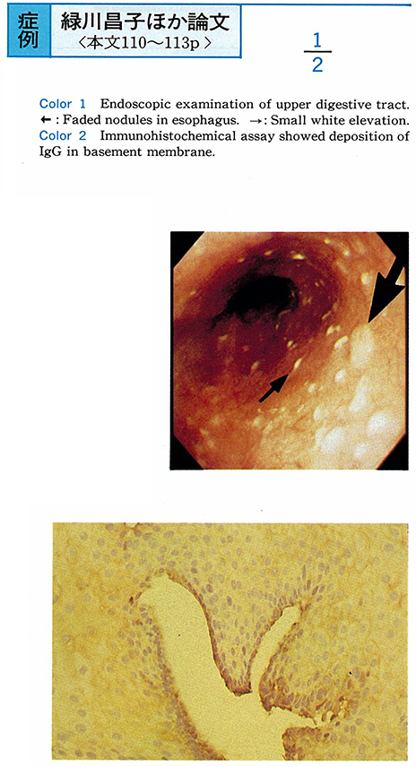
    1994 Volume 44 Pages 110-113
    Published: June 06, 1994
    Released on J-STAGE: May 25, 2015
    JOURNAL FREE ACCESS
    This paper describes esophageal involvement associated with bullous pemphigoid which we have experienced in our hospital.
    The patient is a man of 69 years, who has skin rashes in both legs since November 1992. He came to see a doctor of dermatology of our hospital. As a result of skin biopsy, he was diagnosed as having bullous pemphigoid and was admitted to our hospital.
    No subjective symptoms have been manifested, but endoscopic examination of upper digestive tract, performed to make a close investigation, revealed distended, softened and faded nodules in esophagus. Squamous epithelium was gathered from all strata of biopsy specimens. Just under the squamous epithelium, bullae-like blotch was observed. Moreover, immunohistochemical assay showed deposition of IgG in basement membrane. These results led us to diagnose this case as esophageal involvement associated with bullous pemphigoid.
    Download PDF (929K)
  • Eigo Sato, Tsuneo Kawasaki, Shunro Otsukasa, Masatoshi Jibiki, Togo Ao ...
    1994 Volume 44 Pages 114-118
    Published: June 06, 1994
    Released on J-STAGE: May 25, 2015
    JOURNAL FREE ACCESS
    We have experienced a case of double early gastric cancers which have arisen in the reconstructed stomach tube after radical operation for synchronous double cancers of the esophagus and stomach. Only 17 cases of such early gastric cancer including ours have been reported in Japan so far, and 6 cases were treated by endoscopical intervention.
    An asymptomatic 78-year-old man was pointed out having a polypoid lesion in the reconstructed stomach tube by upper GI series on medical check-up. Double early gastric cancers of IIa and IIb type were found by endoscopical examination, and were successfully treated by EMR and laser irradiation. On postoperative follow-up, it is important to check not only the recurrence of the esophagus cancer but also the existence of gastric cancer in the reconstructed stomach tube.
    Download PDF (1917K)
  • Satoru Shimono, Nobuhiro Takahashi, Toshikazu Sakuyama, Takeo Ohnishi, ...
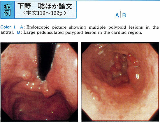
    1994 Volume 44 Pages 119-122
    Published: June 06, 1994
    Released on J-STAGE: May 25, 2015
    JOURNAL FREE ACCESS
    A 32-year-old man visited our hospital because he was diagnosed as having gastric cancer by endoscopic examination. At our endoscopy, multiple polypoid lesions were found in the antral region and several pedunculated polypoid lesions in the cardiac region and in the greater curvature of the gastric body. Large pedunculated polypoid lesion in the cardiac region was endoscopically polypectomized. Histologically, this lesion showed hyperplastic polyp, but multiple polypoid lesions in the antral region showed moderately differentiated tubular adenocarcinoma.
    Since he was diagnosed as IIa type early gastric cancer with multiple hyperplastic polyps, partial gastrectomy was performed. Histological findings showed the widely spreaded IIa type gastric cancer, moderately differenciated tubular adenocarcinoma with submucosal invasion surrounded with gastric juvenile polyps.
    This patient is considered to be a rare case in juvenile polyposis of the stomach complicated with gastric cancer. Therefore, this patient with juvenile polyposis are considered to require the endoscopic follow-up examination.
    Download PDF (1285K)
  • Makoto Nakamura, Takayuki Ishii, Yasuo Konuma, Takeki Yamane, Hiroshi ...
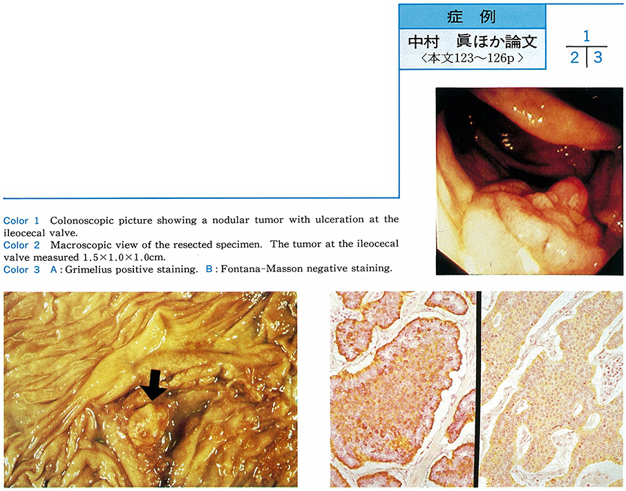
    1994 Volume 44 Pages 123-126
    Published: June 06, 1994
    Released on J-STAGE: May 25, 2015
    JOURNAL FREE ACCESS
    A 58-year-male came to our hospital with a complaint of lower abdominal pain. The barium enema examination showed a protruding lesion at the ileocecum. The colonoscopic examination revealed a nodular tumor with ulceration, measuring 15×10 mm in size, at the ileocecal valve. The result of the biopsy showed carcinoid tumor. No metastatic lesion was detected by the CT scan and the Ga scintigram. No specific result was found on the hormonal study. Then a right hemicolectomy was performed.
    This case was diagnosed as carcinoid tumor of the iliocecal valve. Only four similar cases have been reported in Japan.
    Download PDF (1083K)
  • Kayoko Sasamoto, Toshiyuki Hashimoto, Isao Sasaki, Hanako Kanazawa, Ma ...
    1994 Volume 44 Pages 127-129
    Published: June 06, 1994
    Released on J-STAGE: May 25, 2015
    JOURNAL FREE ACCESS
    A 35-year-old Japanease female was admitted to our hospital in June 1993 because of abdominal pain and nausea of a month's duration. She had been suffering from bronchial asthma since autumn of 1991.
    Laboratory examination on admission revealed leukocytosis, eosinophilia, thrombocytosis, increased erythrocyte sedimentation rate, elevated concentration of plasma immunoglobulin E and positive RA test. Colonoscopy disclosed multiple erosions and map-like, irregular ulcers with reddish mucosa of rectum through descending colon. Histological examination of biopsy specimen showed marked eosinophilic infiltration into ulcerated mucosa. Two weeks after hospitalization, muscle weakness and numbness of extremities due to multiple mononeuropathy occurred.
    After starting steroid pulse therapy, number of leucocytes and eosinocytes became normal and rapid improvement of her symptoms were recognized. Re-examination of colonoscopy and biopsy showed neither ulceration nor eosinophilic infiltration.
    Although reported cases of Churg-Strauss syndrome have been increasing, those with colonic disease are rare and only 7 cases were reported for latest 5 years in Japan.
    Download PDF (331K)
  • Shoji Maruyama, Tsuneo Kawasaki, Eigo Sato, Yousuke Izumi, Tougo Aoi, ...
    1994 Volume 44 Pages 130-135
    Published: June 06, 1994
    Released on J-STAGE: May 25, 2015
    JOURNAL FREE ACCESS
    We report a case of scirrhous type carcinoma of sigmoid colon of a 14-year-old boy and reviewed the cases. He admitted to our hospital because of the swelling of Virchow lymphnode and bloody stool. The biopsy of this LN revealed signet-ring cell carcinoma. After barium enema, sigmoid colon carcinoma was detected but the operation was ended only by resecting primary lesion.
    Now 56 cases of colorectal carcinoma younger than 15 years old including ours have been already reported in Japan. This disease is very rare in children, so in spite of many signs of exsisting colon disease, lack of barium enema and endoscopy made it difficult to find the early stage of this disease.
    Recently, the tendency of increasing colon cancer, we must examine the barium enema or endoscopy even in infant patients. This patient survived only 3 months.
    Download PDF (1682K)
  • Moriaki Tomikawa, Fuminori Horimukai, Yoshitaka Hirahara, Yasuhiko Sai ...
    1994 Volume 44 Pages 136-138
    Published: June 06, 1994
    Released on J-STAGE: May 25, 2015
    JOURNAL FREE ACCESS
    A 56-year-old male visited a hospital in his neighborhood with the complaint of anal bleeding. At that hospital, he was diagnosed as anorectal malignant melanoma on the basis of colonoscopic biopsy. Then he was admitted.
    Barium enema and colonoscopy demonstrated two elevated lesions contacting each other, and having irregular surface. The lesions, however, did not show black, the typical deature of melanoma. The pathological diagnosis of resected specimen was“hypomelanotic malignant melanoma”.
    In case of malignant melanoma, it is adbocated that to perform biopsy is contraindicated because it may promote metastasis. But, as in this case, it is very difficult to diagnose amelanotic or hypomelanotic malignant melanoma precisely by colonoscopy. We must distinguish anorectal malignant melanoma from rectal carcinoma so that we may choose different therapy for them as the case may be. At present, we should depend on biopsy to make differential diagnosis between them.
    Download PDF (663K)
  • Hiroshi Asakawa, Yasuhiro Satou, Tomoko Nakabayashi, Masayuki Kaneki, ...
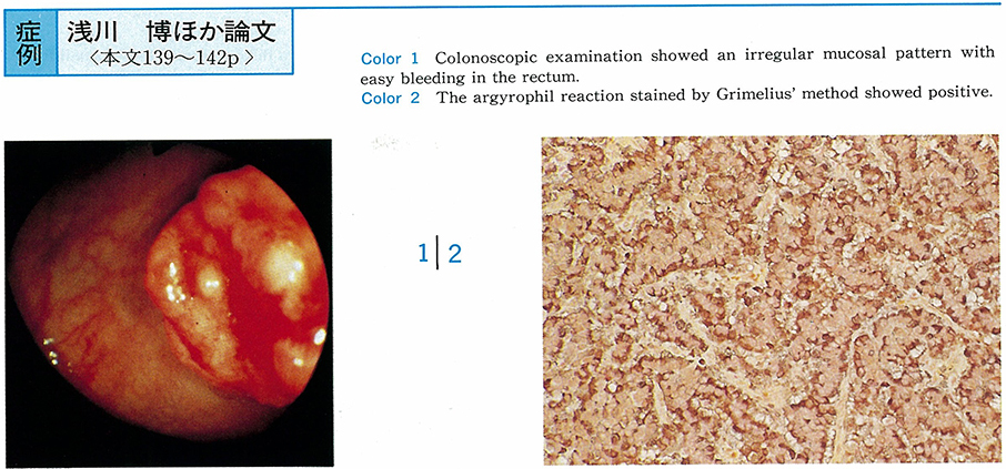
    1994 Volume 44 Pages 139-142
    Published: June 06, 1994
    Released on J-STAGE: May 25, 2015
    JOURNAL FREE ACCESS
    A 40-year-old female visited our hospital with a chief complaint of rectal bleeding. Digital and barium enema exnamination disclosed the elevated lesion with major axis of about 15 mm in the lower part of the rectum. Colonoscopic examination showed an irregular mucosal pattern with easy bleeding.
    Histological study of the biopsy specimens revealed that the lesion was endocrine cell carcinoma and positive for the argyrophil reaction by Grimelius' method. The tumor was removed by surgical operation.
    Endocrine cell carcinoma of the rectum is rare. Furthermore it is difficult to differentiate it from sm-invasive differentiated adenocarcinoma and carcinoid tumor by endoscopy. Endocrine cell carcinoma had worse prognosis than these diseases. It is therefore important to make a correct diagnosis by histological examination.
    Download PDF (1649K)
  • Kenkichi Tokumaru, Norio Ueno, Kiichi Tamada, Masahiko Ichiyama, Takes ...
    1994 Volume 44 Pages 143-146
    Published: June 06, 1994
    Released on J-STAGE: May 25, 2015
    JOURNAL FREE ACCESS
    A 55-year-old female was admitted to our hospital due to epigastric pain. Conventional ultrasonography and computed tomography revealed marked dilatation of the common bile duct (CBD) and CBD stone.
    Endoscopic retrograde cholangiopancreatography (ERCP) showed fusiform dilatation of the CBD and irregularity of the dilated CBD wall. The common channel of the choledochus and the pancreatic duct was twenty millimeters long. The diagnosis of congenital choledochal dilatation accompanied with anomalous arrangement of the pancreaticobiliary ductal system (AAPBDS) was made.
    Percutaneous transhepatic cholangioscopy (PTCS) and intraductal ultrasonography (IDUS) were performed. IDUS demonstrated the union of the choledochus and the pancreatic duct in the pancreatic parenchyma. It meaned the union existed outside of the duodenal wall and the diagnosis AAPBDS was confirmed.
    Although, ERCP alone could show the maljunction in this case, simultaneous IDUS will be useful for the accurate diagnosis of AAPBDS, especially in cases of shorter common channel demonstrated by ERCP.
    Download PDF (1637K)
  • Yoshifumi Takenaka, Tai Ohmori, Shigeo Ono, Keiichi Yoshino, Mayumi Ue ...
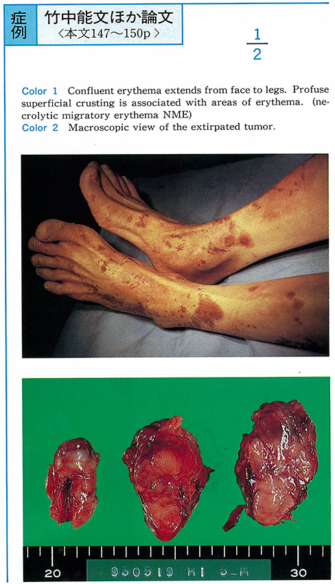
    1994 Volume 44 Pages 147-150
    Published: June 06, 1994
    Released on J-STAGE: May 25, 2015
    JOURNAL FREE ACCESS
    A 53-year-old male with recurrent skin eruption for about 3 years admitted to our hospital because of the pancreatic tumor found by CT.
    Laboratory findings indicated hypoproteinemia (total protein 5.7g/dl) and anemia (hemoglobin 10.0g/dl) . The oral glucose tolerance test (GTT) showed diabetic pattern. Serum glucagon levels were high. The cystic dilatation of the first branch of pancreatic duct was found by endoscopic retrograde pancreaticography. Arteriography showed a hypervascular tumor in the tail of the pancreas.
    Tumorectomy was done because tumor was easily separated from its surroundings. Histopathologically, it was an islet cell tumor and encapsuled by thin fibrous layer. These proliferative cells were positive for glucagon and pancreatic polypeptide. Neither mitotic figure nor vascular invasion was found. After the operation, his glucagon level and GTT became normal, and eruption (so called necrolytic migratory erythema) was cured.
    We found 67 cases of glucagonoma in Japanese literature. Out of these 68 cases including our cases, only 24 cases are accompanied with diabetes mellitus and erythema. Therefore we think this case is rare one.
    Download PDF (1450K)
  • Yasuo Okada, Jo Ariyama, Masafumi Suyama, Kaoru Ogawa, Kazuhiro Satou, ...
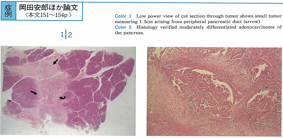
    1994 Volume 44 Pages 151-154
    Published: June 06, 1994
    Released on J-STAGE: May 25, 2015
    JOURNAL FREE ACCESS
    A 42-year-old male was admitted to our hospital due to abdominal discomfort and jaundice. Ultrasound (US) and computed tomography (CT) demonstrated dilatation of common bile duct, but tumor was not depicted.
    Endoscopic ultrasound (EUS) showed hypoechoic mass within the wall of the common bile duct, and acoustic shadow was observed adjacent to the mass. Percutaneous transhepatic biliary drainage (PTBD) revealed stenosis in the mid portion of the common bile duct. Arterial phase of gastroduodenal on angiogram encasement of posterior arcade branches was identified as small pancreatic carcinoma accomapnied with minimal invasion to parenchymal.
    Histology of resected specimen after pancreatoduodenectomy disclosed pancreatic ductal adenocarcinoma arising from peripheral side branch measuring 1.5×1.5×1.0cm. Wirsung, Santorini and the main pancreatic duct were normal. Pathological feature corelates to those of diagnostic imagings with EUS and angiography.
    Download PDF (1720K)
Technology and instrument
  • Shinji Kitamura, Yutaka Nagasawa, Takashi Murakami, Kazunari Iseki, It ...
    1994 Volume 44 Pages 156-157
    Published: June 06, 1994
    Released on J-STAGE: May 25, 2015
    JOURNAL FREE ACCESS
    Newly devised upper gastrointestinal endoscopes, GIF-XQ30 and GIF-P30, were evaluated clinically concerning to insertion, observation and biopsy channel comparing with GIF-XQ20 and GIF-P20.
    Endoscopists who examined the same 55 patients with GIF-XQ30 and GIF-XQ20, or GIF-P30 and GIF-P20 answered a questionnaire. It revealed that by using GIF-XQ30 or GIF-P30 examiners could insert endoscopes more easily to the duodenal bulb, observe more widely especially at the down-observation or at the inverted observation, and insert biopsy forceps more easily through the channel even at the inverted observation than by using GIF-XQ20 or GIF-P20.
    These results indicate that GIF-XQ30 and GIF-P30 are more useful in the endoscopic diagnosis than the previous endoscopes.
    Download PDF (182K)
  • Shinichi Nakamura, Atsushi Mitsunaga, Yukihito Nemoto, Ikuo Ikeda, Sat ...
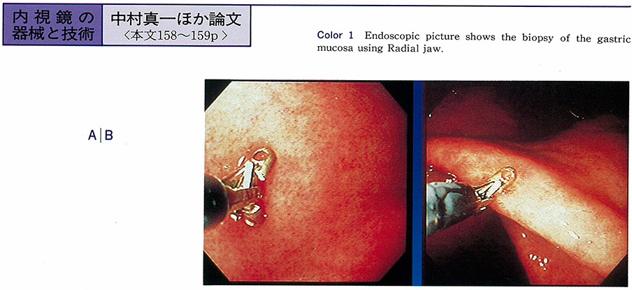
    1994 Volume 44 Pages 158-159
    Published: June 06, 1994
    Released on J-STAGE: May 25, 2015
    JOURNAL FREE ACCESS
    We reported the characteristics and usefulness of the disposable biopsy forceps (Radial jaw) from Microvasive Boston Scientific Corporation. Radial jaw has the tiny teeth around the cup and the wire with sheeth.
    We biopsied the specimens from the antrum in stomach. We discussed the size and the damage of biopsied pathological specimens obtained by Radial jaw and the Olympus standard forceps. We also observed the slip of cup and bleeding tendency after biopsy by both forceps.
    Radial jaw could biopsy 1.9 times larger specimens than standard. The damage of specimens was slight, and both of them were almost same level. Radial jaw was appropriate to perform the exact biopsy from oblique direction because of the cup form, and was useful of biopsy from long distance because of the sheeth. Bleeding tendency after biopsy was trivial in both forceps.
    Recently many kinds of biopsy forceps are used, Radial jaw is suitable for upper GI examination.
    Download PDF (549K)
  • Motoyasu Chibai, Nobuhiro Takahashi, Kimio Isshi, Manabu Yamamoto
    1994 Volume 44 Pages 160-161
    Published: June 06, 1994
    Released on J-STAGE: May 25, 2015
    JOURNAL FREE ACCESS
    Laparoscopic cholecystectomy (LC) is widely accepted as a standard procedure, however, higher complication rates are reported than open cholecystectomy. Aim of this study is to evaluate the usefulness of a portable laparoscopic doppler probe (LapaDop, HADECO, Kawasaki, Japan) for the prevention of arterial injury.
    LapaDop is light and compact equipment (75×280×65 mm, Wt 500g) . It has a frequency of 8 MHz and the probe is 4.8 mm in diameter that fits through a 5 mm laparoscopic port. We used this equipment for 30 patients who underwent LC. Cystic artery and other possible vascular structures were scanned before cauterization or ligation. The blood velocity wave, mean blood velocity speed and the heart rates were also measured and recorded.
    Vascular structures were identified in all cases without any injuries. Mean blood velocity speed of cystic artery was 8.3 cm/sec while the right hepatic artery was 20 cm/sec.
    To prevent the arterial injury, doppler probe was useful because it is easy to use and reliable to identify the vascular structures during LC.
    Download PDF (495K)
Clinical study
  • Masue Mutoh, Eisaku Kondo, Syuichi Yamada, Hiroshi Matsuzaki, Yoshihis ...
    1994 Volume 44 Pages 162-163
    Published: June 06, 1994
    Released on J-STAGE: May 25, 2015
    JOURNAL FREE ACCESS
    An ultrasonic probe (miniture ultrasonic probe, UM-1W, Olympus) was used in this study. It had an external diameter of 3.4mm and a radial scan transducer with a frequency of 7.5MHz. Ultrasonography with the miniture ultrasonic probe was performed in 7 cases before and after endoscopic injection sclerotherapy (EIS) for esophageal varices.
    Our technique of EIS is the para and intra-variceal injection of 1% aethoxysklerol (AS) . Low echo-lumen images absorbed before EIS where changed to high echo-ones from their circumference immediately after EIS. Also we observed that a part of collateral blood flow pathway of the extra walls which visualized as low echo-lumen images disappear.
    The followings were pointed out as important usefulness of a miniture ultrasonic probe.
    1) Observation of the esophageal varices before and after EIS, under endoscopically visual control.
    2) Performed EIS exactly, at the same time.
    3) We could grasped collateral blood flow pathway of the extrawalls noninvasible.
    Download PDF (586K)
  • Hiroshi Miyoshi, Tamami Hanashi, Yuji Hanatani, Tatsuo Asagoe, Tadahik ...
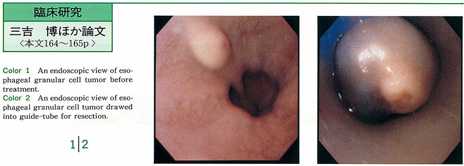
    1994 Volume 44 Pages 164-165
    Published: June 06, 1994
    Released on J-STAGE: May 25, 2015
    JOURNAL FREE ACCESS
    Four cases of esophageal granular cell tumor were treated using method of endoscopic esophageal mucosal resection (EEMR) . Either electric knife or guide-tube was employed with electric snare to complete EEMR method. All the tumors were entirely resected by this method, though one case suffered from fever and pleural effusion after treatment.
    Granular cell tumor sometimes appears as malignant tumor, therefore entire resection of this tumor using EEMR method should be recommended in stead of incomplete resection using endoscopic polypectomy method.
    Download PDF (909K)
Case report
  • Takashi Nakakuma, Atsushi Ihara, Yukihito Yamada, Yoshiki Hiki, Akira ...
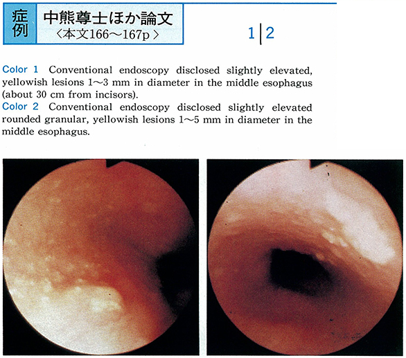
    1994 Volume 44 Pages 166-167
    Published: June 06, 1994
    Released on J-STAGE: May 25, 2015
    JOURNAL FREE ACCESS
    Two cases of sebaceous glands in the esophagus were diagnosed by panendoscopy and biopsy. Endoscopy disclosed a flat elevated round granular, yellowish lesion in the middle esophagus.
    As sebaceous glands is an ectodermal origin, it is very rare that we observe it in the esophagus, an endodermal origin. But we found two new cases with sebaceous glands in the esophagus during a half year. Twenty-one cases including our 2 cases during endoscopy have been reported. We consider that frequency of sebaceous glands in the esophagus will be increasing if we perform endoscopy strictly.
    Download PDF (718K)
  • Shigeo Ishida, Takeo Yamanaka, Yukio Yoshida, Hironari Matsumoto, Mits ...
    1994 Volume 44 Pages 168-169
    Published: June 06, 1994
    Released on J-STAGE: May 25, 2015
    JOURNAL FREE ACCESS
    A 42-year-old male consulted us because of hiccups on eating, which was revealed to be caused by an esophageal cyst. Though it is confirmed to be one of congenital diseases, such matters of importance as clinical classification and indication for operation still remain controversial.
    The therapeutic procedure we made was endoscopic ultrasonogram (EUS) -guided needle aspiration. The echoendoscope, Pentax FG32-UA, is a forward oblique-scanning fiberoptic scope with a convex type array ultrasonic transducer. The needle inserted through the instrumentation channel can be visualized both endoscopically and ultrasonically. This new method enabled us to puncture the esophageal cyst with accuracy and safety, and the lesion apparently decreased in size.
    In general, cystic lesions of gastrointestinal tract may be treated the best by EUS-guided needle aspiration. It is certain that the modality will play a more important role in both diagnosis and treatment in the near future.
    Download PDF (750K)
  • Masayuki Sekine, Motomichi Urabe, Noboru Mizobuchi, Satoshi Matsumura, ...
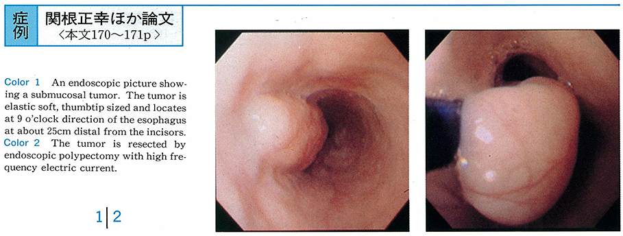
    1994 Volume 44 Pages 170-171
    Published: June 06, 1994
    Released on J-STAGE: May 25, 2015
    JOURNAL FREE ACCESS
    A 55-year-old man visited our clinic with chief complaints of upper abdominal discomfort and tarry stools. An upper endoscopic examination showed a submucosal tumor on the upper intrathracic esophagus and a gastric ulcer stage A2. The tumor was elastic soft, thumbtip sized and located at the 9 o'clock direction of the esophagus at about 25cm distal from the incisors. It showed an equable low echoic lesion that existed at the second layer by an endoscopic ultrasonic examination.
    It was resected by endoscopic polypectomy with high frequency electric current. The resected specimen was 23×13×15mm in size, was resected completely, and the leiomyoma originated from muscularis mucosae by a histopathological examination. Four weeks after polypectomy on the esophagus, the ulcer healed and was covered with normal esophageal epithelium.
    This case was dignosed as leiomyoma of the esophagus originating from muscularis mucosae due to endoscopic ultrasonic findings. Even though its size was over 20mm, it was resected safely and surely by endoscopic polypectomy, and the patient was cured with no complications.
    Download PDF (1194K)
  • Toshihiro Mita, Akira Torii, Masayuki Kaneki, Yasuhiro Satou, Mika Mat ...
    1994 Volume 44 Pages 172-173
    Published: June 06, 1994
    Released on J-STAGE: May 25, 2015
    JOURNAL FREE ACCESS
    A 19-year-old male was admitted to our hospital with chief complaints of epigastralgia and systemic edema. They occured 2 days after taking a non-steroidal anti-inflammatory drug. Serum total protein was 3.1g/dl.
    Endoscopic examination revealed an acute gastric mucosal lesion (AGML) at the area from the middle body to the antrum of the stomach. α1-anti-trypsin gastric clearance markedly increased, indicating that he had a protein-losing gastroenteropathy. He was treated with H2-receptor antagonist for 4 weeks and relieved from the systemic edema.
    Download PDF (495K)
  • Kazuhito Shirato, Mikio Matsuoka, Koichiro Dohmori, Kotaro Matsuyama, ...
    1994 Volume 44 Pages 174-175
    Published: June 06, 1994
    Released on J-STAGE: May 25, 2015
    JOURNAL FREE ACCESS
    A 74-year-old female underwent gastrointestinal endoscopy due to chief complaint of nausea. Endoscopy revealed gastric polyp with stalk below the esophagogastric junction. It had the erosion and the bleeding on the surface of the top. Polypectomy was done due to the suspicious for malignancy through the biopsy. However, the tissue, which was suspected for malignancy was a part of pseudosarcomatous granulation tissue, and the polyp as a whole was the hyperplastic polyp with the erosion of the top.
    Pseudosarcomatous change is sometimes found in the inflammatory reaction, especially the reparative process of the erosion. However, it is said that the ease is very rare.
    Histologically, its change consists of the granulation tissue with giantic and atypical histiocytes, therefore, some cases were misdiagnosed for malignancy in small biopsies.
    Our experience suggests that it is not difficult to deny malignancy, if you examine the polypectomized material, and compair it with a histological features of granulation tissue in a biopsy.
    Download PDF (855K)
  • Takeshi Suzuki, Manabu Asada, Akira Nakamura, Yoshihiko Inaba, Ei Itob ...
    1994 Volume 44 Pages 176-177
    Published: June 06, 1994
    Released on J-STAGE: May 25, 2015
    JOURNAL FREE ACCESS
    A 67-year-old female having been checked of her anemia was referred to upper GI series and endoscopy, and they revealed a huge protruding lesion with multinodular and multigranular appearance at the angulus of the stomach.
    The patient received subtotal gastrectomy, after the biopsy result of well differentiated tubular adenocarcinoma. On the resected specimen, the tumor size was 9.0×7.5cm and its height 2.1cm. In spite of the big size of the tumor, the depth of invasion was limited within mucosal layer.
    Download PDF (954K)
  • Atsushi Kiyohashi, Hideya Sano, Osamu Motohashi, Seiichi Takagi, Youic ...
    1994 Volume 44 Pages 178-179
    Published: June 06, 1994
    Released on J-STAGE: May 25, 2015
    JOURNAL FREE ACCESS
    The case was a 46-year-old male. A duodenal polyp had been diagnosed for the first time 3 years ago when he had had a melena. This time he came to our hospital for a surgical treatment. The upper gastrointestinal endoscopy showed a giant pedunculated polyp with the base at the posterior wall of the duodenal bulb and with an erosion at the top.
    On August 18, 1993 endoscopic polypectomy was performed without any complication. The size of the resected specimen was 4.2×3.5×2.0 cm. This was histologically hyperplasia of the Brunner's gland and one of the greatest endoscopically resected polyps of the Brunner's gland hyperplasia.
    Download PDF (1263K)
  • Masayoshi Odagiri, Norichika Narimiya, Kenji Komiyama, Masahiko Iwasak ...
    1994 Volume 44 Pages 180-181
    Published: June 06, 1994
    Released on J-STAGE: May 25, 2015
    JOURNAL FREE ACCESS
    A 69-year-old male had a hepatocellular carcinoma (HCC) (60 mm in size) in the right hepatic lobe in July 1988, and received transcatheter arterial embolization (TAE) twice and ethanol injection therapy 11 times. When the patient was examined in February 1993, the HCC had occupied the area from the right to the left lobe and part of the tumor had protruded through the inferior plane of the liver.
    The patient received endoscopy in May 1993 because of melena. At that time, the esophagus and the stomach were free of varices, but the duodenal bulb had an elevated lesion with an irregular surface and a hemorrhagic tendency. This lesion was rated as representing direct invasion of the HCC by biopsy and diagnostic imaging.
    Duodenal invasion or metastasis by HCC was less frequent and 9 cases have been reported in Japan (1981-1993) . Our experience with this case indicates that adequate examinations are needed of the duodenum, in addition to examinations of varices and ulcer, when patients with HCC-complicated liver cirrhosis receive emergency endoscopy due to bleeding from the upper gastrointestinal tract.
    Download PDF (1299K)
  • Makoto Okuda, Hideo Matsui, Yo Isobe, Atsushi Kubochi, Shunji Ikeuchi, ...
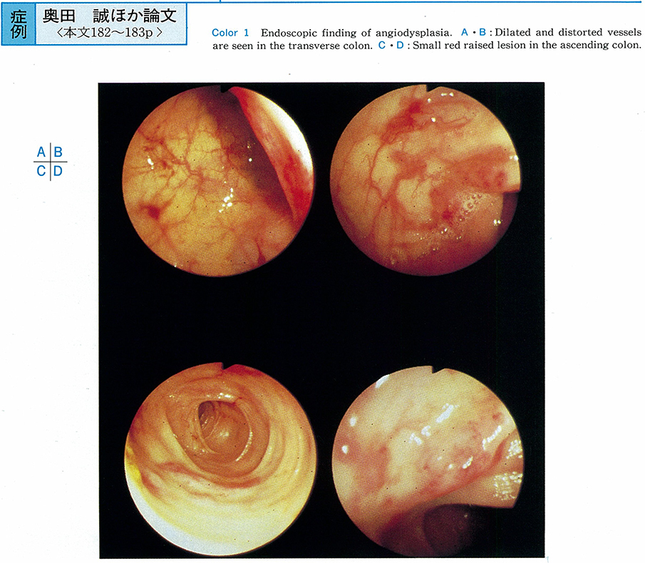
    1994 Volume 44 Pages 182-183
    Published: June 06, 1994
    Released on J-STAGE: May 25, 2015
    JOURNAL FREE ACCESS
    A 74-year-old woman who was treated as iron deficiency anemia for more than 8 years was admitted because of lassitude and palpitation. The Hb concentration was 6.6g/dl, iron was 8µg/dl and occult blood in the stool was positive. Results of barium roentgenographic studies of the stomach, small intestine and large intestine were normal. Result of 99mTc-albumin scan was also normal.
    Total colonoscopy revealed several vascular ectatic areas and small red discreted raised lesion in the sigmoid, descending, transverse, ascending colon and cecum. An arteriogram was performed and revealed a vascular tuft and early filling vein in right-sided colon.
    The descending, transverse, ascending colon and cecum were resected and ileosigmoidostomy was performed. Results of a pathologic examination of the resected colon demonstrated multiple dilated distorted veins in submucosa.
    Because of heigher predictive value, colonoscopy should be the first line investigation in the patients of chronic lower intestinal bleeding. After that, selective angiography should be performed to make accurate diagnosis as angiodysplasia.
    Download PDF (1055K)
  • Tomoko Kobayashi, Masato Nakajima, Keisuke Sasaki, Morihito Igawa, Yum ...
    1994 Volume 44 Pages 184-185
    Published: June 06, 1994
    Released on J-STAGE: May 25, 2015
    JOURNAL FREE ACCESS
    A 60-year-old male patient visited our hospital complaining of abdominal pain. He was hospitalized because he was diagnosed cholecystitis by ultrasonogram. After hospitalization, cholecystitis was recovered with the aid of antibiotics, but abdominal pain continued.
    Barium enema X-ray and colonoscopy revealed a smooth surfaced yellowish semipedunclated submucosal tumor sized 13 mm in diameter in the cecum, and colonoscopic polypectomy was applied.
    Histology with HE staining showed a well demarcaled tumor in the submucosa and revealed the presence of eosinophilic granules in the cytoplasm of the tumor cells. Using immunohistochemical study, the tumor cells were to be positively stained by S-100 protein and neuron specific enolose (NSE) , but negatively stained by Desmin.
    So the diagnosis of granular cell tumor was made. Granular cell tumor of the large bowel in Japan were made a report of 15 inclusive of our case.
    Download PDF (957K)
  • Shigeru Sorimachi, Jun Sasaki, Tsugio Yoshimine, Toushoku Minoo, Hirof ...
    1994 Volume 44 Pages 186-187
    Published: June 06, 1994
    Released on J-STAGE: May 25, 2015
    JOURNAL FREE ACCESS
    A 55-year-old woman was admitted to our hospital for the evaluation of liver masses. Ultrasonography revealed multiple masses of the liver. They appeared as low density areas on a CT scan. The masses were diagnosed as metastatic tumors in the liver. While exploring for the primary site, we observed a flat tumor with a central depression in the rectum, located 5 cm from anal ring. The tumor was small (10 mm) . It was histologically diagnosed as a rectal carcinoid tumor.
    As there were no metastatic lesions other than liver metastasis. Liver biopsy was performed. Histologically, the masses were diagnosed as liver metastasis of rectal carcinoid tumor. It was very rare for a rectal carcinoid tumor, which was smaller than 10mm in diameter. The tumor size and the presence of depression were considered important diagnostic indices associated with malignancy.
    Download PDF (840K)
  • Yasuo Mizumura, Shuji Niki, Kazuya Takeda, Mari Ide, Kazuhiko Miwa, Ka ...
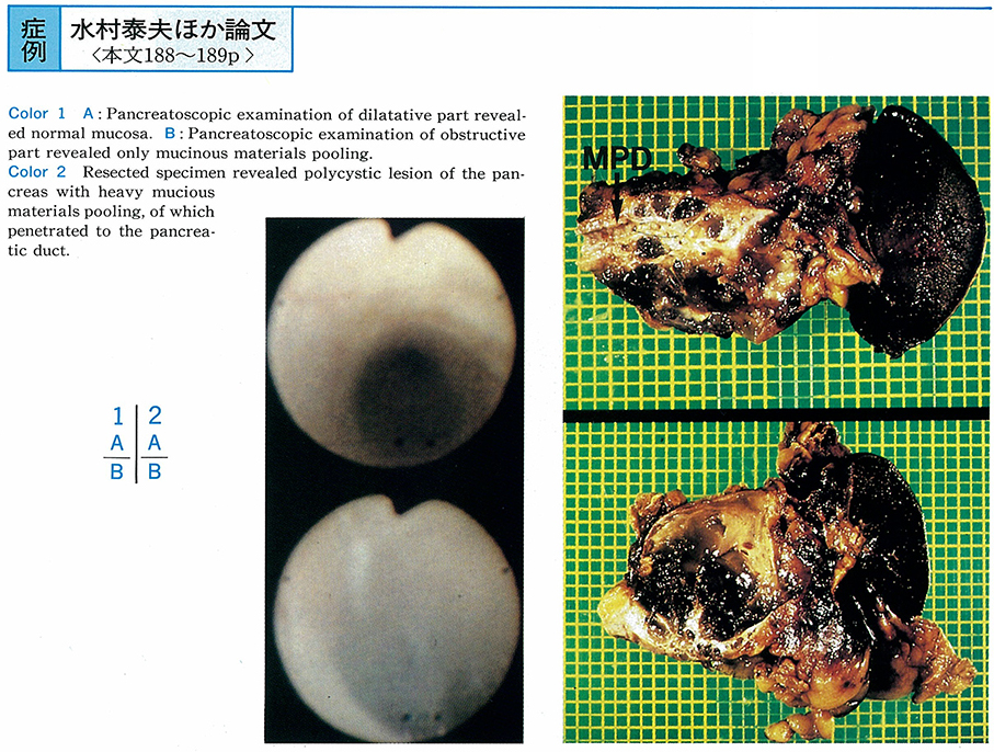
    1994 Volume 44 Pages 188-189
    Published: June 06, 1994
    Released on J-STAGE: May 25, 2015
    JOURNAL FREE ACCESS
    A 72-year-old female who was admitted to nearby hospital because of acute abdominal pain and who was found to have cystic lesion of the pancreas was referred to our hospital for further examination.
    Balloon-ERP demonstrated the cystic dilatation and obstruction, which have the possibility to be induced by tumor, in the main duct of the body of the pancreas. Peroral pancreatoscopy observing the obstructive lesion enabled us to recognized mucious materials pooling in the main duct. Distal pancreatectomy was performed under the diagnosis of mucin producing cystic tumor.
    Resected specimen revealed polycystic lesion of the pancreas with heavy mucinous materials pooling, which penetrated the main duct. Histological examination showed diagnosis of cystadenocarcinoma which have no invasion to the pancreas parenchyma.
    Peroral pancreatoscopy is considered to be an useful tool in such a case as our's.
    Download PDF (974K)
feedback
Top