Abstract
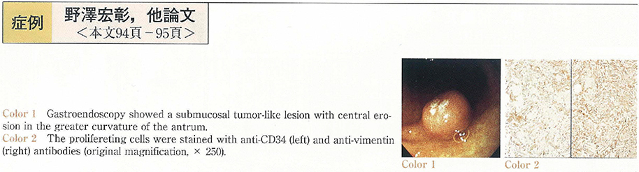
A 53-year-old woman was admitted to our hospital for evaluation and treatment of the abnormality in the gastric antrum revealed by barium fluorography. Endoscopic examination showed a semi-pedunculated submucosal tumor-like lesion with slight central erosion, 1 cm in diameter, in the greater curvature of the antrum. Furthermore, two similar polyps of 5 mm and 3 mm in diameter were found in the anterior wall of the angle. All the biopsy specimen of these polyps showed benign findings (Group I) . Since she was asymptomatic, only the polyp in the antrum was removed by endoscopic mucosal resection. Histopathologically, spindle-shaped cells proliferated chiefly in the submucosal space, which were positive for CD34 and vimentin. The polyp was diagnosed as inflammatory fibroid polyp (IFP) that exhibited unusual histological features ; typical“onion-skin pattern”was not observed, and infiltration of eosinophils was mild. Interestingly, the other polyps remained disappeared by endoscopy 6 months later. The observation seems to support that IFP may be developed through inflammation.