Progress of Digestive Endoscopy(1972)
Online ISSN : 2189-0021
Print ISSN : 0389-9403
Current issue
Displaying 1-37 of 37 articles from this issue
Technology and instrument
  • Yoshiki Kida, Mitsuhiro Kida, Miyoko Takezawa, Kyouko Shirasu, Yoshiki ...
    2000 Volume 57 Issue 2 Pages 20-23
    Published: November 15, 2000
    Released on J-STAGE: November 04, 2014
    JOURNAL FREE ACCESS
    The result of endoscopic diagnosis and treatment for diseases in billiary system dose not only depend heavily on operator's abilities but utilization of the endoscopic implement serving the purpose. Development and improvement of endoscopic implements is always of importance. In this report, we discussed following 5 endoscopic implements in the development stage. 1) ERCP (Endoscopic Retrograde Cholangio-Pancreatography) catheter with side holes (Olympus Co. Ltd.) to obtain bile sample for cytological and genetic diagnosis. 2) Guide wire brushing catheter for selective insertion into the target bile duct brunch. 3) Guide wire basket catheter for selective insertion into the bile duct brunch having bile-stone or Vater's papilla with insertion difficulties. 4) Revolving basket catheter for catching bile duct stone with ease. 5) ERCP catheter with the divisions of a scale for choosing the optimum length of ERBD (Endoscopic Retrograde Billiary Drainage) stent.
    Download PDF (1830K)
Clinical study
  • Shinichi Nakamura, Atsushi Mitsunaga, Yoko Hoshino, Maiko Kishino, Hir ...
    2000 Volume 57 Issue 2 Pages 24-28
    Published: November 15, 2000
    Released on J-STAGE: November 04, 2014
    JOURNAL FREE ACCESS
    The subjects were 120 patients undergoing endoscopic treatments for esophageal variceal bleeding between January 1995 and December 1999. The aim of study was retrospectively to assess the therapeutic results of endoscopic treatments for esophageal variceal bleeding. The patients consisted of 87 males and 33 females with a mean age of 59.5 years. The location of variceal bleeding was esophagus in 96 patients and Lg-c in 24 patients. The initial endoscopic treatment for variceal bleeding was endoscopic variceal ligation (EVL) in 70 patients (58.3%) , endoscopic injection sclerotherapy (EIS) in 32 patients (26.7%) , and Histoacryl injection therapy in 18 patients (15.0%) . EVL was carried out in 67.7% of esophageal bleedings, Histoacryl injection therapy was carried out in 54.1% of Lg-c bleeding. Hemostasis of variceal bleeding was achieved in 118 patients (98.3%) , hemostatic difficulty encountered in 2 patients (1.7%) . No statistical differences were seen in the Kaplan-Meier analysis of the cumulative rebleeding-free rates in endoscopic treatment, liver function, variceal form, and bleeding location. Rebleeding rate was significant higher in the with hepatocellular carcinoma (HCC) group (P<0.05) . Survival rate was significant higher in the EIS group (P<0.05) in the Kaplan-Meier analysis of the cumulative survival rates in endoscopic treatment. Survival rate was significant lower in the Child-Pugh C group of liver function (P<0.05) and in the with HCC group (P<0.05) .
    EVL was carried out in more than 60% patients with variceal bleeding, and has just turned the first choice of endoscopic technique for esophageal variceal bleeding. Histoacryl injection therapy is useful for Lg-c and esophageal variceal bleeding with hemostatic difficulty. The underlying liver function and the concomitant HCC are considerd to be the important factors in rebleeding and outcomes.
    Download PDF (575K)
  • Mari Fujii, Miyako Itou, Masato Katagiri, Shigeru Nakano, Hiroshi Mats ...
    2000 Volume 57 Issue 2 Pages 30-33
    Published: November 15, 2000
    Released on J-STAGE: November 04, 2014
    JOURNAL FREE ACCESS
    The endoscopic ligation therapy for esophageal varices is now widely accepted, and is thought to be less effective on the variceal inflow vessels than other procedures. We have applied eradicative endoscopic variceal ligation (eEVL) to esophageal varices to make up for this disadvantage. We used color-Doppler endoscopic ultra-sonography (CDEUS) to measure the blood flow of the left gastric vein (LGV) . Ten patients, who were not treated previously, were enrolled in this study, and underwent eEVL which involves the application of approximately 40 O-rings for each patient. The LGV flow was measured by ultrasonographic system (EUB 525, HITACHI) and fiberscopic system (FG36UX, PENTAX) before and after eEVL to ligate not only the varices but also the esophago-gastric junction and the intervariceal regions. We measured the diameter, the direction, the velocity and the flow volume of LGV and its anterior and posterior branches. As the result of our investigation, the blood flow of the LGV either decreased or was not detected. And in other cases, the hepatofugal blood flow of post erior branch of LGV continued after therapy. Thus eEVL is an effective theraputic procedure for reducing the blood flow to esophageal varices.
    Download PDF (837K)
  • Takeshi Matsuhisa, Takahiro Hayama, Joji Yoshimura, Nobutaka Yamada
    2000 Volume 57 Issue 2 Pages 34-39
    Published: November 15, 2000
    Released on J-STAGE: November 04, 2014
    JOURNAL FREE ACCESS
    Atrophy and intestinal metaplasia of gastric mucosa in Helicobacter pylori (Hp) non-infected cases were studied comparing with Hp infected cases.
    1) Frequency of endoscopic atrophy at the point of lesser curvature of the lower body in Hp non-infected cases was lower than that of Hp infected cases (each 17.6%, 36.5%) . 17.6% was high score and it was beyond our expectation.
    2) We got a positive correlation between age and endoscopic mucosal score in Hp non-infected cases as well as infected cases. It was same in age and intestinal metaplasia score.
    3) Frequency of glandular atrophy and intestinal metaplasia were higher in Hp infected cases than non-infected cases except intestinal metaplasia of number 2 specimen.
    4) We couldn't find a correlation between age and serum pepsinogen (PG) I/II ratio, PGI/II ratio and endoscopic gastric mucosa in Hp non-infected cases. On the other hand, there were negative correlation between age and PGI/II ratio, PGI/II ratio and endoscopic gastric mucosa in Hp positive cases.
    Download PDF (587K)
  • Noboru Yoshimura, Koji Matsuzaki, Shigeaki Nagao, Atsushi Kawaguchi, J ...
    2000 Volume 57 Issue 2 Pages 40-44
    Published: November 15, 2000
    Released on J-STAGE: November 04, 2014
    JOURNAL FREE ACCESS
    [Background & Amis]Various methods of endoscopic gastric mucosal resection have been developed. EMRC (Endoscopic Mucosal Resection Using a Oblique Trnsparent Cap-Fitted Panendoscopy) is a one of useful techniques for mucosal resection. However, the vertical ranges of resection by this method have not been clearly determined. In this study, we investigated the surgical depth of EMRC specimen, and demonstrated its usefullness and safety.[Methods]Tissue samples were obtained by EMRC fro 42 lesions with early gastric cancer or adenoma with informed consent. We classified 42 lesions into two groups, the elevated type and the flat or depressive type. Each surgical depth was measured by using haematoxylin-eosin staining sections. These sections were observed under a microscope and were measured by using NIH image. We defined two kinds of surgical depth, i. e. m-depth and sm-depth. And we measured the horizontal size of these EMRC specimens.[Results]1) All specimens were resected at the submucosal layer. Of 42 lesions, m-depth was 2.19±0.83mm, and sm-depth was 1.11±0.67mm. (mean±SD) 2) The m-depth of the elevated type was greater than the flat or depressive type, and the sm-depth of the flat or depressive type was greater than the elevated one, although not statistically significant (p=0.09, p=0.21) . 3) Both m-depth and sm-depth appeared to be not correlated with the horizontal size of these specimens.[Conclusion]These results suggest that injection of sufficient volume of saline solution into the submucosal layer is important to prevent perforation, especially, in case of the flat or depressive type. We also demonstrated that if we can inject sufficient volume of saline, the lesions of gastric cancer slightly invading the submucosal layer would be completely resected by EMRC. In conclusion, EMRC is a simple, safe and useful method of tratment for gastric mucosal lesions.
    Download PDF (983K)
  • Terumi Kamisawa, Naoto Egawa, Nobuhiro Sakaki, Jun-ichi Ishiwata, Tosh ...
    2000 Volume 57 Issue 2 Pages 45-49
    Published: November 15, 2000
    Released on J-STAGE: November 04, 2014
    JOURNAL FREE ACCESS
    Point mutations of the K-ras oncogene at codon 12 were examined by enriched-PCR/ELMA method in DNA obtained from pancreatic juice and duodenal aspirate collected endoscopically in patients with 31 pancreatic carcinoma, 26 mucin producing pancreatic tumor and 19 chronic pancreatitis. Incidence of K-ras mutation in pancreatic juice was 73% of patients with pancreatic carcinoma, 70% of those with mucin producing pancreatic tumor, and 40% of those with chronic pancreatitis. Semiquantitative analysis showed that the mutant gene occupied more than 2% of total DNA in 55% of patients with pancreatic carcinoma and 39% of those with mucin producing pancreatic tumor, but only 13% of those with chronic pancreatitis. Analysis of K-ras oncogene in pancreatic juice can be useful for detecting the high risk group of pancreatic carcinoma, and semiquantitative analysis may provide a clinical tool for differential diagnosis of pancreatic carcinoma.
    K-ras mutation was detected in duodenal aspirate with secretin stimulation in 32% of patients with pancreatic carcinoma. This analysis in duodenal aspirate may also be useful for screening the high risk patients of pancreatic carcinoma.
    Download PDF (413K)
  • Takahiro Hayama, Yasuo Inoue, Takeshi Matsuhisa
    2000 Volume 57 Issue 2 Pages 50-51
    Published: November 15, 2000
    Released on J-STAGE: November 04, 2014
    JOURNAL FREE ACCESS
  • Kouji Otsuka, Masahiko Murakami, Takashi Katou, Toshihiro Fujioka, Yos ...
    2000 Volume 57 Issue 2 Pages 52-53
    Published: November 15, 2000
    Released on J-STAGE: November 04, 2014
    JOURNAL FREE ACCESS
    54 cases of acute appendicitis were operated laparoscopically for past 3 years (Three cases were converted to open exploration.) . Patients were divided into three groups according to postoperative pathological findings of appendix. (Group A : catarrhal, Group B : phlegmonous, Group C : gangrenous) . In each groups, operation time, postoperative period of bed rest and NPO (nothing per os) , pyretic term after operation, length of hospital stay and postoperative complications were analyzed to estimate the quality of laparoscopic appendectomy. Three ports were inserted on the abdomen ; under umblication, right upper abdomen (3 or 5 mm) and above left pubic (12 mm in a diameter) . After looking over the whole abdominal cavity, the mesoappendix was divided including an appendix artery by LCS (Laparosonic Coagulating Sheers System, Ethicon) and then, the root of appendix was dissected by Endo-GIA (Auto suture) or ligated by Endo-loop (Ethicon) . Resected appendix stored in a vinyl bag was taken out through a port of above pubic. Abdominal cavity was washed out by saline water (1,000-7,000 ml) . No drainage tubes were applied to all cases.
    In Group C, period of bed rest was longer than Group A. No significant difference was detected in another classification. There were no remarkable complications as wound infection or abdominal abscess after surgery except paralytic ileus in two patients. These results demonstrate laparoscopic surgery is appropriate, and should be standard method for acute appendicitis.
    Download PDF (258K)
  • Takeshi Nagahama, Michio Maruyama, Tatsuya Yoshida
    2000 Volume 57 Issue 2 Pages 54-55
    Published: November 15, 2000
    Released on J-STAGE: November 04, 2014
    JOURNAL FREE ACCESS
    Endoscopic treatment and observation of colon for poorly prepared patients is difficult from poor visibility during procedures. We have experienced transparent cap fitted colonoscopy for five patient who could not tolerate mechanical bowel preparation. Insertion of colonoscopy to the level of cecum was easily attained for colon bleeding and colon volvulus under good visualization. Insertion to the level of stricture was also easily attained for patients with sigmoid colon cancer. With this transparent cap fitted procedure it was easy to maintain a visual field between colonic mucosa and the tip of colonoscope. Therefore adhesion of stool and blood to the surface of the tip could be easily avoided. Maneuverbility of scope during insertion was relatively poor than that without transparent cap as elongation of bending section of endoscopy. However attaining fair visibility is more beneficial for endoscopic observation and treatment.
    We can conclude that transparent cap is a useful device for colonoscope with poor bowel preparation.
    Download PDF (1740K)
  • Yoichiro Fujii, Toshimitsu Murohisa, Yutaka Okamoto, Koji Sudoh, Hidet ...
    2000 Volume 57 Issue 2 Pages 56-57
    Published: November 15, 2000
    Released on J-STAGE: November 04, 2014
    JOURNAL FREE ACCESS
Case report
  • Yuji Hayashi, Yoshihisa Sekita, Masao Tani, Naoya Saito, Tatsuyuki Kaw ...
    2000 Volume 57 Issue 2 Pages 58-60
    Published: November 15, 2000
    Released on J-STAGE: November 04, 2014
    JOURNAL FREE ACCESS
    A 17 years-old woman was admitted with upper abdominal pain and hematemesis. By endoscopic examination, Yamada IV type of protruded tumor (3cm in diameter) with an ulcer and hemorrhage from the ulcer were detected in the stomach. Endoscopic-ultrasonography revealed the muscular layer was intact with no invasion. The tumor was resected endoscopically using a detachable snare successfully.
    The immunohistochemical staining of the tumor was positive only for S-100 protain, and the tumor was diagnosed as gastric schwannoma histologically.
    In some cases, gastric schwannoma tend to have hemorrhagic ulcer. So such case was often found out by melena or hematemesis.
    The cases of gastric schwannoma have been reported almost in middleaged or senior patients. The case of gastric schwannoma developed in a juvenile patient as this case was quite rare.
    Download PDF (1354K)
  • Daigo Onodera, Shigeaki Nagao, Atsushi Kawaguchi, Yukiko Yoshida, Nori ...
    2000 Volume 57 Issue 2 Pages 61-63
    Published: November 15, 2000
    Released on J-STAGE: November 04, 2014
    JOURNAL FREE ACCESS
    Case 1 : In 1999, a 87-year-old female visited our hospital because of appetite loss. The endoscopic examination revealed a giant ulcer with a white plague in the posterior wall of the antrum. The pathological diagnosis was MALT lymphoma with existence of Helicobacter pylori (HP) , and therefore she received the eradication therapy with 1,500mg/day of amoxicillin, 40mg/day of lansoplazole, and 400mg/day of clarithromycin, respectively, for 10 consecutive days. The endoscopic findings were improved and the giant ulcer was disappeared.
    Case 2 : In 1997, a 83-year-old male underwent an endoscopic examination because of epigastralgia. It revealed multiple ulcers in the greater curvature from the upper to lower gastric body. He was pathologically diagnosed as MALT lymphoma. Though HP was not proven by either pathological examination, 13C-urease breath test or by bacterial culture examination, the eradication therapy was done with 1,500mg/day of amoxicillin, 40mg/day of lansoplazole, and 400mg/day of clarithromycin, respectively, for 14 consecutive days. Though the endoscopic findings were not remarkably improved and ulcers still remained, they were not aggravated at least. Many reports have recently suggested the possible relation between MALT lymphoma and HP, but few cases were investigated in the elderly patients. We report two cases of MALT lymphoma in elderly patients who were treated only by eradication therapies, and suggest that eradiation will be one of treatments for MALT lymphoma.
    Download PDF (1098K)
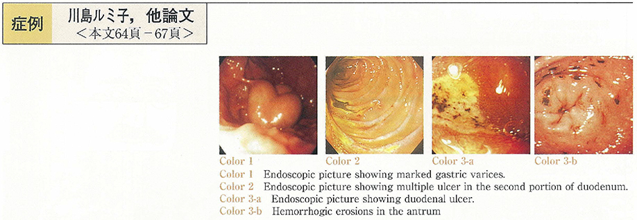
  • Rumiko Kawashima, Yukinori Imai, Shin Arai, Koutarou Kakoi, Yumi Mashi ...
    2000 Volume 57 Issue 2 Pages 64-67
    Published: November 15, 2000
    Released on J-STAGE: November 04, 2014
    JOURNAL FREE ACCESS
    A 81-year-old man with type C liver cirrhosis and hepatocellular carcinoma was admitted to our hospital for the treatment of gastric varices. On Nov. 29th. 2000, B-RTO was performed. From next day, ascites appeared and gradually increased.
    He complained of epigastralgia. Melena was noticed. Upper gastrointestinal fiberscope revealed the presence of esophagitis, multiple gastric hemorrhagic erosions and multiple duodenal ulcers. Esophagitis and gastric erosions healed and duodenal ulcers improved to H1 stage by the treatment with proton pump inhibitor (PPI) for 10 days. After ascites also disappeared by use of diuretics, he discharged.
    Since ascites increased in the beginning of January 2000, he was re-admitted to our hospital on January, 22th. Two days later, he had hematemesis and similar extensive lesions to previous examination were found. These lesions appeared following occurrence of ascites, suggesting that the elevation of portal venous pressure induced by B-RTO might play a causative role in gastro intestinal injuries of this patient.
    Download PDF (1233K)
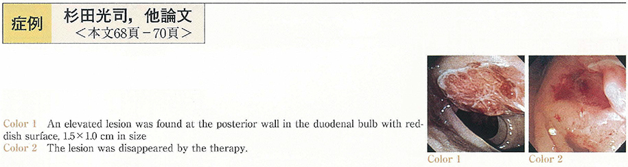
  • Koji Sugita, Yoshitake Kitagawa, Yukiko Yoshida, Hiroyuki Nakajima, No ...
    2000 Volume 57 Issue 2 Pages 68-70
    Published: November 15, 2000
    Released on J-STAGE: November 04, 2014
    JOURNAL FREE ACCESS
    A 55-year-old male was admitted to our hospital due to anemia, who underwent the right total nephrectomy for renal cell cancer (R. C. C.) with curative operation in 1986. Endoscopic examination revealed a submucosal tumor with erosion on the top in duodenal bulb. The diagnosis was made as metastatic R. C. C. in the duodenum from the biopsy specimens. Several other examinations revealed that his metastases of the disease spreaded in the pancreas, the liver, the left shoulder blade, the left lung as well. Three million units of interferon α was administered intramuscularly twice a week for 36 weeks. The endoscopic examination after 16 weeks showed the reduction of the size of the tumor. Tumor was disappeared after 28 weeks, and the pathological findings showed no malignancy.
    Metastases of RCC have been often reported in the lungs, the liver, and the bones, but rarely done in the gastrointestinal tract. The effectiveness of interferon has been reported especially in the case of metastases to the lungs. This is a very rare case of metastatic RCC in the duodenum, which was diagnosed 13 years after curative right nephrectomy and for which interferon therapy was effective.
    Download PDF (944K)
  • Shizuka Nagaba, Kiyonori Kobayashi, Miwa Sada, Shigeru Yoshizawa, Masa ...
    2000 Volume 57 Issue 2 Pages 71-74
    Published: November 15, 2000
    Released on J-STAGE: November 04, 2014
    JOURNAL FREE ACCESS
    A 21-year-old female had been suffering from relapsing symptoms with diarrhea and bloody stool for 6 years from the onset of ulcerative colitis (total colitis) . Her clinical symptoms could not be controlled by the therapies with corticosteroid and immunosuppresive drugs. She was admitted to our hospital because of abdominal pain and bloody stool. Colonoscopic examination showed the rectal tumor occupying hole of the lumen, and histological diagnosis of the biopsied specimens was poorly differentiated adenocarcinoma mixed with signetring cell carcinoma. Total colectomy and Miles' operation was performed. Macroscopic findings of the resected specimen showed that the nodular tumor was widely spread in the rectum, and active inflammation with multiple ulcers were also seen in the entire colon. Histological findings showed that high grade dysplasia were observed in some part of the tumor.
    It is relatively rare to develop the colitic cancer over a short term from the onset of ulcerative colitis. It is necessary to start the surveillance colonoscopy more earlier for the patient with chronic persistent colitis.
    Download PDF (1989K)
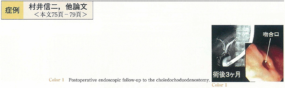
  • Shinji Murai, Kenta Motogami, Kouji Fujita, Shinnichi Handa, Akihiko N ...
    2000 Volume 57 Issue 2 Pages 75-79
    Published: November 15, 2000
    Released on J-STAGE: November 04, 2014
    JOURNAL FREE ACCESS
    Case 1 : A 30-year-old woman visited the hospital for the chief complaint of pain in the right hypochondrium. After close examination, pancreatic-duct-biliary-tract confluence disorder associated with cystic dilatation of the common bile duco was diagnosed. The dilated portion of the common bile duct was subsequently resected under a laparoscope, and common-hepatic-duct-duodenum anastomosis was performed. Case 2 : A 53-year-old woman visited a local clinic for the chief complaint of upper abdominal pain, where US examination revealed a polyp of gallbladder, and she was referred to our hospital. US and EUS examinations revealed protruded lesions (maximum ta) in the gallbladder, which had a maximum diameter of 25 mm. ERCP revealed coexisting confluence disorder of the pancreatic duct and biliary tract without dilatation of the common bile duct. Laparoscopic cholecystectomy was performed, and after identifying a benign tumor by intraoperative rapid examination, laparoscopic choledochectomy, and common-hepatic-duct-duodenum anastomosis were performed. In case 1 and 2, surgery was performed completely under the guidance of a laparoscope, and the patients began to take food 5 days after the procedure, and were discharged 10 days post-operatively. The mean length of surgery was 7 hours 31 minutes. Therefore, this method of surgery, which allows postoperative endoscopic follow-up, can be an excellent and less invasive treatment for pancreatic-duct-biliary-tract confluence disorder, if cases are selected according to indication to this therapy.
    Download PDF (2400K)
  • Mitsunori Hoshino, Hiroshi Ishii, Akio Nakamura, Koichi Takamura, Sho ...
    2000 Volume 57 Issue 2 Pages 80-83
    Published: November 15, 2000
    Released on J-STAGE: November 04, 2014
    JOURNAL FREE ACCESS
    A 69-year-old female was followed up to our second department of medicine by cronic pancreatitis with diffuse irregular narrowing of the pancreatic duct without autoimmune complex since May, 1997. August, 1998, obstructive jaundice caused by stenosis of the lower portion of the bile duct, and treated by ERBD (endoscopic retrograde biliary drainage) and corticosteroid. She occured cholangitis over again after treatment. So she admitted to our department surgery. An abdominal CT showed pancreas swelling, andrecongnized diffuse irregular narrowing of the main pancreatic duct and stenosis of the lower portion of the bile duct by ERCP (endoscopic retrograd cholangio-pancreatography) . We established PTBD (percutaneous transhepatic biliary drainage) , and underwent microtase coaguration therapy via PTCS (pecutaneous transhepatic cholangioscopy) route using coaxal cables having such diameters and probes as 1.8 mm ϕ bullet shaped to the portion bile duct stenosis. Stenosis of the lower portion of the bile duct was improved. After treatment and she had been followed by out patient.
    Download PDF (1790K)
  • Terumi Kamisawa, Masaki Sanaka, Yuyang Tu, Naoto Egawa, Nobuhiro Sakak ...
    2000 Volume 57 Issue 2 Pages 84-87
    Published: November 15, 2000
    Released on J-STAGE: November 04, 2014
    JOURNAL FREE ACCESS
    A 81-year-old female with bile duct carcinoma, being replaced of metallic stent 3 months previously, was readmitted in complaint of jaundice due to obstructed stent by ingrowth of the tumor. PTCD and trimodality treatment with local hyperthermia, chemotherapy (5Fu and CDDP) and radiotherapy (46Gy) were performed. The patency of the stent in the common bile duct was restored, however, she developed shock as a result of hemobilia and intestinal bleeding. Emergent angiography showed rupture of pseudoaneurysms of inferior pancreatoduodenal artery and right hepatic artery and the transcatheter embolization was effective in hemostasis. The pseudoaneurysms might be resulted from the damaged arterial wall by the metallic stent, encroached into the wall of the bile duct and by the trimodality treatment.
    Download PDF (1610K)
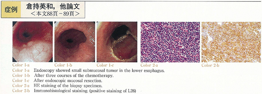
  • Hidekazu Kuramochi, Kazuhiko Hayashi, Hiroko Ide, Yoko Murata, Shigeru ...
    2000 Volume 57 Issue 2 Pages 88-89
    Published: November 15, 2000
    Released on J-STAGE: November 04, 2014
    JOURNAL FREE ACCESS
    A 69-years old man was diagnosed as esophageal malignant lymphoma by regular endoscopic GI check up. Endoscopy showed small submucosal tumor in lower esophagus. EUS revealed low echoic tumor in lpm-sm layer. Three courses of chemotherapy (CHOP) was given from June 1999 to September 1999. The tumor showed no remarkable change in size by endoscopy and EUS after chemotherapy. We performed EMR of the lesion and the pathological finding showed that most of this elevated lesion was consisted of fibrosis and inflamatoric cell, but still there existed a certain amount of viable tumor cells at the edge of the specimen thus we added radiation therapy. In this case, EMR seems to be critical for not only treatment but also the accurate evaluation of chemotherapy.
    Download PDF (877K)
  • Naoto Chihara, Eiji Naito, Akiko Shigeta, Osamu Komine, Toshiaki Komaz ...
    2000 Volume 57 Issue 2 Pages 90-91
    Published: November 15, 2000
    Released on J-STAGE: November 04, 2014
    JOURNAL FREE ACCESS
    Although esophageal lesion of pemphigus vulgalis is rare, its incidence has been increasing with the spread of endoscopes. A 61-year-old man was admitted to our hospital because of idiopatic necrotic head of femur, pyrexia, stomatitis, exanthema and ascites when we followed up esophageal varices and alcoholic cirrhosis. The patient could not ingest because of oral pain. By a biopsy specimen of the erosive skin the patient was diagnosed as pemphigus vulgalis and received local treatment with steroid. It was hard to treat esophageal varices with endoscopy because of detachment of almost entire esophageal mucosa with fresh blood. Therefore, it is important in the follow-up of patients with pemphigus vulgalis to take esophageal lesion into consideration. Furthermore, we suggest that careful attension to endoscopic examination in selected cases of pemphigus vulgalis may reveal a higher incidence of this entity.
    Download PDF (851K)
  • Kumiko Tahara, Toshiharu Mitsuhashi, Izumi Yamazaki, Satoshi Tanabe, H ...
    2000 Volume 57 Issue 2 Pages 92-93
    Published: November 15, 2000
    Released on J-STAGE: November 04, 2014
    JOURNAL FREE ACCESS
    We reported a 58-year-old female with a double pylorus, who had been treated with prednisolone because of rheumatoid arthritis. The first endoscopic examination showed a gastric ulcer on the posterior wall of the prepylorus. Two years later, the second examination showed an accessory opening and gastric ulcer on the posterior wall of the prepylorus. The third examination performed two years later showed an enlarged accessory opening and gastric ulcer on the posterior wall of the prepylorus. One year later at the fourth examination (five years after the initial examination) the double pylorus fused and formed a large single pylorus. Eight years after the initial examination, a gastric ulcer was observed on the same position. Of 118 cases of double pylorus reported in Japan, 15 cases developed a single pylorus. The fistula was closed in 4 cases and fused in 11 cases of 15 cases.
    Download PDF (242K)
  • Hiroaki Nozawa, Hajime Tsukui, Satsuki Kina, Yuichi Takakura, Naoaki H ...
    2000 Volume 57 Issue 2 Pages 94-95
    Published: November 15, 2000
    Released on J-STAGE: November 04, 2014
    JOURNAL FREE ACCESS
    A 53-year-old woman was admitted to our hospital for evaluation and treatment of the abnormality in the gastric antrum revealed by barium fluorography. Endoscopic examination showed a semi-pedunculated submucosal tumor-like lesion with slight central erosion, 1 cm in diameter, in the greater curvature of the antrum. Furthermore, two similar polyps of 5 mm and 3 mm in diameter were found in the anterior wall of the angle. All the biopsy specimen of these polyps showed benign findings (Group I) . Since she was asymptomatic, only the polyp in the antrum was removed by endoscopic mucosal resection. Histopathologically, spindle-shaped cells proliferated chiefly in the submucosal space, which were positive for CD34 and vimentin. The polyp was diagnosed as inflammatory fibroid polyp (IFP) that exhibited unusual histological features ; typical“onion-skin pattern”was not observed, and infiltration of eosinophils was mild. Interestingly, the other polyps remained disappeared by endoscopy 6 months later. The observation seems to support that IFP may be developed through inflammation.
    Download PDF (1146K)
  • Izumi Shimbo, Taketo Yamaguchi, Takeo Odaka, Kazuya Yamaguchi, Takeshi ...
    2000 Volume 57 Issue 2 Pages 96-97
    Published: November 15, 2000
    Released on J-STAGE: November 04, 2014
    JOURNAL FREE ACCESS
    77-year-old woman was referred to our hospital for further evaluation of the stomach lesion. A subpedunculated tumor, 15mm in diameter, was found at the upper anterior part of gastric body by the gastrointestinal endosopic examination. The tumor was well demarcated from the surrounding mucosa, and the surface of the tumor was papillary and lobulated with no ulcer and erosion. The tumor was diagnosed papillary adenocarcinoma by the biopsy. With the upper gastrointestinal radiologic examiniation, the tumor was depicted a round radiolucant mass without wall deformity. From the results of gastrointestinal endoscopy and radiologic examiniation, the tumor was considered to be early gastric carcinoma. However, the low-echoic mass located from the mucosal layer through the proper muscle layer was demonstrated by the endoscopic ultrasonography (EUS) , indicating the carcinoma in advanced stage. Total gastrectomy was performed and the histology of the resected specimen showed gastric carcinoma of the gastric type. In the detailed histopathological examination, differentiation of the tumor was varied ; well differentiated adenocarcinoma in superficial layer and poorly differentiated adenocarcinoma in deep layer which infiltrated the serosa. EUS appeared to be accurate in terms of tumor staging compared with gastrointestinal endosopy or upper gastrointestinal radiologic examination in this case.
    Download PDF (1108K)
  • Kazuko Beppu, Norishige Takemoto, Kouiti Koizumi, Syunkiti Kai, Gouiti ...
    2000 Volume 57 Issue 2 Pages 98-99
    Published: November 15, 2000
    Released on J-STAGE: November 04, 2014
    JOURNAL FREE ACCESS
    A 55-year-old-woman who had a small discolored lesion of the rest stomach, revealed by an endoscopy performed by a local doctor, was admitted to our hospital in May, 1999. Endoscopic examination revealed a small discolored lesion with irregular margin of the rest stomach. The pathological diagnosis was undifferentiated type gastric cancer. Upper GI X-ray examination showed a small irregular depressed lesion of the greater curvature of the rest stomach.
    Then, we diagnosed a minute undifferentiated type gastric cancer of the rest stomach and performed endoscopic mucosal resection.
    The lesion was 3×3mm in size and undifferentiated adenocarcinoma, which located in the mucosal layer. In conclusion, It is important to look for discovered discolored lesions and discolored lesions with very small reddish granules of the middle part of gastric body.
    Download PDF (501K)
  • Hiroaki Hagiwara, Teruo Yoshinaga, Tugio Higuchi
    2000 Volume 57 Issue 2 Pages 100-101
    Published: November 15, 2000
    Released on J-STAGE: November 04, 2014
    JOURNAL FREE ACCESS
    A 69-year-old woman who had underwent endoscopic polypectomy of stomach before 4 years was admitted to my clinic for endoscopy. In endoscopic examination, five polyps classified Yamada's type II were located on greater curvature of antrum, anterior wall of angulus and greater curvature of upper body. Histological findings of biopsied specimens from these polyps revealed hyperplasia of the foveolar epithelium with moderate infiltration of inflammatory cells which was consistent with hyperplastic polyp. These polyps were unchanged during 2 years and Helicobacter pylori (H. pylori) was found by the CLO test and cultures of biopsied specimens.
    She was treated with one week course of lansoprazole, amoxicillin and clarithromycin. About 14 months later, all polyps were disappeared and eradication of H. pylori was confirmed by biopsy, cultures and Urea breath test.
    These result suggests that eradication of H. pylori may be expected as a useful treatment to hyperplastic polyp of stomach.
    Download PDF (766K)
  • Takashi Ohno, Sumito Mizuguchi, Tadashi Kitamura, Kazuya Kawazoe, Teru ...
    2000 Volume 57 Issue 2 Pages 102-103
    Published: November 15, 2000
    Released on J-STAGE: November 04, 2014
    JOURNAL FREE ACCESS
    We reported a rare case of adenoma of the papilla of Vater. A 63-year-old female had been diagnosed to adenoma of the papilla of Vater on ERCP and biopsy before 6 years. The papilla was followed up by endoscopy every 6 months. Recently, the papilla was enlarged and deformed with granular surface. ERCP showed normal findings in biliary system and main pancreatic duct, however accessory pancreatic duct was narrow. We performed endoscopic papillectomy for completely biopsy under the informed consent. Since endoscopic papillectomy is a safe treatment and useful in the final pathological diagnosis of the adenoma diagnosed by biopsy. The tumor was resected completely and the final pathological diagnosis was adenoma. However acute pancreatits, one of the complication of endoscopic papillectomy was occurred. This acute pancreatitis recovered by conservative therapy. Six months after papillectomy there was no sign of recurrence. This case suggested that endoscopic papillectomy is useful for adenoma of the papilla of Vater and that method of prevention from the complication of endoscopic papillectomy will further be investigated.
    Download PDF (528K)
  • Mitsuji Tsurui, Taku Kudo, Shoji Ogiwara, Shigefumi Morita, Hiroyuki O ...
    2000 Volume 57 Issue 2 Pages 104-105
    Published: November 15, 2000
    Released on J-STAGE: November 04, 2014
    JOURNAL FREE ACCESS
    Shönlein-Henoch purpura (SHP) is a common vasculitis affecting the skin, gastrointestinal tract, joints and, occasionally other organs. Gastrointestinal manifestation, such as abdominal pain is common.
    We present a 23-year-old Japanese male with SHP. He was admitted to our hospital with abdominal pain, and purpuric rash on his extremities. Endoscopic examination of upper gastrointestinal tract revealed multiple erosion at the 2nd portion of duodenum. Colonoscopic findings were multiple small ulcers and erosions. We diagnosed SHP from physical findings and examinations, and started to treat with steroid medicine. Gradually, he recovered and discharged our hospital.
    Download PDF (571K)
  • Yasuhiro Onozato, Toshihiko Sagawa, Takehiko Abe, Takeshi Ishikawa, Ry ...
    2000 Volume 57 Issue 2 Pages 106-107
    Published: November 15, 2000
    Released on J-STAGE: November 04, 2014
    JOURNAL FREE ACCESS
    A 44-year-old male complained of abdominal pain was admitted to our hospital. He had been diagnosed as Peutz-Jeghers syndrome and operated three times due to ileus caused by intestinal polyps. His diagnosis was invagination which was treated in the operation with minor wound. Sixty-six polyps ranging in size from 5mm to 40mm were polypectomized or ligated with detachable snare by intraoperative laparoscope-assisted enteroscope.
    Download PDF (1239K)
  • Ken-ichiro Sekigawa, Hiroshi Matsuura, Masaya Matsukawa, Yu-ichi Takak ...
    2000 Volume 57 Issue 2 Pages 108-109
    Published: November 15, 2000
    Released on J-STAGE: November 04, 2014
    JOURNAL FREE ACCESS
    A 50-year-old woman showed a positive result for fecal occult blood test on her general health check-up. Colonoscopic examination at a local hospital showed a Borrmann type 1-like tumor on her ileocecal valve sizing 1.5 cm with a small top depression. Then she was referred to our hospital. Our examination on 12 days later revealed that it had grown to a Borrmann type 2-like lesion having a large ulcer. Histological examination on biopsy specimen showed dense submucosal infiltration of medium to large atypical lymphoid cells, which were positive for LCA and L-26 but negative for CD3 and UCHL-1. No other lymphoid lesions were observed. Thus diagnosis of diffuse medium to large-sized B cell lymphoma originated on the ileocecal valve was made. Ileocecal resection was performed on 50 days after initial examination. However no tumor was observed on the surgical specimen without localized microscopical aggregation of small lymphocytes which were positive for LCA and CD3 but negative for L-26. The result of immunohistochemical stain suggested that they were reactive T cells. Endoscopic and histological observation of disintegration and regression of intestinal malignant lymphoma is quite rare. The clinical course of our patient is unique in view of the spontaneous regression of intestinal MALT lymphoma and reactive lymphoid hyperasia.
    Download PDF (901K)
  • Kazuo Maruta, Tsunehito Oda, Kohei Kawakami, Kenji Ueda, Masako Natsum ...
    2000 Volume 57 Issue 2 Pages 110-111
    Published: November 15, 2000
    Released on J-STAGE: November 04, 2014
    JOURNAL FREE ACCESS
    The patient was a 71-year-old woman. She became aware of her own exertional dyspnea in June 1999, and was referred to our hospital for admission. The patient underwent colonoscopic examination because iron deficiency anemia and positive occult blood reaction in the feces were observed. In the colonoscopic examination, angiodysplasia with an uneven surface was seen in the cecum, which was thought to be the source of bleeding. In addition, abdominal angiography of the patient revealed early venous filling in the region of the cecum., leading to a diagnosis of colonic arteriovenous malformation. Thereafter she was followed up as an ambulatory patient. However, form around November 1999, anemia became pronounced and the patient was again hospitalized.
    Colonoscopic examination conducted during hospitalization showed bleeding from the cecum with arteriovenous malformation. We unsuccessfully performed a hemostatic procedure with a heat probe, resulting in the adoption of laparoscopic ileocecal resection. The postoperative course of the patient has been satisfactory, and no progression of the anemia has been observed. We report our experience of a case with lower gastrointestinal bleeding due to colonic arterioveous malformation.
    Download PDF (978K)
  • Osamu Totsuka, Yoshifumi Tanahashi, Hiroshi Matsumoto, Izumi Takeyoshi ...
    2000 Volume 57 Issue 2 Pages 112-113
    Published: November 15, 2000
    Released on J-STAGE: November 04, 2014
    JOURNAL FREE ACCESS
    A 76-year-old woman visited the emergency room complaining of anal discomfort. Yellowish mucosa was prolapsed from the anus, and it was easily reductable with a hand manipulation. She admitted the hospital to do further examination four months later because of repeated. Barium enema roentogenograms revealed a smooth and well defined tumor, about 4cm in size, in the sigmoid colon 35cm from the anus. Colonoscopic examination showed a smooth and yellowish submucosal tumor with a positive cushion sign. These findings were compatible with a lipoma of the sigmoid colon, and endoscopic mucosal resection of the tumor should not be indicated. Under general anesthesia with muscle relaxation, we successfully drew the tumor to the outside the anus by using a colonoscope, and resected directly without laparotomy or laparoscopy. The patient resumed diet from the next day after surgery. This method may be applicable to a benign lower colonic tumor under general anesthesia with muscle relaxation.
    Download PDF (898K)
  • Toshinari Awai, Mitsuru Kaise, Nobuaki Suzuki, Jun Miwa, Kuniaki Nakao ...
    2000 Volume 57 Issue 2 Pages 114-115
    Published: November 15, 2000
    Released on J-STAGE: November 04, 2014
    JOURNAL FREE ACCESS
    A 58-year-old man with advanced cholangiocarcinoma used diclofenac sodium suppository up to 200 mg in order to relieve right hypochondralgia and fever. One month after continuous NSAID usage, he was hospitalize to our hospital because of bloody diarrhea followed by massive rectal bleeding. Urgent endoscopic examination revealed irregular shaped multiple rectal ulcers, indicating a diagnosis of sodium suppository-induced acute hemorrhagic rectal ulcer. Bowel rest and GeloformTM tamponade achieved temporal hemostasis. However, rectal bleeding recurred and total amount of hemorrhage reached to 6,000 ml. We performed colonoscopy-guided argon plasma coagulation (APC) to the hemorrhagic rectal ulcers two times and complete hemostasis was achieved. Since APC is able to coagulate digestive mucosa of target widely and safely, APC is considered as an effective endoscopic hemosatatic procedure to acute hemorrhagic rectal ulcer.
    Download PDF (238K)
  • Takeshi Shimizu, Yoshihisa Kuzutani, Kouichi Kuroda, Akira Masaka, Kat ...
    2000 Volume 57 Issue 2 Pages 116-117
    Published: November 15, 2000
    Released on J-STAGE: November 04, 2014
    JOURNAL FREE ACCESS
    Bullous pemphigoid is known to be one of the paraneoplastic skin manifestation associated with internal malignancies.
    A 60-year-old male patient suffering from erythema and bulla of the body was diagnosed as bullous pemphigoid in 1991. In August, 1997 he was admitted to our hospital because of bloody stool. Endoscopic examination revealed rectal cancer. Thoracic CT and abdominal CT showed metastatic tumour both in the left lung and the liver. After rectal cancer was resected by Miles'method, both metastatic tumours were also resected. Dermatological symptoms were remarkably reduced after these treatment. Four months later, again bulla appeared simultaneously with the reccurence of liver metastasis. Resection of this lesion lead to an improvement in the skin manifestation.
    It is reported that internal malignancies are found in 5 to 20% of bullous pemphigoid patients. Including this case, only 9 cases of rectal cancer associated with bullous pemphigoid have been reported domestically. The characteristics of these patients are that they are comparatively aged and that the bullas are detected before the cancer is found. Also, 5 out of 9 patients showed improvement in their dermatological symptoms after resecting or chemotherapy treatment against the cancer, as in this case too.
    In conclusion, it is important to examine thoroughly for internal malignancies including lower gastrointestinal tracts, in bullous pemphigoid patients.
    Download PDF (570K)
  • Shoji Maruyama, Masayuki Ando, Shinobu Ohnuma, Nobuyuki Sakaniwa, Hide ...
    2000 Volume 57 Issue 2 Pages 118-119
    Published: November 15, 2000
    Released on J-STAGE: November 04, 2014
    JOURNAL FREE ACCESS
    A rare case of primary malignant lymphoma of the rectum is reported. An 83-years-old woman complained of hemorrhage on defecation. She was diagnosed as malignant lymphoma of the rectum with the colonoscopy and barium enema. Abdominoperineal resection of the rectum was performed. Histological examination of the resected specimen indicated a malignant lymphoma of the rectum, invaded to the proper muscle. Several immunohistochemical studies proved that the lymphoma was diagnosed as the diffuse large B cell type in REAL classification.
    Download PDF (615K)
  • Makoto Yoshida, Shinya Saruya, Kazuhisa Yuasa, Tsuneta Kobayashi, Yuhj ...
    2000 Volume 57 Issue 2 Pages 120-121
    Published: November 15, 2000
    Released on J-STAGE: November 04, 2014
    JOURNAL FREE ACCESS
    A 84-year-old man suffering from aspiration pneumonia on pulumonary emphysema who had taken home oxygen therapy was admitted to our hospital. Because of positive result of the fecal occult blood test on admission, colonoscopy was performed without oxygen administration after the cure of the pneumonia. A sigmoid colon polyp of 10 mm in diameter (ISP type) was easily removed in a short time by the method of saline injection in submucosal layer. On that night a light melena was found without abdominal pain, so sigmoidscopy was performed in the next day. Longitudinal mucosal redness and erosions were found in the sigmoid colon from the anal side of the resected site. Pathological findings of biopsy specimen were compatible with those of ischemic colitis. There were no pathogenic bacterium from liquid stool at sigmoidscopy and no recent history of antibiotics use. Endoscopic findings were improved one week later only by fasting and parenteral nutrition. We think this ischemic colitis was caused by circulatory disturbance due to temporal pressure and stretch of the colon in the colonoscopy procedure. In case colonoscopy or polypectomy is performed to a patient with hypoxemia, we think oxygen supplementation should be considered with extra care to explosion.
    Download PDF (358K)
  • Nobutaka Yasui, Noriaki Kameyama, Atsushi Toizumi, Yoichiro Tamura, Ta ...
    2000 Volume 57 Issue 2 Pages 122-123
    Published: November 15, 2000
    Released on J-STAGE: November 04, 2014
    JOURNAL FREE ACCESS
    We report our experience with four cases of acute hemorrhagic rectal ulcer in recent years. The patients included three men and a woman. Age of the patients ranged from 76 to 86. All of them had some serious underlying disorder, including cerebral vascular disease, senile dementia, renal failure, respiratory failure and myelodysplastic syndrome. They complained of sudden fresh bloody stool without abdominal pain. Emergency colonoscopic examination revealed rectal ulcer localized in the lower rectum in all cases. Two patients were cured of anal bleeding with treatment in the lateral position. The others required treatment to stop bleeding, for example endoscopic hemostasis. Emergency colonoscopic examination is necessary for the diagnosis and rational treatment of acute hemorrhagic rectal ulcers.
    Download PDF (235K)
feedback
Top