ABSTRACT
Objectives: This study investigated the outcomes of the early introduction of a standing program for patients with Duchenne muscular dystrophy (DMD).
Methods: This was a retrospective observational study of 41 outpatients with DMD aged 15–20 years. We introduced the standing program using knee–ankle–foot orthoses (KAFO) to slow the progression of scoliosis when ankle dorsiflexion became less than 0° in the ambulatory period.
Results: Thirty-two patients with DMD were offered the standing program with KAFO; 12 continued the program until the age of 15 years (complete group) and 20 discontinued the program before the age of 15 years (incomplete group). The non-standing program group included 9 patients. The standing program with KAFO was significantly associated with the Cobb angle at the age of 15 years after adjustment for the duration of corticosteroid use and DMD mutation type (P=0.0004). At the age of 15 years, significant correlations were found between the ankle dorsiflexion range of motion (ROM) and non-ambulatory period (P=0.0010), non-ambulatory period and Cobb angle (P<0.0001), Cobb angle and percent predicted forced vital capacity (P=0.0004), and ankle dorsiflexion ROM and Cobb angle (P=0.0066). In the complete group, the age at ambulation loss (log-rank P=0.0015), scoliosis progression (log-rank P=0.0032), and pulmonary dysfunction (log-rank P=0.0006) were significantly higher than in the non-standing program group.
Conclusions: The early introduction of a standing program for DMD patients may prolong the ambulation period and slow the progression of scoliosis and pulmonary dysfunction.
INTRODUCTION
Duchenne muscular dystrophy (DMD) is the most common progressive muscular disorder. It is caused by X-linked recessive mutations in the dystrophin gene, resulting in progressive muscle weakness.1) In Japan, DMD patients progress to a non-ambulatory state at the age of about 10 years in the natural course, followed by the development of scoliosis.2) If left untreated, over 70% of patients will develop scoliosis with a Cobb angle greater than 20°.3,4) The progression of scoliosis leads to decreased sitting tolerance, progression of pulmonary dysfunction, including loss of vital capacity and difficulty with expectoration, and decreased quality of life (QOL).5,6,7) Several treatment modalities have been used to prevent the progression of scoliosis.8) As a medical modality, corticosteroids have been used to reduce the incidence of scoliosis. However, 10%–30% of DMD patients treated with corticosteroids develop scoliosis with a Cobb angle greater than 20°.3,9,10) As a surgical modality, spinal fusion in DMD patients results in improvements in scoliosis, sitting balance, and QOL and prevents pulmonary dysfunction.11,12,13,14,15,16) However, the risk of complications following surgical correction of neuromuscular scoliosis is still estimated to be 33%–62%.17,18,19)
Attempts to prevent scoliosis development using spinal braces and seating systems as rehabilitation modalities have been ineffective.20) However, standing devices have improved contractures and have prevented scoliosis and pulmonary dysfunction.21,22,23) Standing devices are recommended by the standard care guidelines for DMD in the initial non-ambulatory phase.24,25) However, the efficacy of prolonging the ambulatory period, introduction time, and the recommended duration are unclear.26) Only a few studies have focused on this topic, and none have evaluated the effects of outpatient standing programs on DMD in the Japanese population.
In previous studies,22,23) standing programs were introduced after patients had developed difficulty with independent walking. However, leg contractures related to DMD start when patients can still walk independently. There is a direct relationship between the development of lower leg contractures and the loss of independent ambulation during the initial stage of DMD.27) In addition, it has been reported that ambulation loss at an older age is related to later onset of scoliosis.28) Therefore, we hypothesized that initiating a standing program before losing ambulation may prevent lower leg contractures, prolong the ambulation period, and slow the progression of scoliosis. We retrospectively investigated the effects of a standing program with knee–ankle–foot orthoses (KAFO) on ambulatory period, scoliosis, and pulmonary dysfunction.
MATERIALS AND METHODS
Study Design and Recruitment of PatientsWe screened all patients with DMD visiting our rehabilitation facility from April 2005 to August 2018. Subsequently, we selected subjects based on the inclusion and exclusion criteria. The following inclusion criteria were used: 1) date of birth between August 1998 and July 2003; and 2) the DMD diagnosis was confirmed by genetic and/or pathology testing. The following exclusion criteria were used: 1) a lack of essential data; 2) no restriction in ankle dorsiflexion at the age of 10 years; and 3) participation in any clinical trial for other DMD treatments.
Based on whether they had participated in a standing program, participants were classified into the standing program group or the non-standing program group. Patients in the standing program group were divided into two subgroups: the “complete group” who continued the standing program at least once a week until the age of 15 years and the “incomplete group” who discontinued its use before the age of 15 years (Fig. 1). Although we recommended a daily 1-h session, the exact time and frequency were not strictly controlled. Transient interruptions caused by treatment complications were not considered discontinuation, including pneumonia and pressure ulcers. Before visiting our hospital, we asked patients in the non-standing program group about their treatment history, age at ambulation loss, and Cobb angle.

This study was approved by the Medical Ethics Committee of the National Center of Neurology and Psychiatry (A2018-099). Informed consent was obtained from patients or their parents in an opt-out manner.
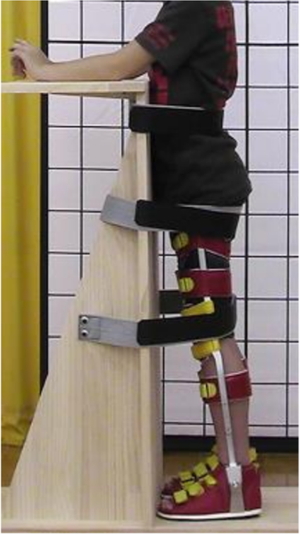
Medical ManagementPatient follow-up occurred at intervals of 1–6 months to evaluate lower leg range of motion (ROM). We offered a standing program with KAFO to patients that could walk independently when ankle dorsiflexion became 0° on at least one side. Patients were instructed to stand on a standing board with bilateral KAFOs, maintaining a symmetrical position of natural thoracic kyphosis and lumbar lordosis, leaning forward to maintain mild extension of the hips, and maintaining knee extension and ankle dorsiflexion as much as possible (Fig. 2). We recommend that patients continue the standing program until the age of at least 15 years, even after ambulation loss, because Japanese children have a pubertal height increase between 10 and 15 years of age.29) Independent of the standing program, we provided instructions to all patients during the observation period for lower leg and trunk stretches, lumbar lordosis in the chair-sitting position, as well as respiratory rehabilitation. We indicated nocturnal ventilation in patients who had any of the following: signs or symptoms of hypoventilation [patients with percent predicted forced vital capacity (%FVC<30%) are at especially high risk], a baseline oxygen saturation (SpO2) of less than 95% and/or blood or end-tidal pCO2 greater than 45 mmHg while awake, an apnea-hypopnea index (AHI) greater than 10 h−1 on polysomnography, or four or more episodes of SpO2 less than 92% or falls in SpO2 of at least 4% per hour of sleep.30)
At each visit, functional measures, including age, body weight, and body mass index (BMI) at ambulation loss, standing program discontinuation, spinal fusion surgery, and initiation of non-invasive positive pressure ventilation (NPPV), were routinely recorded by attending physicians, physiatrists, physiotherapists, and occupational therapists. In addition, the daily duration of the standing program, training frequency per week, and the lower leg range of motion (ROM) were recorded. We adjusted and re-prescribed the KAFO several times as the patients grew. For patients older than 7 years, spirometry and radiography of the spine were performed once or twice per year. We also collected data regarding corticosteroid use.
The endpoints used to assess the efficacy of the standing program were age at the following times: at ambulation loss, when scoliosis became greater than 40°, when the %FVC became less than 40%, and when mechanical ventilation was initiated. These endpoints (Cobb angle >40°; %FVC <40%) were used because patients with a Cobb angle of 40° or more experience sitting difficulties and respiratory interventions are recommended when %FVC drops below 40%.30) Loss of ambulation was defined as patient inability to ambulate alone or full-time use of a wheelchair. Spinal curvature was measured using the Cobb method on anteroposterior spine radiographs recorded while the patients were seated. If the patient was unable to sit, radiographs were taken in the supine position. Spirometry was performed in the sitting position.
Statistical AnalysisContinuous variables are expressed as mean ± standard deviation, and categorical variables are expressed as numbers and percentages. Fisher’s exact test was used for categorical variables, and analysis of variance or the Kruskal–Wallis test was used for continuous variables. Spearman correlation coefficients assessed the correlation between ankle dorsiflexion ROM, non-ambulatory period, Cobb angle, and %FVC at the age of 15 years.
Multiple regression analysis was used to assess the relationship between the Cobb angle at the age of 15 years, status of standing program implementation, duration of corticosteroid use, and DMD mutation type. We set the Cobb angle at the age of 15 years as the dependent variable, and the independent variables were the status of standing program implementation, duration of corticosteroid use, and DMD mutation type. We classified the implementation status into three categories: complete, incomplete, and non-standing program groups. In addition, DMD mutation was categorized into three groups: minor type (patients amenable to skip exon 8 or 44),31,32,33) severe type (patients amenable to skip exon 51 or 53),31) and others (patients not amenable to skip exons 8, 44, 51, 53). The dependent variables were logarithmically transformed to exhibit a normal distribution.
We used Kaplan–Meier analysis to compare the complete, incomplete, and non-standing program groups for ambulation loss, Cobb angle greater than 40°, %FVC less than 40%, and initiation of mechanical ventilation. The median participant age at initiation of the standing program was compared using a log-rank test. Hazard ratios (HR) were expressed with 95% confidence intervals (95% CI). P values less than 0.05 with Bonferroni correction were considered statistically significant. All analyses were performed using JMP version 17.0 software (SAS Institute, Cary, NC, USA).
RESULTS
A total of 103 patients were screened; 40 were excluded because of insufficient data. Among the 63 patients analyzed, 13 were excluded because they had no restrictions in ankle dorsiflexion at the age of 10 years. Nine patients were excluded because they had participated in clinical trials. Finally, 32 patients accepted the standing program (standing program group). Among the complete group, 11 of the 12 patients engaged in the standing program for a minimum of 30 min per session, four times weekly, while they were 15 years old. The remaining patient underwent a weekly standing program. Nine patients who could not stand or walk independently because of severe dorsiflexed ankle contractures at the first visit were not introduced to the standing program (non-standing program group) (Fig. 1). However, these patients had undergone standard treatment before visiting our hospital, including corticosteroid therapy and standard rehabilitation management (lower leg and trunk stretches and lumbar lordosis guidance in the chair-sitting position).
Table 1 shows the characteristics of the three groups. Regarding age at the first visit, patients in the standing program group were significantly younger than those in the non-standing program group. Most patients were on oral corticosteroids during the observation period. Among the three groups, there was no significant difference in corticosteroid use or the age of corticosteroid administration. Most patients in the standing program group had been on corticosteroids before starting the standing program (11/12 patients in the complete group; 17/20 patients in the incomplete group). Two patients in the incomplete group were not using corticosteroids during the observation period. Two remaining patients in the complete group and the incomplete group started corticosteroid therapy 1 year after the standing program started. Corticosteroids were continued without interruption throughout the observation period. When the standing program started, there was no significant difference between the complete and incomplete groups for age or Cobb angle. Although pulmonary function was only measured in 14 of the 32 patients within 1 year of the program starting, the mean %FVC in the complete group was significantly greater than that in the incomplete group.
Table 1. Characteristics among the three groups
| Characteristic | Standing program group (n=32) | Non-standing program group (n=9) | P | Complete vs. non-standing | Incomplete vs. non-standing | Complete vs. incomplete |
| Complete group (n=12) | Incomplete group (n=20) |
| Age at first visit, years | 7.33 (1.83) | 7.75 (1.45) | 14.00 (1.5) | <0.0001* | <0.0001* | <0.0001* | 0.75 |
| Corticosteroid use ≥1 year | 12/12 | 18/20 | 6/9 | 0.086+ | | | |
| Age starting corticosteroids, years | 7.25 (1.91) | 6.28 (1.53) | 6.00 (0.89) | 0.19 | | | |
| Age starting standing program, years | 8.75 (1.42) | 8.40 (1.27) | - | 0.48 | | | |
| Spinal fusion, n (mean age) | 0 | 4 (14.25) | 1 (16) | | | | |
| Cobb angle (degrees) at initiation standing program, (n=31) | 6.00 (4.08) (n=12) | 6.21 (7.04) (n=19) | - | 0.93 | | | |
| FVC% at initiation within ±1 year, (n=14) | 88.96 (10.42) (n=5) | 76.20 (8.80) (n=9) | - | 0.031* | | | |
| Mutation | | | | | | | |
| Exon deletion | 12/12 | 9/20 | 3/9 | | | | |
| Exon duplication | 0/12 | 1/20 | 1/9 | | | | |
| Small mutation | 0/12 | 10/20 | 4/9 | | | | |
| Not available | 0/12 | 0/20 | 1/9 | | | | |
| Minor type | 3 | 0 | 0 | | | | |
| Severe type | 5 | 3 | 0 | | | | |
Data given as mean (standard deviation), number/total number, or number.
* Statistically significant between groups with Bonferroni-corrected P <0.05.
+ Fisher's exact test
Table 2 shows the results for the three groups. At the age of 15 years, the Cobb angle was significantly smaller and the %FVC was significantly greater in the complete program group than in the non-standing program group. However, there was no significant difference among the three groups for the ankle dorsiflexion ROM. More than 50% of patients discontinued the program before 15 years of age (mean age of 11.9 years). The main reasons for discontinuation were increased care burden related to weight increase, change in lifestyle upon entering junior high school, or fear of standing after suffering fractures in previous falls. No pain was caused by the standing exercise. No patients fell during the standing program with the KAFO. The mean age at ambulation loss was significantly older in the complete group, followed by the incomplete group, and then the non-standing program group. No data were available for body weight and BMI in the non-standing program group. The body weight at ambulation loss was significantly greater in the complete group than in the incomplete group, whereas the BMI at ambulation loss was not significantly different between the two groups.
Table 2. Results of the three groups
| Result | Complete group (n=12) | Incomplete group (n=20) | Non-standing program group (n=9) | P | Complete vs. non-standing program | Incomplete vs.
non-standing program | Complete vs. incomplete |
Cobb angle at age
15 years (±1),
degrees | 10.33 (13.68)
(n=12) | 23.20 (21.62)
(n=20) | 48.11 (32.59)
(n=9) | 0.0021* | 0.0007* | 0.0222* | 0.097 |
| %FVC at age 15 years (±1), % | 61.02 (19.39)
(n=12) | 35.95 (17.46)
(n=20) | 39.47 (20.18)
(n=9) | 0.0023* | 0.0326* | 0.886 | 0.0020* |
| Larger ROM of ankle dorsiflexion at 15 years, degrees | −21.55 (6.08)
(n=11) | −35.25 (4.51)
(n=20) | −31.43 (7.63)
(n=7) | 0.21 | | | |
Age at program
discontinuation, years (n=20) | | 11.9 (1.48) | | | | | |
| Age at ambulation loss, years | 13.25 (3.34) | 10.45 (2.04) | 8.89 (1.29) | 0.0023* | 0.0039* | 0.0215* | 0.0226* |
Body weight at
ambulation loss, kg | 45.36 (13.22)
(n=12) | 34.06 (7.46)
(n=15) | Unknown | - | - | - | 0.0212* |
| BMI at ambulation loss, kg/m2 | 22.15 (4.52)
(n=12) | 19.73 (3.48)
(n=15) | Unknown | - | - | - | 0.1436 |
Data given as mean (standard deviation).
Significant difference P<0.05.
Figure 3 shows the results of correlation analysis. At the age of 15 years, there were moderate correlations between ankle dorsiflexion ROM and the non-ambulatory period, non-ambulatory period and Cobb angle, Cobb angle and %FVC, and ankle dorsiflexion ROM and the Cobb angle. Table 3 displays the results of the multiple regression analysis. The status of standing program implementation with KAFO was significantly associated with the Cobb angle at the age of 15 years after adjustment for duration of corticosteroid use and DMD mutation type.
Table 3. Multiple regression analysis for the Cobb angle at age 15 years
| Factor | B | Standard error | β | P value |
| Status of standing program implementation | 21.94 | 5.64 | 0.61 | 0.0004* |
| Duration of corticosteroid use | −1.23 | 1.11 | −0.15 | 0.28 |
| DMD mutation type | −8.00 | 4.97 | −0.25 | 0.12 |
*Significantly different between groups.
B, partial regression coefficient; β, standard partial regression coefficient.
Figures 4 and 5 show the detailed course of scoliosis and %FVC in the three groups. In the complete group, scoliosis was controlled to a Cobb angle mostly under 20°, even after loss of ambulation (Fig. 6A), excluding the patient who only performed the standing program once a week. The remaining patients in the complete group participated in a standing program at least four times a week. In the complete group, pulmonary dysfunction was better controlled than in the incomplete and non-standing program groups, even after loss of ambulation (Fig. 6B).

In the incomplete group, scoliosis was controlled during the period before discontinuation of the standing program, even after ambulation loss (Fig. 6A). However, after discontinuing the program, scoliosis progressed to greater than 40°. In addition, %FVC was mostly maintained before the standing program was discontinued, even after ambulation loss (Fig. 6B). In the non-standing program group, the scoliosis of several patients progressed more severely than those in the incomplete group.
Figure 7 shows the results of the Kaplan–Meier survival analysis. The median ages at ambulation loss were 12.5, 10, and 9 years in the complete, incomplete, and non-standing program groups, respectively. The log-rank test revealed that the index age in the complete group was significantly higher than that in the incomplete and non-standing program groups; additionally, the incomplete group index age was greater than that in the non-standing program group. The median age of scoliosis exceeding 40° was 14 years in the non-standing program group. However, this finding could not be determined in the complete and incomplete groups because over 50% of patients were still alive when the observation period ended. The log-rank test demonstrated that the index age was significantly higher in the completed program group than in the non-standing program group. The median age when %FVC dropped below 40% could not be determined in the complete program group; however, it was 15 and 16 years in the incomplete and non-standing program groups, respectively. The log-rank test revealed that the index age was significantly higher in the completed program group than in the incomplete and non-standing program groups. The median age at ventilation initiation could not be determined in the complete program group; however, it was 18 and 20 years in the incomplete and non-standing program groups, respectively. The log-rank test indicated that there was no significant difference in index age among the three groups.

For the complete vs. non-standing program groups, incomplete vs. non-standing program groups, and complete vs. incomplete groups, the HRs (95% CI) for ambulation loss were 4.49 (1.48–13.66), P=0.0082; 1.05 (0.48–2.33), P=0.899; and 2.45 (1.04–5.74), P=0.0396, respectively. The HRs for scoliosis over 40° were 11.65 (1.38–98.14), P=0.0240; 2.22 (0.74–6.66), P=0.1531; and 4.99 (0.61–40.68), P=0.1332 for the complete vs. non-standing program groups, incomplete vs. non-standing program groups, and complete vs. incomplete groups, respectively. The HRs for %FVC below 40% were 8.23 (1.96–34.51), P=0.0039; 1.22 (0.48–3.11), P=0.6752; and 5.68 (1.54–20.95), P=0.0091 for the complete vs. non-standing program groups, incomplete vs. non-standing program groups, and complete vs. incomplete groups, respectively. For ventilation initiation, the HRs were 4.69 (0.48–46.12), P=0.1856; 1.44 (0.41–5.13), P=0.5700; and 4.42 (0.51–38.05), P=0.1756 for the complete vs. non-standing program groups, incomplete vs. non-standing program groups, and complete vs. incomplete groups, respectively.
DISCUSSION
Main FindingsIn this study, we observed the long-term rehabilitation outcomes of patients with DMD in Japan. We consider that early introduction of a standing program with KAFO is associated with ambulation period and the prevention of scoliosis progression and pulmonary dysfunction.
Possible Mechanisms of the Standing Program for Preventing Scoliosis and Pulmonary DisfunctionIn our study, the Cobb angle and %FVC at the age of 15 years were significantly better in the standing group than in the non-standing program group. Furthermore, the ambulation period was significantly longer in the standing group than in the non-standing group. We considered that stretching the ankle, knee, and hip joints with a KAFO while standing on a standing board may prolong the ambulatory period and maintain postural symmetry, including natural thoracic kyphosis and lumbar lordosis, and prevent the progression of scoliosis, even after loss of ambulation.
Although there was no significant difference in ankle dorsiflexion ROM at the age of 15 years among the complete, incomplete, and non-standing program groups, the ROM was significantly correlated with the non-ambulatory period and the Cobb angle. In addition, there was a significant correlation between the non-ambulatory period and the Cobb angle. The lack of a significant difference in ankle ROM among the groups can be attributed to increased variability because of the presence of ambulatory and non-ambulatory patients in each group at this age. In a previous study, controlling lower extremity contractures using KAFO prolonged the ambulation period.34,35) Furthermore, correcting foot alignment and pelvic obliquity with a foot orthosis improved scoliosis.36) In ambulatory patients with DMD, lumbar lordosis locks the facet joints and prevents scoliosis. However, reduced spine extensibility causes lumbar kyphosis after loss of ambulation, collapsing the facet joint locking; therefore, scoliosis develops rapidly.37) These findings support the hypothesis that the standing program prevents scoliosis.
In our study, patients were instructed to continue the standing program even after loss of ambulation. In addition, scoliosis was controlled while the standing program was continued during the non-ambulatory period. In the standing position, the lumbar region and lower extremities bear the body weight, resulting in better stretching of the hip, knee, and ankle joints than in the sitting position. Therefore, a standing program may help prevent the progression of scoliosis during the non-ambulatory period.
In our study, Cobb angle and %FVC at the age of 15 years were significantly correlated. A standing program with a KAFO may be associated with slowed progression of scoliosis as well as slower declines in vital capacity and peak expiratory flow rate.22) However, there was a significant age delay when %FVC was less than 40% in the complete group when compared with the other groups. However, initiation of ventilation was not affected by the standing program. We introduced NPPV in patients with DMD because it reportedly preserves pulmonary function and improves their prognosis.22) NPPV indications are dependent on pulmonary dysfunction and other factors, including quality of life and the beliefs of patients and their care providers. Therefore, no statistically significant relationship between initiation of NPPV and standing time was observed.
Other Factors that may Affect OutcomesOther factors affecting scoliosis include corticosteroid use and DMD mutation type. Treatment with corticosteroids reduces the risk of early ambulation loss, pulmonary dysfunction, scoliosis, and cardiomyopathy.2,38,39) McDonald et al.39) reported that the median age at ambulation loss in patients on corticosteroids was 13.4 years, whereas Takeuchi et al.2) reported it as 11.0 years in Japanese patients. The age at ambulation loss in Japan is less than those in other countries. This may be caused by differences in ethnic origin, corticosteroid regimens, clinical definitions of DMD, and definitions of ambulation.2) In our study, the median ages at ambulation loss were 12.5, 10, and 9 years in the complete, incomplete, and non-standing program groups, respectively. In the complete group, the median age at ambulation loss was older than that in the study by Takeuchi et al.2) with patients treated only with corticosteroids. Furthermore, DMD mutations may affect outcomes, including scoliosis.31–33) In this study, only patients with minor mutations were in the complete group, which may explain the slightly better %FVC during program initiation in the complete group when compared with the incomplete group. Although the proportion of patients with severe-type mutations was higher in the complete group (five of eight patients) than in the incomplete group (three of eight patients), the standing program appeared to control scoliosis, even in patients with severe-type mutations. In addition, multiple regression analysis demonstrated a significant association between the standing program and scoliosis, even after adjustment for duration of corticosteroid use and DMD mutation type. Therefore, our results suggest that the standing program provides results that are comparable with those of corticosteroid treatment.
The age and body weight at ambulation loss were significantly higher in the complete group than in the incomplete group. However, the BMI was not significantly different between the two groups. Previous reports showed that growth curves for DMD patients treated with corticosteroids show higher weight and greater BMI than males in the general population.40,41) The mean body weight and BMI for Japanese males in the general population were 33.9 kg and 17.78 kg/m2 at 10 years, and 44.7 kg and 18.25 kg/m2 at 13 years.29) In our study, compared with Japanese males in the general population, patients with DMD in the standing program group had slightly greater body weight and BMI at ambulation loss. However, the BMI at ambulation loss was not significantly different between the complete group and the incomplete group. Although we could not compare body weight and BMI at the same age because of limited data, these results may indicate that differences in weight growth were not the primary determining factor for discontinuing the standing program.
During the standing program, scoliosis was controlled even after ambulation loss. In the complete group, only one patient developed severe scoliosis. He performed the standing program only once a week, whereas the remaining patients performed it at least four times a week. Therefore, weekly use of the standing program may not be sufficient to achieve meaningful results. During the standing program, pulmonary function was controlled in much the same manner as scoliosis.
Comparison with Other TreatmentsIn our study, the age at which the complete treatment group lost their brace-free ambulation ability was 3.5 years older than the non-standing program group. Prolonged ambulation for 2–4 years using KAFOs has been reported.34,35) The standing program may be effective in prolonging the ambulatory period, similar to the findings of previous studies.34,35) However, ambulation loss was prolonged by 1–2 years among patients that received foot and ankle surgical interventions when compared with other patients.34,42,43,44) However, patients who underwent Achilles tendon and/or posterior tibialis lengthening had severe ankle/foot contractions a year postoperatively.45) Therefore, to prevent the recurrence of contractions, patients need a posterior tibial tendon transfer or a sustained passive stretching program conducted by physiotherapists combined with postoperative orthotic use.46) In Japan, the facilities performing contraction-release surgeries are limited. Foot or ankle surgical intervention was not performed in any patient in our study.
Spinal bracing is ineffective for preventing scoliosis. Furthermore, it may reduce vital capacity,47) and the DMD clinical care guidelines do not recommend them.24,25) Therefore, we do not recommend spinal bracing for our patients. In our study, only one patient in the non-standing program group was prescribed spinal bracing at the age of 12 years before visiting our hospital. His scoliosis developed from a Cobb angle of 66° at the age of 14 years to 86° a year later.
One of the limitations of our standing program was its high discontinuation rate. In this study, more than half of the patients discontinued the standing program at a mean age of 11.9 years. Common reasons for discontinuation were that weight growth made it difficult for parents to assist the children and that study as well as other school activities made it difficult to find time for the standing program.
In this study, we found that scoliosis progressed if patients discontinued the standing program before the adolescent height increase. A study in Boston, Massachusetts, reported that the factors limiting adherence were device malfunction, family vacations, ankle pain, long school days limiting home time, and decreased motivation to participate in a standing program.21) The following ideas may improve adherence: powered standing wheelchairs to reduce the burden on assisting parents, implementing standing programs in a school-based setting, more frequent monitoring (>1 times/month), admission for rehabilitation during vacations, and identifying incentives to motivate patients. In addition, it may be effective to show new functional activities as a short-term goal to motivate patients; for example, accessing the freezer for frozen snacks, accessing higher shelves in the refrigerator, or standing urination. Although no patients fell while using the KAFO in this study, some could not stand with the KAFO for several weeks after suffering fractures from earlier falls. Because leg contractures progressed during this period, these patients could not return to the standing program.
Study LimitationsThis study has several limitations. First, many patients discontinued the standing exercise program. Therefore, we cannot rule out that only patients with minor impairments could continue the standing program. Second, the study is subject to limitations associated with its retrospective design. Yamashita et al.48) reported that the vital capacity and Cobb angle at the age of 10 years and age at ambulation loss predicted the progression of spinal deformity. However, we could not control these risk factors because the age at first visit in the non-standing program group was over 10 years. Radiographic data for determination of the Cobb angle were obtained from both sitting and supine positions. The Cobb angle in the sitting position is greater than that in the supine position.49) In this study, spinal curvature was measured in the sitting position when the patients could sit and in the supine position when they could not; therefore, most patients underwent radiography in the supine position when the Cobb angle exceeded 40°. In addition, the study includes a small number of cases because we excluded many patients because of missing data. Finally, in the non-standing program group or the incomplete group, the scoliosis of some patients progressed mildly. Wilkins and Gibson37) reported two natural scoliosis patterns: an unstable pathway (severe scoliosis) and a stable pathway (minor scoliosis). Oda et al.50) reported that the minor scoliosis type was present in 15.7% (7/45) of patients. Because of these limitations, caution should be exercised when interpreting our results.
CONCLUSIONS
Early introduction of a standing program with KAFO before loss of ambulation may be associated with a prolonged ambulation period and slower progression of scoliosis and pulmonary dysfunction. Further prospective studies with improved protocols are required to confirm the effectiveness of this program.
ACKNOWLEDGMENTS
This research was supported by an Intramural Research Grant (2–4) for Neurological and Psychiatric Disorders from the National Center of Neurology and Psychiatry. The authors thank Narihiro Minami for genetic analyses of DMD, Shinya Hirakawa for statistical analyses, and Takuya Watanabe for physical therapy. We thank all physiotherapists and occupational therapists involved in this study.
CONFLICTS OF INTEREST
Outside the scope of this study, Hirofumi Komaki has received support from Nippon Shinyaku Co., Ltd. and has received grants from Taiho Pharmaceutical Co., Ltd., Sarepta Therapeutics, Inc., and Pfizer, Inc. The authors declare no conflict of interest.
REFERENCES
- 1. Hutter OF: The membrane hypothesis of Duchenne muscular dystrophy: quest for functional evidence. J Inherit Metab Dis 1992;15:565–577. PMID:1528017, DOI:10.1007/BF01799615
- 2. Takeuchi F, Yonemoto N, Nakamura H, Shimizu R, Komaki H, Mori-Yoshimura M, Hayashi YK, Nishino I, Kawai M, Kimura E, Takeda S: Prednisolone improves walking in Japanese Duchenne muscular dystrophy patients. J Neurol 2013;260:3023–3029. PMID:24057148, DOI:10.1007/s00415-013-7104-y
- 3. Alman BA, Raza SN, Biggar WD: Steroid treatment and the development of scoliosis in males with Duchenne muscular dystrophy. J Bone Joint Surg Am 2004;86:519–524. PMID:14996877, DOI:10.2106/00004623-200403000-00009
- 4. Sussman M: Duchenne muscular dystrophy. J Am Acad Orthop Surg 2002;10:138–151. PMID:11929208, DOI:10.5435/00124635-200203000-00009
- 5. Galasko CS, Delaney C, Morris P: Spinal stabilisation in Duchenne muscular dystrophy. J Bone Joint Surg Br 1992;74:210–214. PMID:1544954, DOI:10.1302/0301-620X.74B2.1544954
- 6. Hsu JD: The natural history of spine curvature progression in the nonambulatory Duchenne muscular dystrophy patient. Spine 1983;8:771–775. PMID:6665578, DOI:10.1097/00007632-198310000-00014
- 7. Kurz LT, Mubarak SJ, Schultz P, Park SM, Leach J: Correlation of scoliosis and pulmonary function in Duchenne muscular dystrophy. J Pediatr Orthop 1983;3:347–353. PMID:6874933, DOI:10.1097/01241398-198307000-00014
- 8. Gloss D, Moxley RT, III, Ashwal S, Oskoui M: Practice guideline update summary: corticosteroid treatment of Duchenne muscular dystrophy: report of the Guideline Development Subcommittee of the American Academy of Neurology. Neurology 2016;86:465–472. PMID:26833937, DOI:10.1212/WNL.0000000000002337
- 9. Biggar WD, Harris VA, Eliasoph L, Alman B: Long-term benefits of deflazacort treatment for boys with Duchenne muscular dystrophy in their second decade. Neuromuscul Disord 2006;16:249–255. PMID:16545568, DOI:10.1016/j.nmd.2006.01.010
- 10. Biggar W, Politano L, Harris VA, Passamano L, Vajsar J, Alman B, Palladino A, Comi LI, Nigro G: Deflazacort in Duchenne muscular dystrophy: a comparison of two different protocols. Neuromuscul Disord 2004;14:476–482. PMID:15336688, DOI:10.1016/j.nmd.2004.05.001
- 11. Eagle M, Bourke J, Bullock R, Gibson M, Mehta J, Giddings D, Straub V, Bushby K: Managing Duchenne muscular dystrophy—the additive effect of spinal surgery and home nocturnal ventilation in improving survival. Neuromuscul Disord 2007;17:470–475. PMID:17490881, DOI:10.1016/j.nmd.2007.03.002
- 12. Modi HN, Suh SW, Hong JY, Cho JW, Park JH, Yang JH: Treatment and complications in flaccid neuromuscular scoliosis (Duchenne muscular dystrophy and spinal muscular atrophy) with posterior-only pedicle screw instrumentation. Eur Spine J 2010;19:384–393. PMID:19885687, DOI:10.1007/s00586-009-1198-z
- 13. Suk KS, Lee BH, Lee HM, Moon SH, Choi YC, Shin DE, Ha JW, Song KM, Kim HS: Functional outcomes in Duchenne muscular dystrophy scoliosis: comparison of the differences between surgical and nonsurgical treatment. J Bone Joint Surg Am 2014;96:409–415. PMID:24599203, DOI:10.2106/JBJS.M.00777
- 14. Suk KS, Baek JH, Park JO, Kim HS, Lee HM, Kwon JW, Moon SH, Lee BH: Postoperative quality of life in patients with progressive neuromuscular scoliosis and their parents. Spine J 2015;15:446–453. PMID:25301022, DOI:10.1016/j.spinee.2014.09.030
- 15. Watanabe K, Lenke LG, Daubs MD, Watanabe K, Bridwell KH, Stobbs G, Hensley M: Is spine deformity surgery in patients with spastic cerebral palsy truly beneficial?: a patient/parent evaluation. Spine 2009;34:2222–2232. PMID:19752709, DOI:10.1097/BRS.0b013e3181948c8f
- 16. Yang JH, Kim KS, Lee GH, Kim HS: Comparison of survival analysis between surgical and non-surgical treatments in Duchenne muscular dystrophy scoliosis. Spine J 2020;20:1840–1849. PMID:32535073, DOI:10.1016/j.spinee.2020.06.004
- 17. Mohamad F, Parent S, Pawelek J, Marks M, Bastrom T, Faro F, Newton P: Perioperative complications after surgical correction in neuromuscular scoliosis. J Pediatr Orthop 2007;27:392–397. PMID:17513958, DOI:10.1097/01.bpb.0000271321.10869.98
- 18. Gau YL, Lonstein JE, Winter RB, Koop S, Denis F: Luque-Galveston procedure for correction and stabilization of neuromuscular scoliosis and pelvic obliquity: a review of 68 patients. J Spinal Disord 1991;4:399–410. PMID:1810562, DOI:10.1097/00002517-199112000-00001
- 19. Benson ER, Thomson JD, Smith BG, Banta JV: Results and morbidity in a consecutive series of patients undergoing spinal fusion for neuromuscular scoliosis. Spine 1998;23:2308–2317. PMID:9820912, DOI:10.1097/00007632-199811010-00012
- 20. Smith PE, Calverley PM, Edwards RH, Evans GA, Campbell EJ: Practical problems in the respiratory care of patients with muscular dystrophy. N Engl J Med 1987;316:1197–1205. PMID:3553943, DOI:10.1056/NEJM198705073161906
- 21. Townsend EL, Bibeau C, Holmes TM: Supported standing in boys with Duchenne muscular dystrophy. Pediatr Phys Ther 2016;28:320–329. PMID:27008581, DOI:10.1097/PEP.0000000000000251
- 22. Galasko CS, Williamson JB, Delaney CM: Lung function in Duchenne muscular dystrophy. Eur Spine J 1995;4:263–267. PMID:8581525, DOI:10.1007/BF00301031
- 23. Minato J, Minato J, Minato M. The management of Duchenne muscular dystrophy: establishing an early straight or hyperextended spine [in Japanese]. Jpn J Rehabil Med 2006;43:438–445. DOI:10.2490/jjrm1963.43.438
- 24. Birnkrant DJ, Bushby K, Bann CM, Apkon SD, Blackwell A, Brumbaugh D, Case LE, Clemens PR, Hadjiyannakis S, Pandya S, Street N, Tomezsko J, Wagner KR, Ward LM, Weber DR, DMD Care Considerations Working Group: Diagnosis and management of Duchenne muscular dystrophy, part 1: diagnosis, and neuromuscular, rehabilitation, endocrine, and gastrointestinal and nutritional management. Lancet Neurol 2018;17:251–267. PMID:29395989, DOI:10.1016/S1474-4422(18)30024-3
- 25. Birnkrant DJ, Bushby K, Bann CM, Alman BA, Apkon SD, Blackwell A, Case LE, Cripe L, Hadjiyannakis S, Olson AK, Sheehan DW, Bolen J, Weber DR, Ward LM, DMD Care Considerations Working Group: Diagnosis and management of Duchenne muscular dystrophy, part 2: respiratory, cardiac, bone health, and orthopaedic management. Lancet Neurol 2018;17:347–361. PMID:29395990, DOI:10.1016/S1474-4422(18)30025-5
- 26. Pedlow K, McDonough S, Lennon S, Kerr C, Bradbury I: Assisted standing for Duchenne muscular dystrophy. Cochrane Database Syst Rev 2019;10:CD011550. PMID:31606891
- 27. Scott OM, Hyde SA, Goddard C, Dubowitz V: Prevention of deformity in Duchenne muscular dystrophy. A prospective study of passive stretching and splintage. Physiotherapy 1981;67:177–180. PMID:7301976
- 28. Kinali M, Main M, Eliahoo J, Messina S, Knight RK, Lehovsky J, Edge G, Mercuri E, Manzur AY, Muntoni F: Predictive factors for the development of scoliosis in Duchenne muscular dystrophy. Eur J Paediatr Neurol 2007;11:160–166. PMID:17257866, DOI:10.1016/j.ejpn.2006.12.002
- 29. Ministry of Health, Labour, and Welfare: Part 2: health and hygiene. In: Health statistics handbook. Ministry of Health, Labour, and Welfare. 2018. https://www.mhlw.go.jp/toukei/youran/indexyk_2_1.html. Accessed 22 October 2019.
- 30. Birnkrant DJ, Bushby KM, Amin RS, Bach JR, Benditt JO, Eagle M, Finder JD, Kalra MS, Kissel JT, Koumbourlis AC, Kravitz RM: The respiratory management of patients with Duchenne muscular dystrophy: a DMD care considerations working group specialty article. Pediatr Pulmonol 2010;45:739–748. PMID:20597083, DOI:10.1002/ppul.21254
- 31. Brogna C, Coratti G, Pane M, Ricotti V, Messina S, D’Amico A, Bruno C, Vita G, Berardinelli A, Mazzone E, Magri F, Ricci F, Mongini T, Battini R, Bello L, Pegoraro E, Baranello G, Previtali SC, Politano L, Comi GP, Sansone VA, Donati A, Bertini E, Muntoni F, Goemans N, Mercuri E, on behalf of the International DMD group: Long-term natural history data in Duchenne muscular dystrophy ambulant patients with mutations amenable to skip exons 44, 45, 51 and 53. PLoS One 2019;14:e0218683. PMID:31237898, DOI:10.1371/journal.pone.0218683
- 32. Wang RT, Barthelemy F, Martin AS, Douine ED, Eskin A, Lucas A, Lavigne J, Peay H, Khanlou N, Sweeney L, Cantor RM, Miceli MC, Nelson SF: DMD genotype correlations from the Duchenne Registry: endogenous exon skipping is a factor in prolonged ambulation for individuals with a defined mutation subtype. Hum Mutat 2018;39:1193–1202. PMID:29907980, DOI:10.1002/humu.23561
- 33. Bello L, Morgenroth LP, Gordish-Dressman H, Hoffman EP, McDonald CM, Cirak S, CINRG investigators: DMD genotypes and loss of ambulation in the CINRG Duchenne Natural History Study. Neurology 2016;87:401–409. PMID:27343068, DOI:10.1212/WNL.0000000000002891
- 34. Vignos PJ, Jr, Wagner MB, Karlinchak B, Katirji B: Evaluation of a program for long-term treatment of Duchenne muscular dystrophy. Experience at the university hospitals of Cleveland. J Bone Joint Surg Am 1996;78:1844–1852. PMID:8986661, DOI:10.2106/00004623-199612000-00007
- 35. Main M, Mercuri E, Haliloglu G, Baker R, Kinali M, Muntoni F: Serial casting of the ankles in Duchenne muscular dystrophy: can it be an alternative to surgery? Neuromuscul Disord 2007;17:227–230. PMID:17303425, DOI:10.1016/j.nmd.2006.12.002
- 36. Lee JG, Yun YC, Jo WJ, Seog TY, Yoon YS: Correlation of radiographic and patient assessment of spine following correction of nonstructural component in juvenile idiopathic scoliosis. Ann Rehabil Med 2018;42:863–871. PMID:30613080, DOI:10.5535/arm.2018.42.6.863
- 37. Wilkins KE, Gibson DA: The patterns of spinal deformity in Duchenne muscular dystrophy. J Bone Joint Surg Am 1976;58:24–32. PMID:1249109, DOI:10.2106/00004623-197658010-00004
- 38. Kim S, Zhu Y, Romitti PA, Fox DJ, Sheehan DW, Valdez R, Matthews D, Barber BJ, MD STARnet: Associations between timing of corticosteroid treatment initiation and clinical outcomes in Duchenne muscular dystrophy. Neuromuscul Disord 2017;27:730–737. PMID:28645460, DOI:10.1016/j.nmd.2017.05.019
- 39. McDonald CM, Henricson EK, Abresch RT, Duong T, Joyce NC, Hu F, Clemens PR, Hoffman EP, Cnaan A, Gordish-Dressman H, CINRG Investigators: Long-term effects of glucocorticoids on function, quality of life, and survival in patients with Duchenne muscular dystrophy: a prospective cohort study. Lancet 2018;391:451–461. PMID:29174484, DOI:10.1016/S0140-6736(17)32160-8
- 40. Lamb MM, West NA, Ouyang L, Yang M, Weitzenkamp D, James K, Ciafaloni E, Pandya S, DiGuiseppi C, Muscular Dystrophy Surveillance, Research, and Tracking Network (MD STARnet): Corticosteroid treatment and growth patterns in ambulatory males with Duchenne muscular dystrophy. J Pediatr 2016;173:207–213. PMID:27039228, DOI:10.1016/j.jpeds.2016.02.067
- 41. Goto M, Komaki H, Takeshita E, Abe Y, Ishiyama A, Sugai K, Sasaki M, Goto Y, Nonaka I: Long-term outcomes of steroid therapy for Duchenne muscular dystrophy in Japan. Brain Dev 2016;38:785–791. PMID:27112384, DOI:10.1016/j.braindev.2016.04.001
- 42. Bach JR, McKeon J: Orthopedic surgery and rehabilitation for the prolongation of brace-free ambulation of patients with Duchenne muscular dystrophy. Am J Phys Med Rehabil 1991;70:323–331. PMID:1742004
- 43. Forst J, Forst R: Surgical treatment of Duchenne muscular dystrophy patients in Germany: the present situation. Acta Myol 2012;31:21–23. PMID:22655513
- 44. Weiß C, Stoltenburg C, Bayram D, Funk J, Lebek S: Positive effect of the combination of multilevel contracture release and glucocorticoid treatment in Duchenne muscular dystrophy. J Child Orthop 2020;14:349–352. PMID:32874370, DOI:10.1302/1863-2548.14.200033
- 45. Greene WB: Transfer versus lengthening of the posterior tibial tendon in Duchenne’s muscular dystrophy. Foot Ankle 1992;13:526–531. PMID:1478583, DOI:10.1177/107110079201300907
- 46. Seeger BR, Caudrey DJ, Little JD: Progression of equinus deformity in Duchenne muscular dystrophy. Arch Phys Med Rehabil 1985;66:286–288. PMID:4004517
- 47. Noble-Jamieson CM, Heckmatt JZ, Dubowitz V, Silverman M: Effects of posture and spinal bracing on respiratory function in neuromuscular disease. Arch Dis Child 1986;61:178–181. PMID:3954442, DOI:10.1136/adc.61.2.178
- 48. Yamashita T, Kanaya K, Kawaguchi S, Murakami T, Yokogushi K: Prediction of progression of spinal deformity in Duchenne muscular dystrophy: a preliminary report. Spine 2001;26:e223–e226. PMID:11389405, DOI:10.1097/00007632-200106010-00001
- 49. Ramirez N, Padilla J, Villarin S, Irizarry F, Iriarte I, Sawyer J: Impact of patient position on coronal Cobb angle measurement in non-ambulatory myelodysplastic patients. Eur J Orthop Surg Traumatol 2019;29:25–29. PMID:29915954, DOI:10.1007/s00590-018-2264-1
- 50. Oda T, Shimizu N, Yonenobu K, Ono K, Nabeshima T, Kyoh S: Longitudinal study of spinal deformity in Duchenne muscular dystrophy. J Pediatr Orthop 1993;13:478–488. PMID:8370781, DOI:10.1097/01241398-199307000-00012


