抄録
Introduction: The clinical entity of cervical spondylotic amyotrophy (CSA) is characterized by severe muscle atrophy in the upper extremities with insignificant sensory deficits in patients with cervical spondylosis. However, the pathogenesis of CSA is still unclear.
Methods: We assessed electrophysiological motor conduction through the corticospinal tract and ulnar and tibial nerves, which do not supply the deltoid or biceps muscles, of 18 patients with CSA, 12 patients with compressive cervical myelopathy, and 18 control subjects with cervical spondylotic radiculopathy. Motor evoked potentials following transcranial magnetic stimulation and M-waves and F-waves following electrical stimulation were measured from the bilateral abductor digiti minimi muscles (ADMs) and abductor hallucis muscles (AHs). The peripheral conduction time (PCT) was calculated from the latencies of the CMAPs and F-waves as follows: (latency of CMAPs + latency of F-waves - 1) / 2. The central motor conduction time (CMCT) was calculated by subtracting the PCT from the onset latency of the MEPs.
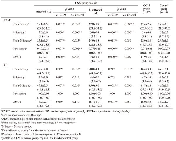
Results: The M-wave (M) latency and minimum F-wave (Fmin) latency from the ADM, and Fmin-M latency from the ADM/AH were significantly longer in the CSA group than in the other groups, on both the affected (p = 0.000-0.007) and unaffected sides (p = 0.000-0.033). F-wave persistence from the bilateral ADMs was significantly lower in the CSA group than in the other groups (p = 0.000-0.002). Among the CSA patients, there were no significant differences in these parameters between the affected and unaffected sides. The CMCT showed no significant differences between the CSA and control groups, but significant differences between the CSA and CCM groups (p = 0.000-0.004).
Conclusions: CSA patients with unilateral deltoid muscle atrophy had subclinical impairments of lower motor neurons and/or peripheral axons in the ulnar nerve, and subclinical impairments of peripheral axons in the tibial nerve. These motor impairments may have originally existed in these individuals before the onset of CSA.