Spine Surgery and Related Research
Online ISSN : 2432-261X
ISSN-L : 2432-261X
3 巻, 3 号
選択された号の論文の13件中1~13を表示しています
REVIEW ARTICLE
  • Tatsunori Ikemoto, Kenji Miki, Takako Matsubara, Norimitsu Wakao
    2019 年 3 巻 3 号 p. 199-206
    発行日: 2019/07/27
    公開日: 2019/07/27
    [早期公開] 公開日: 2018/10/10
    ジャーナル オープンアクセス

    Studies have indicated that chronic low back pain (LBP) should be approached according to its morphological basis and in consideration of biopsychosocial interventions. This study presents an updated review on available psychological assessments and interventions for patients with chronic LBP. Psychosocial factors, including fear-avoidance behavior, low mood/withdrawal, expectation of passive treatment, and negative pain beliefs, are known as risk factors for the development of chronic LBP. The Örebro Musculoskeletal Pain Questionnaire, STarT Back Screening Tool, and Brief Scale for Psychiatric Problems in Orthopaedic Patients have been used as screening tools to assess the development of chronicity or identify possible psychiatric problems. The Pain Catastrophizing Scale, Pain Self-Efficacy Questionnaire, and Injustice Experience Questionnaire are also widely used to assess psychosocial factors in patients with chronic pain. With regard to interventions, the placebo effect can be enhanced by preferable patient-clinician relationship. Reassurance to patients with non-specific pain is advised by many guidelines. Cognitive behavioral therapy focuses on restructuring the negative cognition of the patient into realistic appraisal. Mindfulness may help improve pain acceptance. Self-management strategies with appropriate goal setting and pacing theory have proved to improve long-term pain-related outcomes in patients with chronic pain.

ORIGINAL ARTICLE
  • Hayato Enoki, Toshikazu Tani, Kenji Ishida
    2019 年 3 巻 3 号 p. 207-213
    発行日: 2019/07/27
    公開日: 2019/07/27
    [早期公開] 公開日: 2019/01/15
    ジャーナル オープンアクセス

    Introduction: Leg spasticity in degenerative compression myelopathy causes impairment of fast and rapid repetitive movements, which tends to appear despite the disproportionate paucity of clinical weakness. As clinically useful measures used to quantify the slowness of voluntary leg movements in this pathological condition, we compared the foot tapping test (FTT) with the simple walking test, which is now considered the gold standard in this field.

    Methods: We compared the FTT with the simple walking test, the grip-and-release test, and the functional scales of Nurick and the Japanese Orthopedic Association (JOA) in 77 patients with cervical compression myelopathy and 56 age-matched healthy subjects. The FTT was conducted on both sides separately, and the subject, while being seated on a chair, moved his/her toes up and down repeatedly to tap the floor as fast and as vigorously as possible for 10 sec with his/her heels planted on the floor.

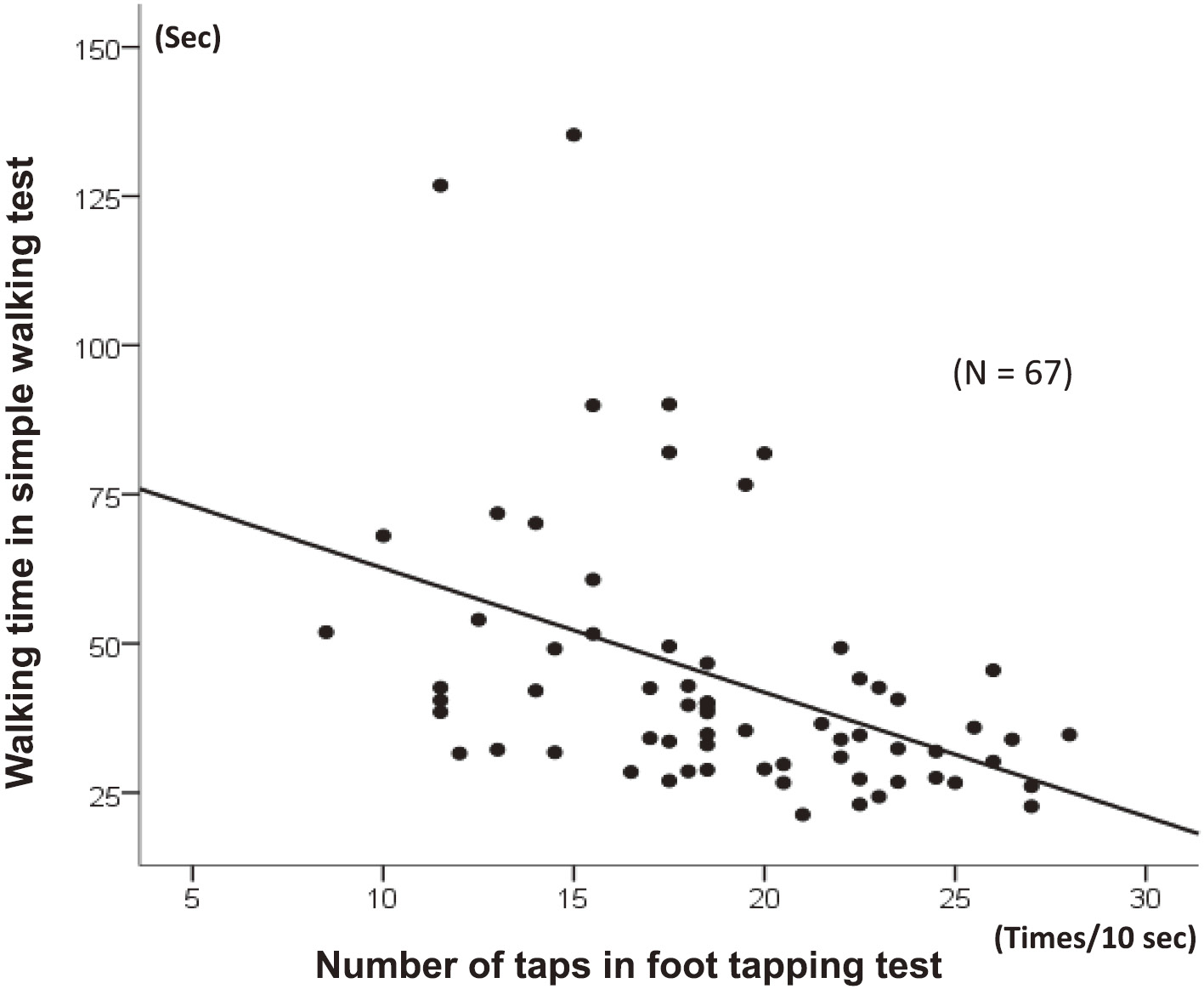
    Results: The number of 10-sec foot tapping in the patient group significantly correlated with the Nurick grades (r = −0.566; P < 0.0001), the JOA scores (r = 0.520; P < 0.0001), and the grip-and-release rates (r = 0.609; P < 0.0001). It also significantly correlated with the 30-m walking time (r = −0.507; P < 0.0001) and the number of steps taken (r = −0.494; P < 0.0001). Assessments of wheelchair-dependent patients and side-to-side comparison, in which the simple walking test plays no role, revealed significantly fewer FTT taps in wheelchair-bound patients than in the ambulatory patients and a significant trend for cervical compression myelopathy to dominantly affect the upper and lower limbs on the same side.

    Conclusions: This study contributes to the reassessment of the currently underutilized FTT as part of a routine neurologic examination of degenerative compression myelopathy.

  • Ryo Sugawara, Katsushi Takeshita, Yasushi Inomata, Yasuhisa Arai, Masa ...
    2019 年 3 巻 3 号 p. 214-221
    発行日: 2019/07/27
    公開日: 2019/07/27
    [早期公開] 公開日: 2018/12/01
    ジャーナル オープンアクセス

    Introduction: The Japanese Scoliosis Society (JSS) created a longitudinal complication survey of spinal deformity surgery and established the Morbidity and Mortality (M&M) Committee in 2012. The purpose of this study was to analyze the results of the complication survey in 2014 and to report the differences in the complication rates between the years 2012 and 2014.

    Methods: A request to participate in this survey was mailed to all JSS members. The questionnaires were sent through e-mail to the members who took part in this survey, and the responses were returned through the same. Diagnosis was grouped into idiopathic scoliosis, congenital scoliosis, neuromuscular scoliosis, spondylolisthesis, pediatric kyphosis and adult spinal deformity. Complication was grouped into death, blindness, neurological deficit (motor or sensory deficit), infection, massive bleeding, hematoma, pneumonia, cardiac failure, DVT/PE, gastrointestinal perforation and instrumentation failure.

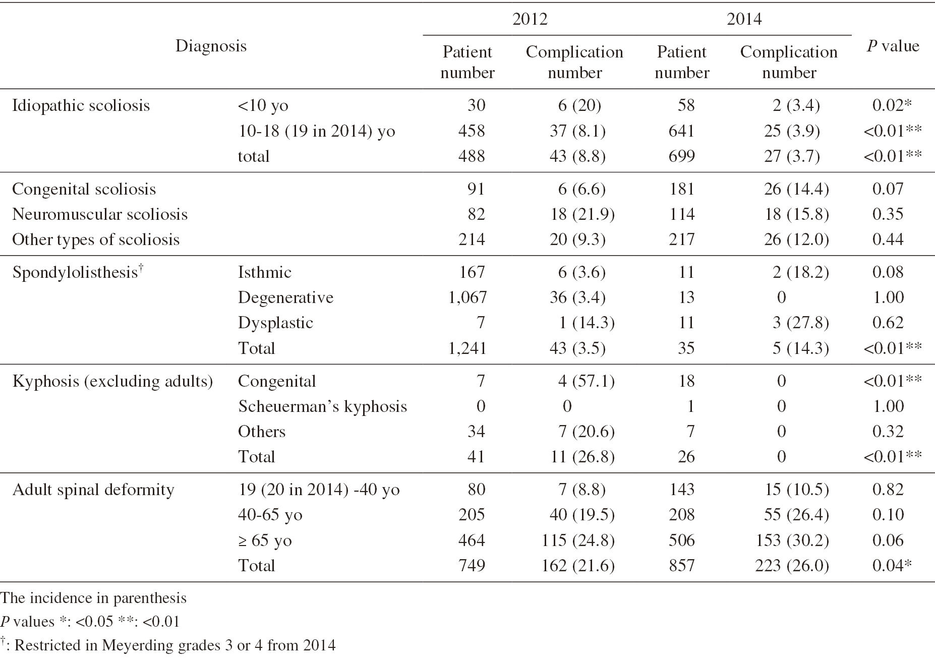
    Results: A total of 2,012 patients were reported from 71 institutes. Overall, complications were observed in 326 patients, and the complication rate increased from 10.4% in 2012 to 15.3% in 2014. The complication rate decreased from 8.8% to 3.7% in idiopathic scoliosis, 21.9% to 15.8% in neuromuscular scoliosis and 26.8% to 0% in kyphosis. The complication rate increased from 6.6% to 14.4% in congenital scoliosis, 9.3% to 12.0% in other types of scoliosis, 3.5% to 14.3% in spondylolisthesis and 21.6% to 26.0% in adult spinal deformity. The rate of neurological deficit, especially in motor deficit, increased from 3.2% to 7.7% in older patients with adult spinal deformity. Instrumentation failure was also more common in patients with adult spinal deformity (5.2% to 5.8%), especially in patients aged 40-65 years (4.4% to 9.1%).

    Conclusions: The major complication trends were an increasing rate of neurological deficit and instrumentation failure, especially in adult spinal deformity.

  • Tsutomu Akazawa, Masahiro Iinuma, Shingo Kuroya, Yoshiaki Torii, Tasuk ...
    2019 年 3 巻 3 号 p. 222-228
    発行日: 2019/07/27
    公開日: 2019/07/27
    [早期公開] 公開日: 2018/12/01
    ジャーナル オープンアクセス

    Introduction: The purpose of this study is to investigate thoracic deformity correction and pulmonary function changes in patients with adolescent idiopathic scoliosis (AIS) five years or more after undergoing posterior spinal fusion with thoracoplasty for correction of a thoracic deformity.

    Methods: Subjects were 57 patients with AIS who underwent posterior spinal fusion between 2004 and 2010. 24 patients who had undergone thoracoplasty at least five years earlier agreed to participate in this research. X-rays, pulmonary function tests, and thoracic cage computed tomography (CT) were performed, and the Scoliosis Research Society Outcomes Questionnaire (SRS-22) was administered. CT axial images were used at the apex of the main thoracic (MT) curve. Apical vertebral rotation was evaluated using rotation angle to the sagittal plane (RAsag). Thoracic deformities were evaluated using the rib hump index (RHi) and the posterior hemithoracic symmetry ratio (PHSr).

    Results: There were no significant differences between the preoperative and the final observation forced vital capacity (FVC) or the preoperative and the final observation %FVC. The forced expiratory volume in 1 s (FEV1) and %FEV1 were significantly improved at the final observation: FEV1 (preoperative: 1.88 L, final observation: 2.05 L, p = 0.045) and %FEV1 (preoperative: 57.1%, final observation: 66.2%, p = 0.001). FEV1/FVC was also significantly improved at the final observation (preoperative: 83.0%, final observation: 86.4%, p = 0.019). The peak expiratory flow (PEF) was significantly improved at the final observation (preoperative: 3.67 L/s, final observation: 4.38 L/s, p = 0.029). On the CT assessment for thoracic deformities, there were no significant changes in RAsag or RHi. PHSr was significantly increased at the final observation compared with the preoperative period.

    Conclusions: With posterior spinal fusion in combination with thoracoplasty for AIS, although the correction of deformities was limited, the pulmonary function testing demonstrated the preservation of vital capacity (VC) and improvements in the forced expiratory volume in 1 s and expiratory flow.

  • Mitsunori Yoshimoto, Noriyuki Iesato, Yoshinori Terashima, Katsumasa T ...
    2019 年 3 巻 3 号 p. 229-235
    発行日: 2019/07/27
    公開日: 2019/07/27
    [早期公開] 公開日: 2018/12/01
    ジャーナル オープンアクセス
    電子付録

    Introduction: There have been several reports on surgical techniques involving microendoscopy or percutaneous endoscopy for treating lumbar foraminal stenosis (LFS). However, no studies have assessed the mid-term clinical results of endoscopic techniques in spite of their relatively long history. In this study, we report 20 consecutive cases of LFS treated by our microendoscopic technique focusing on clinical results with a follow-up of at least two years.

    Methods: Twenty consecutive cases of LFS treated with microendoscopic decompression were followed up at 1, 2, 6, and 12 months postoperatively and annually thereafter. The patients were 14 males and 6 females, and the mean age at the time of surgery was 64.7 years. The Japanese Orthopaedic Association (JOA) score was used as the clinical outcome index.

    Results: Of the 20 patients, 16 were monitored successfully for more than 2 years. The follow-up rate was 80.0%, and the mean follow-up period was 66.3 months. The JOA score improved from 13.8 points before surgery to 24.6 points at final follow-up. Revision fusion surgeries were performed in two cases for LFS recurrence.

    Conclusions: The microendoscopic technique effectively treats LFS.

  • Shigeto Ebata, Hiroki Oba, Tetsuro Ohba, Jun Takahashi, Shota Ikegami, ...
    2019 年 3 巻 3 号 p. 236-243
    発行日: 2019/07/27
    公開日: 2019/07/27
    [早期公開] 公開日: 2019/01/25
    ジャーナル オープンアクセス

    Introduction: We recently demonstrated that pelvic incidence (PI) decreases after long fusion using iliac screws (ISs) and plays a role in good sagittal balance postoperatively. By contrast, the IS loosening rate may cause reversion, increasing the PI and causing loss of sagittal balance. The aim of this study was to determine the effect of the number of ISs inserted into the iliac bone for long fusion to correct adult spinal deformities (ASDs) on the frequency of IS loosening, postoperative PI, and surgical outcomes.

    Methods: We included data from 70 consecutive patients. Cases in which two ISs were inserted bilaterally comprised the dual IS group (Group D), whereas cases in which one IS was inserted bilaterally comprised the single IS group (Group S).

    Results: IS loosening was observed in four patients in Group D (9%) and 14 patients in Group S (61%). Both early and one-year postoperative PI were significantly smaller in Group D (P < 0.001). The sagittal vertical axis (SVA) one-year postoperatively was significantly smaller in Group D (P = 0.003).

    Conclusions: The loosening rate of dual ISs was as low as about one-seventh that of single ISs. Using dual ISs, postoperative PI can be kept small, possibly resulting in a smaller SVA.

  • Yawara Eguchi, Munetaka Suzuki, Takashi Sato, Hajime Yamanaka, Hiroshi ...
    2019 年 3 巻 3 号 p. 244-248
    発行日: 2019/07/27
    公開日: 2019/07/27
    [早期公開] 公開日: 2019/01/25
    ジャーナル オープンアクセス

    Introduction: To investigate the risk of epidural hematoma after spinous process-splitting laminectomy (SPSL).

    Methods: A total of 137 cases (mean age, 72.4 years; 68 men) of SPSL were included. Of these, there were instances (3.7%; mean age, 70.5 years; all male) of postoperative development of new neurologic deficit due to epidural hematoma requiring reoperation. The 133 subjects (72.5 years; 64 men) with normal postoperative course were used as controls, and comparisons were made between both groups using chi-squared and Student's t-tests. Regarding our investigation of risk factors for epidural hematoma, logistic regression was conducted with presence or absence of hematoma as our primary outcome variable, and age, gender, disease duration, number of laminectomies, which levels were decompressed, blood loss, length of case, drain output, coagulopathy, and whether or not there was an intraoperative dural tear were our explanatory variables.

    Results: All cases of hematoma were single-level laminectomies; there was one case of T9-10 and 3 cases of L2-3. In our direct comparison of both groups (hematoma versus control), the proportion of men was significantly higher in the hematoma group (100% versus 48%, p < 0.05); levels decompressed were also significantly higher (p < 0.05) in the hematoma group, and drain outputs were significantly lower (113 mL versus 234 mL, p < 0.05). From our logistic regression analysis, the levels were significantly higher (χ2 = 15, p = 0.0001) and the drain outputs were smaller (χ2 = 4.6, p = 0.03) in the hematoma group.

    Conclusions: Single-level decompression higher than the L2-3 level and reduced drain output were risk factors for spinal epidural hematoma. With this method of spinous process suturing and reconstruction there is less decompression compared with more conventional methods; therefore, the effect of hematoma may be more pronounced at higher vertebral levels with reduced canal width, and drain failure may also occur with this limited space.

  • Soichiro Masuda, Eijiro Onishi, Satoshi Ota, Satoshi Fujita, Tatsuya S ...
    2019 年 3 巻 3 号 p. 249-254
    発行日: 2019/07/27
    公開日: 2019/07/27
    [早期公開] 公開日: 2019/01/25
    ジャーナル オープンアクセス

    Introduction: In general, osteoporotic vertebral fractures with neurological deficits require surgery. However, the ideal surgical method remains controversial. We evaluated the efficacy of combining posterior instrumented fusion and vertebroplasty using allograft bone chips.

    Methods: Twelve patients (five men, seven women; age 68-84 years, mean age 75.9 years) with osteoporotic vertebral fractures with neurological deficits were reviewed retrospectively. They underwent posterior instrumented fusion and vertebroplasty, using allograft bone, at our institution between January 2007 and June 2016. We assessed the surgical results, radiologically and neurologically, after a mean follow-up of 37.3 months.

    Results: The mean local kyphosis angle was 10° before surgery, −3.3° immediately after surgery, and 4.4° at follow-up. The average spinal canal compromise was 26.9% before surgery and 19.5% at follow-up. All patients achieved bony fusion and none needed additional surgery. All patients improved by at least one grade on the modified Frankel grading system.

    Conclusions: Combining vertebroplasty, using allograft bone chips, and posterior instrumented fusion appears to be an effective option for osteoporotic vertebral fractures with neurological deficits.

  • Katsuhito Yoshioka, Hideki Murakami, Satoru Demura, Satoshi Kato, Hiro ...
    2019 年 3 巻 3 号 p. 255-260
    発行日: 2019/07/27
    公開日: 2019/07/27
    [早期公開] 公開日: 2019/03/22
    ジャーナル オープンアクセス

    Introduction: Global spinal balance and its relationship to the pelvis have received much attention, and various formulae have been used to predict postoperative spinopelvic alignment for spinal surgery. However, previous studies had limitations because no consideration was given to the dynamic factor.

    Methods: Fifteen healthy adults without any lumbar disorder (group A) and 9 L4-spondylolisthesis patients (Group B) volunteered to participate in the study. Sequential images were captured with the subjects in the standing position with maximal forward bending followed by backward bending using a dynamic flat panel detector system. Spinopelvic parameters (LL: lumbar lordosis, SA: sacrofemoral angle, SS: sacral slope, PI: pelvic incidence, DP: distance of the horizontal movement of the pelvis) were evaluated. We also investigated the relationship between LL and SA (lumbar/hip [L/H] ratio) as the spinopelvic rhythm.

    Results: In group A, the mean change in LL was 83.2 ± 9.5°; change in SA, 45.4 ± 16.6°; SS, 42.6 ± 8.9°; PI, 43.2 ± 7.7°; DP, 15.7 ± 3.4 cm, and L/H ratio, 3.6 ± 2.7. However, spinopelvic rhythm changed over time, because the change in LL was larger than the change in SA from the middle of the rising motion to the upright position. In group B, the mean change in LL was 50.3 ± 8.0°; SA, 56.9 ± 16.0°; SS, 27.5 ± 13.5°; PI, 47.4 ± 10.4°; DP, 12.7 ± 6.8 cm; and L/H ratio, 1.0 ± 0.5.

    Conclusions: When compared with the change in LL, individual differences were largely noted in the change in SA. These results demonstrated that the range of hip joint motion under physiological conditions, unlike anatomical motion, differed substantially between individuals. Therefore, spinopelvic rhythm is dependent on the change in SA.

  • Ai Mazaki, Sumihisa Orita, Kazuhide Inage, Miyako Suzuki, Kohki Abe, Y ...
    2019 年 3 巻 3 号 p. 261-266
    発行日: 2019/07/27
    公開日: 2019/07/27
    [早期公開] 公開日: 2019/04/05
    ジャーナル オープンアクセス

    Introduction: Causes of pain due to spinal metastases have been insufficiently investigated. Tumor necrosis factor-α (TNF-α) and interleukin-6 (IL-6) were the focus of this study. Both are known as proinflammatory cytokines associated with the pathophysiology of pain syndromes1). It is well known that cancer cells produce these cytokines, but whether osteoclasts produce them as well remains unclear. We hypothesize that osteoclasts produce these cytokines; in other words, pain from spinal metastasis is stronger than pain from the primary tumor.

    Methods: We made a rat spinal metastasis model of breast cancer (metastasis group) and models with a hole in the vertebrae (puncture group) and resected the vertebrae. Tartrate-resistant acid phosphatase (TRAP) staining was performed to reconfirm that osteoclasts increase in vertebrae with spinal metastasis. We then evaluated TNF-α and IL-6 expression using immunohistochemistry and real-time polymerase chain reaction (PCR).

    Results: The results of TRAP staining showed that osteoclasts increase in metastatic vertebrae. The osteoclasts in the puncture models were TNF-α negative but were TNF-α positive in the metastasis model. The osteoclasts in the puncture models and metastasis model were both IL-6 positive. According to the real-time PCR results, TNF-α in vertebrae increased in the metastasis model, but IL-6 did not increase in the metastasis model compared with in the puncture model.

    Conclusions: The number of osteoclasts is higher in the metastasis model. While TNF in the osteoclasts increased in the spinal metastasis model, IL-6 did not. This probably means that breast cancer affects TNF production in osteoclasts. This increase of TNF-α may lead to pain from spinal metastasis.

CASE REPORT
  • Toshio Nakamae, Kiyotaka Yamada, Yasuyuki Tsuchida, Nobuo Adachi, Yosh ...
    2019 年 3 巻 3 号 p. 267-269
    発行日: 2019/07/27
    公開日: 2019/07/27
    [早期公開] 公開日: 2018/07/25
    ジャーナル オープンアクセス

    Introduction: Spinal lesions in synovitis, acne, pustulosis, hyperostosis, and osteitis (SAPHO) syndrome generally have a good prognosis and rarely cause structural destruction or neurological deterioration. We described a surgical case of posterior instrumented surgery without anterior reconstruction and bone graft in a patient with SAPHO syndrome with destructive spondylitis and reviewed the literature on surgical treatment for this entity.

    Case Report: We describe the case of a 73-year-old male who presented with palmoplantar pustulosis. He experienced progressive low back and leg pain for the past 3 months. Destructive spondylitis and lumbar canal stenosis were detected with magnetic resonance imaging (MRI), and aspiration biopsy was used to exclude pyogenic spondylitis and spinal tumors. He underwent posterior decompression and fixation surgery without anterior reconstruction and bone grafting. Low back and leg pain improved after surgery. Postoperative radiography and computed tomography showed boney bridge between vertebral bodies, and MRI showed the decrease of bone marrow edema.

    Conclusions: Posterior fusion without anterior reconstruction produced a bony bridge between the vertebral bodies. Taking the pathophysiology of SAPHO syndrome into consideration, anterior reconstructed fusion for patients with SAPHO syndrome might not be needed.

CLINICAL CORRESPONDENCE
feedback
Top