Abstract
Skin aging is a complex physiological process driven by both endogenous and exogenous factors, involving various mechanisms such as oxidative stress, inflammatory responses, and cellular senescence. As natural bioactive macromolecules, plant polysaccharides possess properties including antioxidant, anti-inflammatory, and free radical scavenging abilities, and have recently shown significant potential in anti-skin aging research. This article systematically reviews the molecular mechanisms of skin aging, with a focus on elucidating how plant polysaccharides exert anti-aging effects by regulating key signaling pathways. Additionally, it summarizes strategies for the extraction, purification, and structural modification of plant polysaccharides, as well as their structure–activity relationships. The aim is to provide a theoretical foundation and research directions for both fundamental studies and product development of plant polysaccharides in the field of anti-skin aging.
1. INTRODUCTION
The primary hallmark of skin aging is the loss of skin elasticity, accompanied by laxity and wrinkling,1) This is closely associated with cellular senescence in the epidermal and dermal layers of the skin, as well as reduced synthesis of hyaluronic acid and collagen.2) The causes of skin aging can be categorized into extrinsic and intrinsic factors. Extrinsic factors are primarily dominated by UV radiation, smoking, air pollution, sleep deprivation, and exposure to harmful chemicals,3) The most prominent intrinsic factor lies in histological changes, particularly those occurring in the basal cell layer. With the accumulation of time and age, the proliferation rate of basal cells slows down, leading to epidermal thinning and a further decline in the proliferative capacity of basal cells.1)
With the advancement of biology, medicine, and pharmacology, significant progress has been achieved in both the research on skin aging mechanisms and the development of anti-skin aging bioactive ingredients. Polysaccharides are biological macromolecules composed of monosaccharide units linked by glycosidic bonds, with molecular weights ranging from tens of thousands to several million,4) and they are widely distributed in plants, animals, and microorganisms, playing a crucial role in the growth and development processes of organisms. Studies have demonstrated that natural plant polysaccharides exhibit anti-inflammatory, free radical scavenging, and antioxidant effects, and their anti-aging potential for the skin has gradually garnered widespread attention.
This article aims to elucidate the mechanisms underlying the skin aging process, outline the extraction and purification methods for plant polysaccharides and their structure–activity relationships in skin aging, summarise key signalling pathways involved in skin aging, and review recent research advances concerning the role of natural plant polysaccharides in improving skin aging. It seeks to provide guidance for the development of polysaccharide-based products targeting skin aging.
2. MECHANISMS OF SKIN AGING
Skin aging is classified into two types: extrinsic aging and intrinsic aging,5) extrinsic aging is mainly influenced by environmental factors. Specifically, UV radiation can penetrate the skin, inducing the degradation of cutaneous collagen and elastic fibers. This not only impairs the adhesion, proliferation, and contraction of fibroblasts to collagen but also increases collagen susceptibility to matrix metalloproteinase-1 (MMP-1), thereby accelerating the loss of extracellular matrix (ECM) function.6) Environmental pollutants such as smoke and dust can induce oxidative stress, thereby impairing the skin. Unhealthy living habits, repetitive facial expressions, and sleep deprivation can also impair the skin’s self-repair capacity, thereby accelerating skin aging.
Numerous factors are primarily responsible for intrinsic aging,7) the core mechanism of cellular senescence is DNA damage, among which the DNA damage response (DDR) is most prominent in the context of UV-induced skin DNA damage. By influencing the inflammatory and endocrine signaling components involved in the aging process, DDR exerts multiple effects on age-related alterations in local and systemic signaling mechanisms8); abnormalities in telomere function can also induce the DDR, thereby triggering skin cell senescence9); mitochondria are organelles that regulate energy and metabolic homeostasis, and they are also the primary source of reactive oxygen species (ROS). Excessive ROS can damage mitochondria, leading to cellular senescence and apoptosis, and mitochondrial dysfunction is closely associated with aging-related10); in addition, senescent cells secrete pro-inflammatory factors, a phenomenon termed the senescence-associated secretory phenotypes (SASP). SASP can promote the occurrence of chronic inflammation and drive normal cells into the senescent process. Specifically, SASP secreted by skin senescent cells significantly disrupts the cutaneous microenvironment, exacerbating inflammation and impairing the epidermal barrier function.11)
These factors ultimately induce structural and functional alterations in components such as cells, fibers, and ECM within the epidermal and dermal layers, thereby accelerating skin aging. From the cellular perspective, fibroblast senescence is a major driver of the skin aging process, fibroblasts produce the dermal ECM and maintain its homeostasis, and they regulate their morphology and function by adhering to the ECM. During the aging process, the ECM is gradually degraded upon the activation of MMPs, this degradation alters the mechanical forces within the dermal ECM and disrupts the interactions between fibroblasts and the ECM, thereby inducing a senescent fibroblast phenotype.12) After keratinocyte senescence, their proliferative and differentiative capacities are significantly impaired, the secretion of ECM is reduced, and intercellular adhesion is decreased, accompanied by the production of a large number of pro-inflammatory factors. These changes further contribute to epidermal aging.13) The crosstalk between immune cells and the matrix in the skin is also a crucial contributor to chronic inflammatory responses, thereby causing physical aging of the skin.14)
From the protein and matrix perspectives, mature elastic fibers are mainly composed of elastin, fibrillin microfibrils, and elastic fiber-associated proteins.15) The overexpression of microfibril-associated protein 5 (MFAP5) is a key factor in the thickening, disorganisation and aggregation of elastic fibres during intrinsic dermal ageing.16) During the aging process, the elasticity and flexibility of elastic fibers decrease, the elastic fiber network fractures, their quantity reduces, and elastin undergoes structural denaturation, ultimately leading to the loss of skin elasticity.17) Collagen is the most abundant structural protein in the skin, playing a crucial role in maintaining skin structure and elasticity.18) After skin aging, the originally compact helical structure of collagen undergoes fragmentation or unwinding, resulting in structural disorganization. This further impairs skin stretchability and elasticity, leading to the loss of skin firmness; increased collagen cross-linking impairs skin moisture absorption, leading to skin laxity and wrinkle formation. Some studies have shown that skin aging can be delayed by maintaining the expression of collagen XVII to promote stem-cell competition.19) In addition, glycosaminoglycans (GAGs) and proteoglycans (PGs) are also structural components of the ECM.20) They play a crucial role in skin physiology by regulating keratinocyte proliferation and differentiation, the occurrence of inflammatory responses, as well as the composition and quality of the ECM.21) After skin aging occurs, changes take place in the content and structure of GAGs and PGs, leading to a decrease in skin moisture-retaining capacity and resulting in the loss of its original firmness and elasticity.
3. EXTRACTION, PURIFICATION, AND MODIFICATION STRATEGIES FOR PLANT POLYSACCHARIDES
The efficacy of plant polysaccharides in mitigating aging is significantly influenced by their purity and structural characteristics, which in turn depend on extraction, purification, and structural modification processes. To enhance the yield, bioavailability, and biological activity of these macromolecular compounds, various strategies have been developed (Fig. 1).
3.1. Extraction MethodsNumerous methods exist for extracting plant polysaccharides, primarily encompassing traditional and modern extraction techniques. Traditional methods predominantly employ hot water, dilute acids and alkalis, and organic solvents for extraction. These remain widely adopted owing to their simplicity of operation and low cost.22) However, modern techniques are increasingly favoured for their higher efficiency and ability to preserve polysaccharide integrity. Ultrasound-assisted extraction (UAE) employs cavitation to disrupt plant cell walls, significantly enhancing extraction efficiency while minimising thermal damage to polysaccharide structures. For instance, UAE optimised for seawater Arthrospira platensis polysaccharides (APPs) demonstrated superior yield and antioxidant activity compared to conventional heating methods.23) Similarly, microwave-assisted extraction (MAE) rapidly heats water within plant matrices via electromagnetic radiation, achieving efficient cell wall disruption and polysaccharide release. A study on Salvia miltiorrhiza polysaccharides (SMPs) demonstrated that MAE not only shortens extraction time but also yields extracts with enhanced antioxidant activity.24) Enzyme-assisted extraction (EAE) employs enzymes such as cellulase and pectinase to decompose cell wall components, offering a highly specific and gentle processing method particularly suited to heat-sensitive polysaccharides. Chestnut flower polysaccharides extracted via EAE demonstrated potent hypoglycaemic effects.25) Furthermore, combining multiple methods enhances polysaccharide extraction efficiency. For instance, ultrasonic-assisted enzymatic extraction of silver ear polysaccharides yielded significantly higher extraction rates compared to single-method approaches.26)
3.2. Purification TechniquesRoughly extracted polysaccharides require simple purification to remove impurities such as proteins, pigments, and small molecules. A typical purification workflow includes deproteinisation, decolourisation, and dialysis to remove salts and small molecules. Subsequent purification employs methods such as column chromatography, membrane separation, and fractionated ethanol precipitation. Polysaccharides from Polygonatum were purified via column chromatography, yielding three homogeneous fractions from its aqueous extract.27) The acid polysaccharide SSP2-2 was purified from thermal water via column chromatography of Suaeda salsa.28) Purification of Undaria pinnatifida polysaccharides through ethanol precipitation demonstrated superior antioxidant activity.29) Integrated membrane separation processes have been employed to purify polysaccharides from Ulva lactuca, enhancing their hygroscopicity and moisturising properties.30)
3.3. Structural ModificationStructural modification of polysaccharides involves altering their molecular backbone, substituent groups, or degree of polymerisation through chemical, physical, or biological means, with the core objective being to optimise their physicochemical properties and biological activity. Chemical modification remains the most prevalent approach, encompassing techniques such as sulfation, hydroxymethylation, and selenation. These methods significantly enhance polysaccharide water solubility, antioxidant capacity, and anti-inflammatory activity. For instance, sulfated polysaccharides extracted from Saussurea costus via the chlorosulfonic acid-pyridine method demonstrated superior immunomodulatory effects on macrophages compared to unmodified polysaccharides.31) Selenium-modified Codonopsis pilosula polysaccharides synthesised via the micro-wave-assisted method exhibited markedly enhanced antioxidant activity.32) Physical modification employs non-chemical approaches such as ultrasonication, high pressure, or radiation to disrupt intermolecular hydrogen bonds or alter aggregate structures, thereby improving polysaccharide solubility and rheological properties. This method carries no chemical reagent residues and offers high safety, though its modifying effects are relatively mild and susceptible to parameter variations. Hydrophilic polysaccharides isolated and purified from the tuberous roots of Codonopsis pilosula were found to enhance the proliferation and phagocytic capacity of RAW264.7 macrophages with increasing ultrasonic intensity. They also significantly elevated antioxidant activity and immune function in the body.33) Moreover, bio-modification relies on enzyme specificity to precisely regulate polysaccharide structures, preserving their natural architecture and activity under mild conditions. Enzymatic modification of red ginseng polysaccharides using α-amylase and amyloglucosidase enhanced their regulatory effects on the intestinal immune system.34)
4. STRUCTURE–ACTIVITY RELATIONSHIP OF PLANT POLYSACCHARIDES IN SKIN AGING
The anti-aging effects of plant polysaccharides depend upon their specific chemical structures. Key structural characteristics include molecular weight, monosaccharide composition, glycosidic linkage patterns, chain conformation, and the presence of functional groups, which collectively determine their biological activity.
4.1. Effect of Molecular WeightThe molecular weight of polysaccharides significantly influences their solubility, viscosity, and bioavailability. Typically, polysaccharides with lower molecular weight exhibit superior antioxidant properties and free radical scavenging capacity. This is primarily due to their structural advantages, including shorter molecular chains with reduced entanglement, which lead to higher aqueous solubility and dispersibility. Their looser spatial conformation allows for more extensive exposure of active sites, thereby significantly increasing the accessible surface area for interaction with free radicals.35) Two low molecular weight polysaccharides, with molecular weights of 6.31 and 14.07 kDa, respectively, were purified from Ganoderma lucidum spores. Research indicates that polysaccharides with lower molecular weights exhibit stronger antioxidant activity and superior free radical scavenging efficacy.36) In polysaccharides from Enoki mushrooms degraded by ultrasonication, low-molecular-weight polysaccharides demonstrated outstanding anti-inflammatory activity, inhibiting the release of inflammatory cytokines and reducing the transcriptional levels of pro-inflammatory pathways.37)
4.2. The Role of MonosaccharideThe composition of polysaccharides, along with the types of glycosidic bonds, the type of monosaccharide involved, and its linkage pattern, collectively determine the primary structure and biological specificity of these molecules. For instance, α-glycosidic bonds constitute the core linkage pattern in pectin-type berry polysaccharides, primarily comprising α-(1→4) and α-(1→5) glycosidic bonds, endowing them with exceptional free radical scavenging efficacy.38) The primary polysaccharides in Undaria pinnatifida comprise alginic acid, fucoidan, and kelp polysaccharides. Fucoidan, predominantly composed of L-fucose linked via α-(1→3) glycosidic bonds, enhances skin antioxidant capacity, mitigates oxidative damage, and concurrently exhibits moisturising properties alongside collagen-promoting functions.39) Meanwhile, the principal polysaccharide components of Auricularia auricula-judae include mannose, galactose, and glucose. It inhibits the production of intracellular ROS in human fibroblasts induced by UVB radiation and also accelerates skin wound healing.40) Moreover, the polysaccharides of Suaeda salsa are predominantly linked by β-glycosidic bonds, with their monosaccharides chiefly comprising galacturonic acid. Their pronounced acidic nature confers exceptional water solubility and charge properties, establishing the structural foundation for binding to cell surface receptors and regulating inflammatory signaling pathways.28)
4.3. The Influence of SubstituentsThe presence and type of substituents constitute one factor influencing the biological activity of polysaccharides. By altering the polysaccharide’s charge properties, water solubility, spatial conformation, and interaction patterns with biomolecules, they affect its biological functions such as antibacterial activity, immunomodulation, antioxidant properties, and antitumour effects. For instance, sulphate groups confer strong negative charges to polysaccharides, enabling them to efficiently scavenge cationic free radicals and disrupt the binding of pro-inflammatory cytokines to their receptors. The potent anti-photoaging effects of brown seaweed polysaccharides primarily stem from their sulphate content, which reduces UV-induced ROS expression by inhibiting collagen degradation.41) Acetyl groups can optimise spatial conformation by altering polysaccharide amphiphilicity, thereby enhancing antioxidant properties. A nanoemulsion gel formulation developed by acetylating hawthorn polysaccharides demonstrated superior efficacy to native hawthorn polysaccharides in treating UVB-induced skin photoaging in mice.42)
5. RELATED PATHWAYS OF PLANT POLYSACCHARIDES IN IMPROVING SKIN AGING
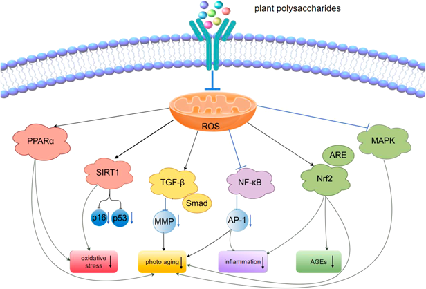
As a natural active ingredient, plant polysaccharides exhibit significant efficacy in ameliorating skin aging. They can mitigate the occurrence of skin aging-related damage by regulating multiple signaling pathways (Fig. 2, Table 1), thereby playing a crucial role in maintaining skin health.
Table 1. Plant Polysaccharides Modulate Signaling Pathways to Improve Skin Aging
| Signaling pathways | Plant polysaccharides | Mechanism of action | References |
|---|
| SIRT1 | Tremella fuciformis polysaccharides | Upregulating SIRT1 expression reduces the level of oxidative stress | 51) |
| Dendrobium officinale polysaccharides | Increasing SIRT1 protein expression exerts an anti-aging effect | 52) |
| Undaria pinnatifida polysaccharides | Activating the SIRT1 signaling pathway decreases the production of ROS | 53) |
| TGF-β/Smad | Fermented P. notoginseng polysaccharides | Upregulated Smad2/3 expression and downregulated Smad7 expression alleviate cellular oxidative damage | 60) |
| Dendrobium officinale polysaccharides | Exerts anti-aging effects through the TGF-β/Smad signaling pathway | 52) |
| Trehalose | Activating the TGF-β/Smad pathway increases the synthesis of type I procollagen | 61) |
| NF-κB | Fermented P. notoginseng polysaccharides | Downregulating the expression of NF-κB and iNOS delays cellular senescence | 70) |
| Premna microphylla Turcz pectin | Decreased levels of NF-κB and COX-2 repair skin photoaging damage | 71) |
| Tremella fuciformis polysaccharides | Inhibiting the activation of Akt, p38, and NF-κB alleviates the cellular oxidative stress response | 72,73) |
| Trehalose | Blocking the signaling transduction of MAPK, AP-1, and NF-κB inhibits MMPs expression | 61) |
| Ophiopogon japonicus polysaccharides | Reducing the level of NF-κB exerts antioxidant activity and delays aging | 74) |
| Nrf2/ARE | Lycium barbarum polysaccharides | Activating the Nrf2/ARE pathway scavenges ROS and prevents photo-damage | 84) |
| Tremella fuciformis polysaccharides | Upregulating Nrf2 expression and downregulating Keap1 expression effectively alleviates skin damage | 85) |
| Premna microphylla Turcz pectin | Increasing the protein level of Nrf2 and decreasing the protein level of Keap1 | 71) |
| Agaricus blazei Murrill acidic polysaccharides | Regulating the Keap1-Nrf2/ARE signaling pathway reduces the occurrence of oxidative stress responses | 86) |
| Ganoderma lucidum polysaccharides | Activating Nrf2 and inhibiting its negative regulator Keap1 | 87) |
| Cistanche deserticola polysaccharides | Alleviating the formation of ROS through the Nrf2/HO-1 pathway | 88) |
| Aloe polysaccharides | Inhibit apoptosis and activate the expression of Keap1, Nrf2, and others. | 89) |
| Bletilla striata polysaccharide | Activation of the Nrf2/HO-1 signalling pathway restores antioxidant capacity in mouse skin | 90) |
| MAPK | Sophora japonica flower bud polysaccharides | Downregulating the expression of MAPK proteins protects the skin | 85) |
| Ganoderma lucidum polysaccharides | Inhibiting the PKA and MAPK signaling pathways activated by UVB | 92) |
| Lycium barbarum polysaccharides | Reduced AP-1 protein expression diminishes photodamage in mouse skin. | 93) |
| PPARα | Saussurea involucrata polysaccharides | Activating the PPARα pathway resists skin photo-damage | 95) |
inducible nitric oxide synthase (iNOS); cyclooxygenase-2 (COX-2).
SIRT1, also known as silent mating type information regulation 2 homolog 1, is a conserved family member of class III histone deacetylases that is dependent on nicotinamide adenine dinucleotide (NAD+). In mammals, there are seven sirtuins (SIRT1 to SIRT7) that are involved in maintaining genomic stability, regulating energy metabolism, and delaying aging.43) Currently, SIRT1 has been extensively studied. Numerous studies have demonstrated that SIRT1 exerts deacetylation modification on histone tails through histoneacetyltransferases (HAT) with different activities, thereby inhibiting cell apoptosis, regulating metabolism, and suppressing inflammation to attenuate the senescence of skin cells.44,45) SIRT1 can also regulate the proliferation and differentiation of major skin cell types, exert anti-aging effects mediated by p53 and p16 proteins, and participate in the maintenance of genomic stability, oxidative stress responses, ECM turnover, and cell cycle regulation.46) In addition, SIRT1 can regulate the expression of filaggrin (FLG); dysregulation of FLG expression leads to epidermal barrier dysfunction, thereby contributing to skin aging.47)
At the molecular level, a typical hallmark of aging is mitochondrial dysfunction.48) Mitochondria produce ROS through oxidative reactions during normal physiological metabolism of cells. However, excessive production of ROS in cells causes mitochondrial damage, reduces mitochondrial ATP generation, decreases its membrane potential, triggers a chain reaction, and accelerates cellular senescence.49) Excessive ROS can damage the structure of DNA, resulting in impaired and disrupted cellular function.50) ROS accumulation can induce oxidative stress by inhibiting SIRT1 activity, leading to damage to lipids, proteins, nucleic acids, and organelles, thereby triggering cellular senescence. This mechanism also constitutes one of the core pathways mediating skin aging.47) Therefore, regulating the SIRT1 signaling pathway helps reduce the occurrence of oxidative stress, thereby achieving the effect of delaying cellular senescence.
Plant polysaccharides can exert their anti-aging effects by activating the SIRT1 signaling pathway. Tremella fuciformis polysaccharide (TFPS) exerts a protective effect against damage to normal human dermal fibroblasts (NHDF). TFPS promotes the expression of SIRT1 in damaged fibroblasts and regulates cellular senescence by activating protein kinases, extracellular signal-regulated kinase (ERK) and protein kinase B (Akt), whilst concurrently reducing the levels of p16, p21, p53, and caspase-3.51) Through efficient enzymatic depolymerisation of Dendrobium officinale, a functional polysaccharide named mannooligosaccharide (DOMOS) was obtained. In vitro studies revealed that DOMOS upregulates ECM gene expression levels and increases SIRT1 protein expression. Following topical application to subjects’ faces, significant improvements in skin radiance, firmness, and wrinkle appearance were observed, indicating DOMOS promotes skin elasticity and moisture retention.52) Purified brown algae polysaccharides from Undaria pinnatifida (UPF) alleviate mitochondrial dysfunction by activating the AMP activated protein kinase (AMPK)/SIRT-1/Peroxisome Proliferator-Activated Receptor Gamma Coactivator-1 alpha (PGC-1α) signalling pathway, thereby reducing ROS production and protecting human immortalised keratinocytes and human foreskin fibroblasts (HFF), thereby improving UV-induced skin aging.53)
5.2. Transforming Growth Factor-β (TGF-β)/Smad Signaling PathwayTGF-β signaling transduction plays important biological functions in regulating cell proliferation, differentiation, migration, immune-inflammatory responses, and angiogenesis.54) Type I collagen is one of the major structural proteins of the ECM in the dermis, and the activity of TGF-β1 can affect the synthesis of type I procollagen (the precursor of type I collagen).In dermal fibroblasts, TGF-β1 can regulate collagen production through the Smad pathway.55) In contrast, Smad7 exerts an inhibitory effect on TGF-β signaling transduction and can suppress the production of type I procollagen in the dermis, ultimately inhibiting the synthesis of type I collagen.
UV radiation is a crucial cause of accelerated skin aging,56) During skin photoaging,57) TGF-β is upregulated and activated, inducing excessive production of MMPs, promoting overexpression of inflammatory cytokines and prolonged infiltration of neutrophils. This leads to progressive collagen degradation and abnormal elastic fiber damage, resulting in the destruction of the ECM in the dermal connective tissue of the skin and subsequent aging-related damage to the skin.58,59)
Polysaccharides can exert anti-skin aging effects by activating the TGF-β/Smad signaling pathway. You et al. observed that Panax notoginseng polysaccharides activate the TGF-β/Smad pathway, inhibiting collagen and elastin loss alongside fibroblast apoptosis, thereby mitigating cellular oxidative damage.60) Subjects applying Dendrobium officinale found its manno-oligosaccharides exert anti-aging effects via the TGF-β/Smad signalling pathway.52) Trehalose can eliminate UVB-induced ROS accumulation, increase endogenous antioxidant factors superoxide dismutase (SOD) and glutathione (GSH) in keratinocytes, and enhance type I procollagen synthesis by activating the TGF-β/Smad pathway.61)
5.3. Nuclear Factor Kappa-B (NF-κB) Signaling PathwayNF-κB is a key nuclear transcription factor in cells, which is involved in inflammatory responses and immune responses of the organism, regulates cell apoptosis, and stimulates oxidative stress responses. Aberrant activation of NF-κB is closely associated with the occurrence of numerous inflammation-related diseases. On the one hand, excessive free radicals can activate the NF-κB signaling pathway, leading to an increase in tumor necrosis factor-α (TNF-α) levels, while promoting the increased expression of activator protein-1 (AP-1) and MMPs. These serial changes ultimately result in the degradation of the ECM, accelerating the rate of skin aging.62) On the other hand, NF-κB plays a crucial role in the SASP. Under senescence-inducing signals, the phosphorylated NF-κB p65/Rel A subunit translocates to the nucleus, where it binds to the promoters of SASP genes to regulate cellular senescence. Research indicates that GATA4, functioning as an upstream activator of NF-κB, exhibits an increased abundance in the skin after exposure to high-dose ionizing radiation (IR) that induces cellular senescence.63) Therefore, targeting GATA4 or its upstream regulators may aid in inhibiting NF-κB activation and suppressing the induction of SASP, thereby delaying the skin aging process.64)
A key cause of skin cell senescence is chronic inflammation, which can stimulate senescent fibroblasts and keratinocytes to secrete large amounts of the SASP,65) UV irradiation can induce inflammation in skin cells, leading to increased levels of MMPs and degradation of collagen. These changes ultimately result in skin cell relaxation and wrinkling,66) the SASP can be regarded as a key characteristic of cellular aging.67) Another factor contributing to skin aging is the non-enzymatic glycosylation reaction,68) UV irradiation can exacerbate skin glycosylation by promoting the formation of advanced glycationend products (AGEs), aggravate oxidative stress, and ultimately lead to the accumulation of AGEs.69) Therefore, the NF-κB signaling pathway is affected by inflammatory responses and the accumulation of glycated proteins, which tends to accelerate the process of cellular senescence and reduce collagen content. Inhibition of NF-κB signaling pathway activation can be regarded as one of the important strategies for preventing and delaying the senescence of skin cells.
Polysaccharides can exert their anti-aging effects by inhibiting the NF-κB signaling pathway. Feng et al. found that Panax notoginseng polysaccharides reduced the expression of NF-κB and inducible nitric oxide synthase (iNOS), increased SOD activity, elevated collagen content, and delayed skin aging.70) Chen et al. investigated the protective effects of tofu-cha pectin against skin aging in mice, finding reduced levels of inhibitor kappa B kinase β (IKKβ), NF-κB, and cyclooxygenase-2 (COX-2) levels in mouse skin were reduced. This indicates that tofu-cha pectin repairs inflammation-mediated photoaging damage by inhibiting the NF-κB signaling pathway.71) Furthermore, tremella fuciformis polysaccharides,72,73) trehalose61) and ophiopogon japonicus polysaccharide74) can block NF-κB signalling to inhibit UVB-induced MMP expression, reduce skin cell apoptosis, and increase hyaluronic acid content in aged skin, thereby delaying skin aging.
5.4. Nuclear Factor-Erythroid 2-Related Factor 2 (Nrf2)/Antioxidant Response Element (ARE) Signaling PathwayNrf2, as an antioxidant transcription factor, plays a crucial role in cellular antioxidant responses.75) The interaction between Nrf2 and Kelch-like ECH-associated protein-1 (Keap1) is mediated through the N-terminal Nrf2-ECH homology 2 domain (Neh2) domain of Nrf2. Binding of Neh2 to Keap1 exerts a negative regulatory effect on Nrf2 function.76) When cells are subjected to stress, the accumulation of ROS disrupts the Keap1-Nrf2 interaction, allowing newly synthesized Nrf2 to translocate into the nucleus. Nrf2 then promotes the transcription of its target genes by binding to the ARE in their promoter regions.77) The ARE is a cis-acting element that enhances gene transcription, thereby regulating genes involved in cellular protective adaptive responses. It is characterized by its ability to increase the expression of antioxidant enzymes and reduce oxidative damage. Additionally, ARE can activate the expression of antioxidant enzyme genes regulated by Nrf2.78) The proper function of Nrf2 in the skin is crucial for resisting oxidative stress and photoaging.79) In addition, when UV irradiation induces substantial accumulation of ROS within the skin, it activates the Nrf2/ARE signalling pathway. Dermal fibroblasts enhance the antioxidant capacity of keratinocytes by regulating the Nrf2 signaling pathway,80) thereby elevating levels of antioxidant enzymes and factors such as SOD and GSH, thus mitigating photoaging of the skin.81)
Beyond direct oxidative stress, the accumulation of AGEs constitutes another pivotal factor in skin ageing.82) Acting as endogenous photosensitising agents, AGEs promote ROS production upon UV exposure, while ROS simultaneously accelerate AGE formation, establishing a positive feedback loop between AGEs and ROS. Notably, the Nrf2/ARE pathway is similarly activated during AGEs degradation, functioning as a defence mechanism against skin ageing.83) Consequently, targeted activation of the Nrf2/ARE pathway offers a dual-strategy approach to counteract skin ageing: alleviating oxidative stress while mitigating the detrimental effects of AGEs.
Polysaccharides can exert their anti-aging effects by activating the Nrf2/ARE signaling pathway. Li et al. found that Lycium polysaccharides activate the Nrf2/ARE pathway, scavenge ROS, and reduce DNA damage, thereby protecting skin from photodamage.84) Fu et al. observed that silver ear mushroom polysaccharides upregulate Nrf2 and downregulate Keap1 expression, mediating Nrf2 nuclear localisation and transcriptional activation. This induces expression of heme oxygenase-1 (HO-1) and quinone oxidoreductase 1 (NQO1) in fibroblasts, effectively alleviating skin damage.85) Additionally, Premna microphylla Turcz pectin,71) maitake mushroom acid polysaccharides,86) Ganoderma lucidum polysaccharides,87) Cistanche deserticola polysaccharides,88) aloe polysaccharides,89) and bai ji polysaccharides90) also significantly reduce ROS levels, activate the key regulatory factor Nrf2, and inhibit the negative regulator Keap1. This promotes the expression of downstream antioxidant enzyme genes, thereby exerting a protective effect against skin aging.
5.5. Other Signaling PathwaysSkin aging is also closely associated with the mitogen-activated protein kinase (MAPK) signaling pathway and the peroxisome proliferator activated receptor α (PPARα) signaling pathway. MAPK is one of the important intracellular signal transduction pathways, involved in regulating various biological processes such as cell growth, differentiation, and metabolism. Sophora japonica flower bud polysaccharides,91) Ganoderma lucidum polysaccharides,92) and Lycium barbarum polysaccharides93) can reduce the expression of phosphorylated p38 MAPK protein, protect mitochondria from UVB damage, inhibit ROS production, mitigate oxidative damage, and safeguard the skin. PPARα is a subtype of PPARs. It can inhibit the increase of MMPs and ROS, suppress UVB-induced inflammatory cytokines in skin fibroblasts, and exert functions such as improving inflammation and skin barrier function.94) Hydrogels containing Saussurea involucrata polysaccharide applied to aging mice were found to reduce UVB-induced oxidative stress and DNA damage while enhancing keratinocyte differentiation and lipid production. Analysis revealed significantly elevated PPARα levels, demonstrating that Saussurea involucrata polysaccharides activate the PPARα pathway to mitigate skin aging.95)
6. SAFETY AND TOXICOLOGICAL CONSIDERATIONS
Substantial research evidence indicates that many plant polysaccharides exhibit favorable safety profiles during use, with a low incidence of irritant or systemic adverse reactions. For instance, the pectin-like polysaccharide extracted from Cucumis melo pulp demonstrates moisturizing effects on the skin without causing irritation.96) Marine algal polysaccharides, which possess functions such as moisturizing, anti-aging, and repairing damaged barriers, have garnered widespread recognition from the public. Additionally, they feature excellent biocompatibility and a low risk of side effects.97) These advantages provide the foundation for their use as cosmetic ingredients.98) Moreover, the application of plant polysaccharides as components in skincare products may offer safer and functionally superior benefits compared to various synthetic ingredients.99)
Although plant polysaccharides are generally considered safe due to their natural origins, comprehensive toxicological and safety evaluations are imperative before any novel plant polysaccharides are applied to the skin.100) Future research should also prioritize systematic in vivo studies to assess the potential for skin irritation, sensitization reactions, and long-term systemic toxicity induced by novel polysaccharide extracts and their derivatives.
7. CONCLUSION AND FUTURE PERSPECTIVES
Skin aging is a complex physiological and pathological process driven by both endogenous and exogenous factors, involving multiple mechanisms such as oxidative stress, inflammatory responses, and cellular senescence. This review systematically elucidates the immense potential and application rationale of plant polysaccharides as natural active ingredients in intervening against skin aging. Firstly, the preparation process forms the foundation: from extraction and purification to modification, each step profoundly influences the physicochemical properties and biological activity of the final product. Secondly, the core principle is that polysaccharide structure determines function; a clear structure–activity relationship provides the intrinsic basis for their precise regulation of signalling pathways. Ultimately, this manifests in multi-pathway synergistic intervention; for instance, silver ear mushroom polysaccharides can simultaneously activate the SIRT1 and Nrf2 pathways, cooperatively delaying skin aging.
Although plant polysaccharides show great promise in combating skin aging, future research must achieve breakthroughs in several areas. Firstly, in-depth investigation of structure–activity relationships and target sites is crucial. Currently, most studies remain confined to correlating the macrostructure of polysaccharides with their macroscopic activity. Future work should integrate modern analytical techniques to elucidate their precise active centres and modes of action at the molecular level, while identifying direct interaction targets with specific cellular receptors or signalling proteins. For instance, future research may further explore the details of how plant polysaccharides improve skin aging through subcellular mechanisms such as mitochondrial-lysosomal crosstalk and regulation of lipid metabolism.101,102) Secondly, the transdermal absorption efficiency of plant polysaccharides represents a bottleneck constraining their efficacy.103) Future efforts should focus on developing advanced delivery systems based on nanotechnology or transdermal enhancers to enhance skin targeting, stability, and bioavailability.104) A vast array of plant polysaccharide resources remain unexplored in nature. On one hand, high-throughput screening techniques should be employed to discover novel polysaccharide resources with unique structures and potent activities. Concurrently, in-depth exploration of synergistic combinations between plant polysaccharides and other bioactive compounds—extending even to non-plant-derived polysaccharides such as GAGs101)—is crucial for developing highly effective anti-aging formulations. Finally, integrating artificial intelligence with big data analytics enables the prediction of polysaccharide structure-function relationships, thereby accelerating the design and discovery of high-performance molecules.
In summary, through continuous advancement in the aforementioned areas, driven by multidisciplinary integration and technological progress, the core value of plant polysaccharides in skin health will be further harnessed. This will provide a robust theoretical foundation and innovative direction for developing polysaccharide-based anti-aging products grounded in well-defined molecular mechanisms.
Acknowledgments
This work was supported by the Shandong Province Key R&D Program (Innovation Capacity Enhancement Project for Technology-Based Small and Medium-Sized Enterprises, 2025TSGCCZZB0800) and Shandong Provincial Key R&D Program (Competitive Platform Project, 2025CXPT145).
DECLARATION
Conflict of InterestThe authors declare no conflict of interest.
REFERENCES
- 1) Zhang S, Duan E. Fighting against skin aging: the way from bench to bedside. Cell Transplant., 27, 729–738 (2018).
- 2) Shin JW, Kwon SH, Choi JY, Na JI, Huh CH, Choi HR, Park KC. Molecular mechanisms of dermal aging and antiaging approaches. Int. J. Mol. Sci., 20, 2126 (2019).
- 3) Csekes E, Račková L. Skin aging, cellular senescence and natural polyphenols. Int. J. Mol. Sci., 22, 12641 (2021).
- 4) Yu Y, Shen M, Song Q, Xie J. Biological activities and pharmaceutical applications of polysaccharide from natural resources: A review. Carbohydr. Polym., 183, 91–101 (2018).
- 5) Xie JH, Jin ML, Morris GA, Zha XQ, Chen HQ, Yi Y, Li JE, Wang ZJ, Gao J, Nie SP, Shang P, Xie MY. Advances on bioactive polysaccharides from medicinal plants. Crit. Rev. Food Sci. Nutr., 56 (Suppl. 1), S60–S84 (2016).
- 6) Asari A, Sakaue H, Kumagai T, Ozeki M, Mizuno K, Sato T. Qualitative changes in collagen induced by UV irradiation affect its interaction with cells. Biol. Pharm. Bull., 48, 1611–1620 (2025).
- 7) Cao C, Xiao Z, Tong H, Liu Y, Wu Y, Ge C. Oral intake of chicken bone collagen peptides anti-skin aging in mice by regulating collagen degradation and synthesis, inhibiting inflammation and activating lysosomes. Nutrients, 14, 1622 (2022).
- 8) Schumacher B, Pothof J, Vijg J, Hoeijmakers JHJ. The central role of DNA damage in the ageing process. Nature, 592, 695–703 (2021).
- 9) Zhu Y, Liu X, Ding X, Wang F, Geng X. Telomere and its role in the aging pathways: telomere shortening, cell senescence and mitochondria dysfunction. Biogerontology, 20, 1–16 (2019).
- 10) Chapman J, Fielder E, Passos JF. Mitochondrial dysfunction and cell senescence: deciphering a complex relationship. FEBS Lett., 593, 1566–1579 (2019).
- 11) Li X, Li C, Zhang W, Wang Y, Qian P, Huang H. Inflammation and aging: signaling pathways and intervention therapies. Signal Transduct. Target. Ther., 8, 239 (2023).
- 12) Fisher GJ, Wang B, Cui Y, Shi M, Zhao Y, Quan T, Voorhees JJ. Skin aging from the perspective of dermal fibroblasts: the interplay between the adaptation to the extracellular matrix microenvironment and cell autonomous processes. J. Cell Commun. Signal., 17, 523–529 (2023).
- 13) Lee YI, Choi S, Roh WS, Lee JH, Kim TG. Cellular senescence and inflammaging in the skin microenvironment. Int. J. Mol. Sci., 22, 3849 (2021).
- 14) Kaur A, Ecker BL, Douglass SM, et al. Remodeling of the collagen matrix in aging skin promotes melanoma metastasis and affects immune cell motility. Cancer Discov., 9, 64–81 (2019).
- 15) Kong J, Liu CZ. Research progress of elastin damage and its mechanism during skin aging. Dermatology and Venereology, 44, 209–212 (2022).
- 16) Sato F, Oku T, Nishigaki Y, Suzuki M, Sakai H, Takeshima H, Kato Y. Microfibril-associated protein 5 contributes to the elastic fiber abnormalities in aged skin. Biol. Pharm. Bull., 48, 450–456 (2025).
- 17) Qi YQ, Liu QZ. Elastic fibers and skin aging. Int. J. Dermatol. Venereol., 40, 97–100 (2014).
- 18) Shi J, Tian SF, Ma SP, Jiang YF, Luo YK, Zhao CY. Research progress on the efficacy and mechanism of collagen peptides in delaying skin aging. Food Science, 45, 316–322 (2024).
- 19) Liu N, Matsumura H, Kato T, Ichinose S, Takada A, Namiki T, Asakawa K, Morinaga H, Mohri Y, De Arcangelis A, Geroges-Labouesse E, Nanba D, Nishimura EK. Stem cell competition orchestrates skin homeostasis and ageing. Nature, 568, 344–350 (2019).
- 20) Lee DH, Oh JH, Chung JH. Glycosaminoglycan and proteoglycan in skin aging. J. Dermatol. Sci., 83, 174–181 (2016).
- 21) Wang ST, Neo BH, Betts RJ. Glycosaminoglycans: sweet as sugar targets for topical skin anti-aging. Clin. Cosmet. Investig. Dermatol., 14, 1227–1246 (2021).
- 22) A Hamid MH, Md Yusoff MH, Rosazlina R, Shafie MH. A review on Moringa oleifera polysaccharides: extraction, purification, structure-activity, bioactivities and application. Int. J. Biol. Macromol., 323, 147089 (2025).
- 23) Wang N, Qin J, Chen Z, Wu J, Xiang W. Optimization of ultrasonic-assisted extraction, characterization and antioxidant and immunoregulatory activities of Arthrospira platensis polysaccharides. Molecules, 29, 4645 (2024).
- 24) Meng H, Wu J, Shen L, Chen G, Jin L, Yan M, Wan H, He Y. Microwave assisted extraction, characterization of a polysaccharide from Salvia miltiorrhiza Bunge and its antioxidant effects via ferroptosis-mediated activation of the Nrf2/HO-1 pathway. Int. J. Biol. Macromol., 215, 398–412 (2022).
- 25) Sun L, Yan X, Niu K, Du B, Yu Z, Yang Y, Peng F. Chemical characteristics and bioactivities of Chestnut flower polysaccharides: insights into the impact of extraction methods. J. Food Sci., 90, e70340 (2025).
- 26) Hou F, Song S, Yang S, Wang Y, Jia F, Wang W. Study on the optimization, extraction kinetics and thermodynamics of the ultrasound-assisted enzymatic extraction of Tremella fuciformis polysaccharides. Foods, 13, 1408 (2024).
- 27) Wang Y, Niu H, Ma Y, Yuan G. Isolation, purification, fractionation, and hepatoprotective activity of polygonatum polysaccharides. Molecules, 29, 1038 (2024).
- 28) Liu XX, Liu F, Zhao S, Guo B, Ling PX, Han GY, Cui Z. Purification of an acidic polysaccharide from Suaeda salsa plant and its anti-tumor activity by activating mitochondrial pathway in MCF-7 cells. Carbohydr. Polym., 215, 99–107 (2019).
- 29) Lee JH, Kim JH, Kim SM, Kim JY, Kim JH, Eom SJ, Kang MC, Song KM. The antioxidant activity of Undaria pinnatifida Sporophyll extract obtained using ultrasonication: a focus on crude polysaccharide extraction using ethanol precipitation. Antioxidants, 12, 1904 (2023).
- 30) Shao W, Zhang H, Duan R, Xie Q, Hong Z, Xiao Z. A rapid and scalable integrated membrane separation process for purification of polysaccharides from Enteromorpha prolifera. Nat. Prod. Res., 33, 3109–3119 (2019).
- 31) Yu Y, Shen M, Wang Z, Wang Y, Xie M, Xie J. Sulfated polysaccharide from Cyclocarya paliurus enhances the immunomodulatory activity of macrophages. Carbohydr. Polym., 174, 669–676 (2017).
- 32) Peng K, Yue L, Song X, Zhang Q, Wang Y, Cui X. Preparation, characterization and evaluation of microwave-assisted synthesized selenylation Codonopsis pilosula polysaccharides. Int. J. Biol. Macromol., 273, 133228 (2024).
- 33) Li H, Liu Z, Liu Q, Zhang X, Li S, Tang F, Zhang L, Yang Q, Wang Q, Yang S, Huang L, Ba Y, Du X, Yang F, Feng H. Extraction of polysaccharides from root of Pseudostellaria heterophylla (Miq.) Pax. and the effects of ultrasound treatment on its properties and antioxidant and immune activities. Molecules, 29, 142 (2023).
- 34) Kim H, Kim HW, Yu KW, Suh HJ. Polysaccharides fractionated from enzyme digests of Korean red ginseng water extracts enhance the immunostimulatory activity. Int. J. Biol. Macromol., 121, 913–920 (2019).
- 35) Khedmat L, Izadi A, Mofid V, Mojtahedi SY. Recent advances in extracting pectin by single and combined ultrasound techniques: A review of techno-functional and bioactive health-promoting aspects. Carbohydr. Polym., 229, 115474 (2020).
- 36) Gao X, Qi J, Ho CT, Li B, Xie Y, Chen S, Hu H, Chen Z, Wu Q. Purification, physicochemical properties, and antioxidant activities of two low-molecular-weight polysaccharides from Ganoderma leucocontextum fruiting bodies. Antioxidants, 10, 1145 (2021).
- 37) Zhang J, Chen X, Zhan Q, Wang Y, Meng K, Hu Q, Zhao L. Studying on the structure–activity relationship of Flammulina velutipes polysaccharides via ultrasonic degradation: insights into molecular weight, chain conformation, and anti-inflammatory activity. Int. J. Biol. Macromol., 302, 140480 (2025).
- 38) Yang J, Song Y, Yu Y, Yang X, Zhang X, Zhang W. Research progress on extraction techniques, structure–activity relationship, and biological functional mechanism of berry polysaccharides: A review. Int. J. Biol. Macromol., 282, 137155 (2024).
- 39) Zhang R, Zhang X, Tang Y, Mao J. Composition, isolation, purification and biological activities of Sargassum fusiforme polysaccharides: a review. Carbohydr. Polym., 228, 115381 (2020).
- 40) Mapoung S, Umsumarng S, Semmarath W, Arjsri P, Thippraphan P, Yodkeeree S, Limtrakul Dejkriengkraikul P. Skin wound-healing potential of polysaccharides from medicinal mushroom Auricularia auricula-judae(Bull.). J. Fungi, 7, 247 (2021).
- 41) Wang L, Jayawardena TU, Kim YS, Wang K, Fu X, Ahn G, Cha SH, Kim JG, Lee JS, Jeon YJ. Anti-melanogenesis and anti-photoaging effects of the sulfated polysaccharides isolated from the brown seaweed Padina boryana. Polymers, 15, 3382 (2023).
- 42) Ain NU, Akhlaq MH, Zhu K, Ji W, Yi L, Li D, Zhang Z. A nanoemulgel formulation of acetylated Crataegus pinnatifida polysaccharide for the treatment of skin photoaging in mice. Int. J. Biol. Macromol., 303, 140649 (2025).
- 43) Chang HC, Guarente L. SIRT1 and other sirtuins in metabolism. Trends Endocrinol. Metab., 25, 138–145 (2014).
- 44) Wątroba M, Dudek I, Skoda M, Stangret A, Rzodkiewicz P, Szukiewicz D. Sirtuins, epigenetics and longevity. Ageing Res. Rev., 40, 11–19 (2017).
- 45) Zhang XB, Liu HY, Li NH, Gu JH, Zeng YQ. Advancement on the SIRT1 in skin photoaging. Zhongguo Pifu Xingbingxue Zazhi, 34, 945–949 (2020).
- 46) Bielach-Bazyluk A, Zbroch E, Mysliwiec H, Rydzewska-Rosolowska A, Kakareko K, Flisiak I, Hryszko T. Sirtuin 1 and skin: implications in intrinsic and extrinsic aging—a systematic review. Cells, 10, 813 (2021).
- 47) Naidoo K, Hanna R, Birch-Machin MA. What is the role of mitochondrial dysfunction in skin photoaging? Exp. Dermatol., 27, 124–128 (2018).
- 48) Rinnerthaler M, Bischof J, Streubel MK, Trost A, Richter K. Oxidative stress in aging human skin. Biomolecules, 5, 545–589 (2015).
- 49) Letsiou S. Tracing skin aging process: a mini-review of in vitro approaches. Biogerontology, 22, 261–272 (2021).
- 50) Gu Y, Han J, Jiang C, Zhang Y. Biomarkers, oxidative stress and autophagy in skin aging. Ageing Res. Rev., 59, 101036 (2020).
- 51) Shen T, Duan C, Chen B, Li M, Ruan Y, Xu D, Shi D, Yu D, Li J, Wang C. Tremella fuciformis polysaccharide suppresses hydrogen peroxide-triggered injury of human skin fibroblasts via upregulation of SIRT1. Mol. Med. Rep., 16, 1340–1346 (2017).
- 52) Yang L, Zi C, Chen D, Li J, He R, Hu JM. Target acquisition of anti-aging manno-oligosaccharide that triggers ECM process via TGF-β/Smads-SIRT1 signalling pathway. Carbohydr. Polym., 302, 120380 (2023).
- 53) Jing R, Guo K, Zhong Y, Wang L, Zhao J, Gao B, Ye Z, Chen Y, Li X, Xu N, Xuan X. Protective effects of fucoidan purified from Undaria pinnatifida against UV-irradiated skin photoaging. Ann. Transl. Med., 9, 1185 (2021).
- 54) Batlle E, Massagué J. Transforming growth factor-β signaling in immunity and cancer. Immunity, 50, 924–940 (2019).
- 55) Park B, Hwang E, Seo SA, Cho JG, Yang JE, Yi TH. Eucalyptus globulus extract protects against UVB-induced photoaging by enhancing collagen synthesis via regulation of TGF-β/Smad signals and attenuation of AP-1. Arch. Biochem. Biophys., 637, 31–39 (2018).
- 56) Battie C, Jitsukawa S, Bernerd F, Del Bino S, Marionnet C, Verschoore M. New insights in photoaging, UVA induced damage and skin types. Exp. Dermatol., 23 (Suppl. 1), 7–12 (2014).
- 57) Ryu J, Park SJ, Kim IH, Choi YH, Nam TJ. Protective effect of porphyra-334 on UVA-induced photoaging in human skin fibroblasts. Int. J. Mol. Med., 34, 796–803 (2014).
- 58) Lee H, Hong Y, Kim M. Structural and functional changes and possible molecular mechanisms in aged skin. Int. J. Mol. Sci., 22, 12489 (2021).
- 59) Ke Y, Wang XJ. TGFβ signaling in photoaging and UV-Induced skin cancer. J. Invest. Dermatol., 141 (4S), 1104–1110 (2021).
- 60) You S, Shi X, Yu D, Zhao D, An Q, Wang D, Zhang J, Li M, Wang C. Fermentation of Panax notoginseng root extract polysaccharides attenuates oxidative stress and promotes type I procollagen synthesis in human dermal fibroblast cells. BMC Complement. Med. Ther., 21, 34 (2021).
- 61) Xiao ZB, Yang ST, Chen JL, Li CY, Zhou CX, Hong PZ, Sun SL, Qian ZJ. Trehalose against UVB-induced skin photoaging by suppressing MMP expression and enhancing procollagen I synthesis in HaCaT cells. J. Funct. Foods, 74, 104198 (2020).
- 62) Chen T, Hou H, Fan Y, Wang S, Chen Q, Si L, Li B. Protective effect of gelatin peptides from pacific cod skin against photoaging by inhibiting the expression of MMPs via MAPK signaling pathway. J. Photochem. Photobiol. B, 165, 34–41 (2016).
- 63) Kang C, Xu Q, Martin TD, Li MZ, Demaria M, Aron L, Lu T, Yankner BA, Campisi J, Elledge SJ. The DNA damage response induces inflammation and senescence by inhibiting autophagy of GATA4. Science, 349, aaa5612 (2015).
- 64) Wang Y, Wang L, Wen X, Hao D, Zhang N, He G, Jiang X. NF-κB signaling in skin aging. Mech. Ageing Dev., 184, 111160 (2019).
- 65) Pająk J, Nowicka D, Szepietowski JC. Inflammaging and immunosenescence as part of skin aging-a narrative review. Int. J. Mol. Sci., 24, 7784 (2023).
- 66) Eassa HA, Eltokhy MA, Fayyaz HA, Khalifa MKA, Shawky S, Helal NA, Eassa HA, Youssef SF, Latz IK, Nounou MI. Current topical strategies for skin-aging and inflammaging treatment: science versus fiction. J. Cosmet. Sci., 71, 321–350 (2020).
- 67) Wang J, Wang JQ, Chen H, Qiu XY, Guo XQ, Zhou X. Progress of skin aging-related biomarkers and their mechanisms. Journal of Tissue Engineering and Reconstructive Surgery, 19, 586–590 (2023).
- 68) Chen J, Waqas K, Tan RC, Voortman T, Ikram MA, Nijsten TEC, de Groot LCPGM, Uitterlinden AG, Zillikens MC. The association between dietary and skin advanced glycation end products: the Rotterdam Study. Am. J. Clin. Nutr., 112, 129–137 (2020).
- 69) Pageon H, Zucchi H, Ricois S, Bastien P, Asselineau D. UVA Exposure combined with glycation of the dermis are two catalysts for skin aging and promotes a favorable environment to the appearance of Elastosis. J. Aging Res., 2021, 6647773 (2021).
- 70) Feng S, Cheng H, Xu Z, Shen S, Yuan M, Liu J, Ding C. Thermal stress resistance and aging effects of Panax notoginseng polysaccharides on Caenorhabditis elegans. Int. J. Biol. Macromol., 81, 188–194 (2015).
- 71) Chen Y, Liu X, Lei X, Lei L, Zhao J, Zeng K, Ming J. Premna microphylla Turcz pectin protected UVB-induced skin aging in BALB/c-nu mice via Nrf2 pathway. Int. J. Biol. Macromol., 215, 12–22 (2022).
- 72) Ruan Y, Li H, Pu L, Shen T, Jin Z. Tremella fuciformis polysaccharides attenuate oxidative stress and inflammation in macrophages through miR-155. Anal. Cell. Pathol., 2018, 5762371 (2018).
- 73) Xu XF, Zhong XW, Zhao MY, He RQ. Research on the improvement effects of collagen peptides and Tremella fuciformis polysaccharides on the skin texture of aging mice and its mechanism. Science and Technology of Food Industry, 43, 357–364 (2022).
- 74) Yang Y, Shi YR, Ji L, Liu W. Effects of Ophiopogon japonicus polysaccharide on skin morphology and serotonin expression in aging rats. Shanxi Medical Journal, 51, 1488–1491 (2022).
- 75) Baird L, Yamamoto M. The molecular mechanisms regulating the Keap1-Nrf2 pathway. Mol. Cell. Biol., 40, e00099-20(2020).
- 76) Li H, Yang L. Molecular regulatory mechanism of Nrf2 antioxidant. Chinese Journal of Bioinformatics, 16, 1–6 (2018).
- 77) Hiebert P, Martyts A, Schwestermann J, Janke K, Hafner J, Boukamp P, Mazza E, Werner S. Activation of Nrf2 in fibroblasts promotes a skin aging phenotype via an Nrf2-miRNA-collagen axis. Matrix Biol., 113, 39–60 (2022).
- 78) Heiss EH, Schachner D, Zimmermann K, Dirsch VM. Glucose availability is a decisive factor for Nrf2-mediated gene expression. Redox Biol., 1, 359–365 (2013).
- 79) Ben Yehuda Greenwald M, Frušić-Zlotkin M, Soroka Y, Ben Sasson S, Bitton R, Bianco-Peled H, Kohen R. Curcumin protects skin against UVB-induced cytotoxicity via the Keap1-Nrf2 pathway: the use of a microemulsion delivery system. Oxid. Med. Cell. Longev., 2017, 5205471 (2017).
- 80) Tsuruta Y, Katsuyama Y, Okano Y, Ozawa T, Yoshimoto S, Ando H, Masaki H, Ichihashi M. Possible involvement of dermal fibroblasts in modulating Nrf2 signaling in epidermal keratinocytes. Biol. Pharm. Bull., 46, 725–729 (2023).
- 81) Zhu S, Zhao ZY, Wang ZJ, Yang Y, Liu T, Wang X, Xia QM, Zhang H, Li N. Progress in molecular mechanism of skin aging and prevention of skin aging by traditional Chinese medicine. Journal of Tianjin University of Traditional Chinese Medicine, 40, 431–439 (2021).
- 82) Chen CY, Zhang JQ, Li L, Guo MM, He YF, Dong YM, Meng H, Yi F. Advanced glycation end products in the skin: molecular mechanisms, methods of measurement, and inhibitory pathways. Front. Med., 9, 837222 (2022).
- 83) Xue M, Rabbani N, Momiji H, Imbasi P, Anwar MM, Kitteringham N, Park BK, Souma T, Moriguchi T, Yamamoto M, Thornalley PJ. Transcriptional control of glyoxalase 1 by Nrf2 provides a stress-responsive defence against dicarbonyl glycation. Biochem. J., 443, 213–222 (2012).
- 84) Li H, Li Z, Peng L, Jiang N, Liu Q, Zhang E, Liang B, Li R, Zhu H. Lycium barbarum polysaccharide protects human keratinocytes against UVB-induced photo-damage. Free Radic. Res., 51, 200–210 (2017).
- 85) Fu H, You S, Zhao D, An Q, Zhang J, Wang C, Wang D, Li M. Tremella fuciformis polysaccharides inhibit UVA-induced photodamage of human dermal fibroblast cells by activating up-regulating Nrf2/Keap1 pathways. J. Cosmet. Dermatol., 20, 4052–4059 (2021).
- 86) Guo X, Ye YJ, Liu XZ, Sheng Y, Yu J, Yang YY, Gu ML, Lin R, Wang BH, An LP, Lu XC. Effects of Agaricus blazei acidic polysaccharide on the aging of mice through keap1-Nrf2/ARE and MAPKs signal pathway. Electron. J. Biotechnol., 57, 31–41 (2022).
- 87) Shi X, Cheng W, Wang Q, Zhang J, Wang C, Li M, Zhao D, Wang D, An Q. Exploring the protective and reparative mechanisms of G. lucidum polysaccharides against H2O2-induced oxidative stress in human skin fibroblasts. Clin. Cosmet. Investig. Dermatol., 14, 1481–1496 (2021).
- 88) Takaya K, Asou T, Kishi K. Cistanche deserticola polysaccharide reduces inflammation and aging phenotypes in the dermal fibroblasts through the activation of the NRF2/HO-1 pathway. Int. J. Mol. Sci., 24, 15704 (2023).
- 89) Yuan L, Duan X, Zhang R, Zhang Y, Qu M. Aloe polysaccharide protects skin cells from UVB irradiation through Keap1/Nrf2/ARE signal pathway. J. Dermatolog. Treat., 31, 300–308 (2020).
- 90) Wang M, Zhang Y, Jiao J, Pourzand C, Yi L, Lin C, Cui J, Zhang Z, Zhong JL. Bletilla striata polysaccharide protects against UVA-induced skin damage by activating Nrf2/HO-1 signaling and inhibiting ferroptosis. Carbohydr. Polym., 370, 124360 (2025).
- 91) Li L, Huang T, Lan C, Ding H, Yan C, Dou Y. Protective effect of polysaccharide from Sophora japonica L. flower buds against UVB radiation in a human keratinocyte cell line (HaCaT cells). J. Photochem. Photobiol. B, 191, 135–142 (2019).
- 92) Hu S, Huang J, Pei S, Ouyang Y, Ding Y, Jiang L, Lu J, Kang L, Huang L, Xiang H, Xiao R, Zeng Q, Chen J. Ganoderma lucidum polysaccharide inhibits UVB-induced melanogenesis by antagonizing cAMP/PKA and ROS/MAPK signaling pathways. J. Cell. Physiol., 234, 7330–7340 (2019).
- 93) Neves LMG, Parizotto NA, Tim CR, Floriano EM, Lopez RFV, Venâncio T, Fernandes JB, Cominetti MR. Polysaccharide-rich hydrogel formulation combined with photobiomodulation repairs UV-induced photodamage in mice skin. Wound Repair Regen., 28, 645–655 (2020).
- 94) Jeon Y, Jung Y, Youm JK, Kang KS, Kim YK, Kim SN. Abietic acid inhibits UVB-induced MMP-1 expression in human dermal fibroblast cells through PPARα/γ dual activation. Exp. Dermatol., 24, 140–145 (2015).
- 95) Wang L, Yang K, Jing R, Zhao W, Guo K, Hu Z, Liu G, Xu N, Zhao J, Lin L, Gao S. Protective effect of Saussurea involucrata polysaccharide against skin dryness induced by ultraviolet radiation. Front. Pharmacol., 14, 1089537 (2023).
- 96) Sripisut T, Teja A, Pengsuwan N, Tree-udom T, Chanpirom S. Pectin-like polysaccharide extracted from Cucumis melo pulp: physicochemical, antioxidant, and skin hydration efficacy. Nat. Prod. Res., 2024, 1–9 (2024).
- 97) Lu SY, Zhou T, Shabbir I, Choi J, Kim YH, Park M, Aweya JJ, Tan K, Zhong S, Cheong KL. Marine algal polysaccharides: multifunctional bioactive ingredients for cosmetic formulations. Carbohydr. Polym., 353, 123276 (2025).
- 98) Albuquerque PBS, de Oliveira WF, Dos Santos Silva PM, Dos Santos Correia MT, Kennedy JF, Coelho LCBB. Skincare application of medicinal plant polysaccharides—a review. Carbohydr. Polym., 277, 118824 (2022).
- 99) Yao YY, Xu BJ. Skin health promoting effects of natural polysaccharides and their potential application in the cosmetic industry. Polysaccharides, 3, 818–830 (2022).
- 100) Liu J, Song J, Chen W, Sun L, Zhao Y, Zong Y, He Z, Du R. Assessment of cytotoxicity, acute, subacute toxicities and antioxidant activities (in vitro) of Sanghuangporus vaninii crude polysaccharide. J. Ethnopharmacol., 319, 117284 (2024).
- 101) Sun T, Shao HR, Wang R, Zeng QK, Shao XT, Liu F, Li G, Ling PX. Single-organelle visualization tracking natural glycosaminoglycans within mitochondria-lysosome crosstalk for inflammatory homeostasis. Int. J. Biol. Macromol., 303, 140362 (2025).
- 102) Yu ZJ, Shao HR, Shao XT, Yu LY, Gao YA, Ren YX, Liu F, Meng CC, Ling PX, Chen QX. In situ visualization of the cellular uptake and sub-cellular distribution of mussel oligosaccharides. J. Pharm. Anal., 14, 100932 (2024).
- 103) Ofuchi Y, Setoyama H, Miyoshi T, Kawano K, Hattori Y, Obata Y. Effect of alcohols on the skin permeation of various drugs. Chem. Pharm. Bull., 73, 291–297 (2025).
- 104) Shiota K, Hama S, Yoshitomi T, Nagasaki Y, Kogure K. Prevention of UV-induced melaninproduction by accumulation of redox nanoparticles in the epidermal layer via iontophoresis. Biol. Pharm. Bull., 40, 941–944 (2017).