Abstract
Cardiac arrest, though not common during coronary angiography, is increasingly occurring in the catheterization laboratory because of the expanding complexity of percutaneous interventions (PCI) and the patient population being treated. Manual chest compression in the cath lab is not easily performed, often interrupted, and can result in the provider experiencing excessive radiation exposure. Mechanical cardiopulmonary resuscitation (CPR) provides unique advantages over manual performance of chest compression for treating cardiac arrest in the cardiac cath lab. Such advantages include the potential for uninterrupted chest compressions, less radiation exposure, better quality chest compressions, and less crowded conditions around the catheterization table, allowing more attention to ongoing PCI efforts during CPR. Out-of-hospital cardiac arrest patients not responding to standard ACLS therapy can be transported to the hospital while mechanical CPR is being performed to provide safe and continuous chest compressions en route. Once at the hospital, advanced circulatory support can be instituted during ongoing mechanical CPR. This article summarizes the epidemiology, pathophysiology and nature of cardiac arrest in the cardiac cath lab and discusses the mechanics of CPR and defibrillation in that setting. It also reviews the various types of mechanical CPR and their potential roles in and on the way to the laboratory. (Circ J 2016; 80: 1292–1299)
Cardiac arrest occurring in the cardiac catheterization laboratory is a rare but devastating event. Usually, it is related to underlying serious systemic illness, but occasionally it can result from a catastrophic complication during the procedure, such as acute vessel occlusion or perforation of a vessel or chamber. Cardiac arrest in the cath lab can be a very stressful and emotional experience. Correcting the underlying cause of the cardiac arrest (ie, re-opening an acutely occluded left main coronary) becomes an important opportunity but also a very personal responsibility for the operator. The usual response is to attempt to fix the acute problem quickly, but one must at the same time also provide systemic circulatory support, with particular attention given to providing adequate brain blood flow. Though most recognize the need for both therapies, providing them simultaneously can be difficult, with one effort often impeding the other.
Incidence of Cardiac Arrest in the Catheterization Laboratory
Death as a consequence of coronary angiography (CAG) remains a rare event. In the 1960 s, mortality associated with diagnostic angiography was reported to be approximately 1%. With improved techniques, equipment, and contrast media, the procedural mortality has declined dramatically. According to statistics from the Society for Cardiovascular Angiography and Interventions (SCAI), the mortality rate for all comers is now approximately 0.1%. This, however, is subject to baseline characteristics. Patients with heart failure, left main or multivessel coronary disease, valvular disease and renal failure tend to have worse outcomes. In the higher risk subsets, the risk of death or major complications is 2.5%.1
Death associated with percutaneous coronary interventions (PCI) is higher when compared with diagnostic angiography, because of the generally sicker population and the use of stiffer catheters and intracoronary wires. The first National Heart Lung Blood Institute (NHLBI) multicenter angioplasty registry1
reported outcomes between 1979 and 1982 and included data from 1,500 patients. It reported PCI-associated mortality at 1.1%. Although technical expertise and equipment have greatly improved since those early days, the overall mortality rate has remained similar because of the greater complexity of patients undergoing PCI. Multiple subsequent data sets, including the second NHLBI registry, and the New York and New England registries, reported that between 1991 and 1997 the mortality rate remained at 0.9%.2
Singh et al3
from Mayo Clinic reported 25-year data outcomes of more than 24,000 PCI procedures from 1979 to 2004. Over that time period, patient age, comorbidities, and lesion complexity increased; however, the procedural mortality went from 3.0% to 1.8%. The need for emergency CABG and major adverse outcome rates also decreased. From those data, a risk prediction model was developed.
Current Trends
Brennan et al4
reported outcomes from 1,208,137 PCI procedures performed between 2009 and 2011 at more than 1,200 NCDR Cath PCI registry sites. The overall hospital mortality rate was 1.4%, ranging from 0.2% for elective cases to over 65% for the highest risk patients (i.e. patients with shock and ongoing cardiac arrest;
Figure 1). The criticism of this data set is that only inpatient outcomes can be tracked, some data may be missing and outcomes for patients transferred to other facilities can be confounding. Nevertheless, the size of the dataset and the inclusion of a wide variety of clinical settings, from standalone catheterization laboratories to tertiary care centers, offers the most comprehensive view of the current status of PCI outcomes.
Elective vs. Emergency
Elective PCI involves stable patients who often have been adequately treated for their underlying comorbidities. In the NCDR database, approximately 45% of the PCIs were elective and in this group, the in-hospital mortality was predictably the lowest at 0.2%. The mortality rates for urgent and emergency PCI without the presence of shock were 0.6% and 2.3%, respectively. In patients with transient shock, the mortality rate was 15%, while the highest risk subset were patients with persistent shock or salvage status and an in-hospital mortality rate >65%. A total of 193,247 PCIs were performed for ST-elevation myocardial infarction; the mortality rate in this group was 5.6%. The presence of a chronic total occlusion, subacute stent thrombosis or left main disease was an angiographic predictor of worse outcomes. Based on these data, a bedside risk prediction model was developed that had excellent correlation with PCI outcomes and was validated in this large dataset.4
SCAI released a web-based risk calculator, which is now available as an app.5
Cardiac Arrest During PCI
Data on the incidence of cardiac arrest during PCI are not robust. Addala et al6
reported outcomes from a single-center PCI database consisting of more than 19,000 patients. Cardiac arrest or ventricular fibrillation (VF) occurred in 164 cases (0.84%). Time to defibrillation was within 1 min and all patients were successfully resuscitated. Mehta et al7
reported the incidence of VT and VF during PCI among 3,065 patients enrolled in the Primary Angioplasty in Myocardial Infarction (PAMI) trials. Cardiac arrest occurred in 4.3% of the patients. Among this group of post-MI patients, risk factors for VF included right coronary territory infarct, delayed presentation, initial TIMI 0 flow, lack of β-blockers pre-procedure and history of smoking. Even in that study, the mortality rate of patients who suffered cardiac arrest during the PCI was similar to that of the rest of the cohort. The American Heart Association guidelines for cardiopulmonary resuscitation (CPR) recommend rapid defibrillation and high-quality CPR to ensure good outcomes.8
However, CPR during PCI can be very challenging because of the competing priorities of fixing the acute cause of cardiac arrest while simultaneous providing adequate systemic circulation to maintain organ function, particularly the central nervous system.
High-Risk PCI
Multivessel and left main PCI is associated with a higher risk of shock and cardiac arrest. Maintenance of systemic circulation during such procedures is crucial to survival of patients. Historically, intra-aortic balloon counterpulsation was the only available device for such situations. Newer devices now available include axial flow pumps (Impella®), left atrial to femoral artery bypass pumps (TandemHeartTM), and extracorporeal membrane oxygenation systems, all of which provide increasing degrees of hemodynamic support. With increasing numbers of high-risk PCI procedures, these devices have gained widespread acceptance. However, prospective clinical data are still limited.9
Treatment of Cardiac Arrest in the Catheterization Laboratory
The first step in treating cardiac arrest in the cath lab is to determine whether the underlying rhythm is shockable or not. VF is the most desirable rhythm because it can respond to defibrillation and has the most promising prognosis. Unfortunately, Wagner et al have shown that only a minority of cath lab cardiac arrests are caused by shockable rhythms.10,11
VF
VF cardiac arrest has 3 distinct phases: an electrical phase, a hemodynamic phase and a late metabolic phase.12
During the first several minutes (electrical phase), immediate defibrillation is key and the correct therapy; during the next 5–15 min (hemodynamic phase), circulatory support with chest compressions should precede defibrillation; and after 15 min, the late metabolic phase occur, for which the new modalities such as mechanical circulatory support devices are usually required.12
VF in the Cath Lab
VF during PCI is rare, occurring in approximately one-third of the 1.3% of PCI cases of patients experiencing cardiac arrest.13
Even though cardiac arrest can occur during both elective and emergency procedures, emergency cases have the higher incidence, especially in patients in cardiogenic shock or other high-risk features such as those with decreased left ventricular ejection fraction, multivessel coronary disease, and chronic ischemic cardiomyopathy with prior MI. VF should be readily recognized and promptly treated with defibrillation during the electrical phase. If repeated attempts to defibrillate fail, circulatory support should be started with adequate chest compressions.
Defibrillation
According to the recommendations from the 2015 update to the Guidelines 2010 for CPR and Emergency Cardiovascular Care, defibrillators using biphasic waveforms are preferred to monophasic defibrillators for treatment of VF, based on their greater success in arrhythmia termination. The guidelines state that it is reasonable to use the manufacturer’s recommended energy dose for the first shock; if this is not known, defibrillation at the maximal dose may be considered.14
If the first shock is not successful in terminating VF, a 2-min period of chest compressions as opposed to immediate successive shocks for persistent VF is recommended in the 2010 guidelines, and was unchanged in the 2015 update. The rationale for this is that providing some blood flow to the myocardium may improve the response to defibrillation efforts.
Reversible causes should be suspected when VF is persistent or recurrent and refractory to defibrillation. Rather than continue to attempt further futile defibrillation, treatment of the precipitating cause appears more successful. Wagner et al recommend correcting the underlying etiology of the refractory VF before attempting additional defibrillation.15
Abrupt vessel re-occlusion must be re-canalized, no-reflow overcome with restoration of microvascular perfusion, perforation with pericardial tamponade relieved by emergency pericardiocentesis, and continued bleeding controlled. Once accomplished, defibrillation is much more likely. The real question is how to correct the underlying etiology of the cardiac arrest while simultaneously providing adequate systemic perfusion of the myocardium and central nervous system to achieve optimal outcomes.
Manual CPR
The cath lab during PCI is often thought to be an ideal place for a cardiac arrest. There are plenty of well-trained staff who are often experienced in performing CPR, the underlying cause of the arrest is often known, and the opportunity exists to treat and even correct the underlying issue (ie, acute coronary occlusion, etc). Manual chest compressions can be performed immediately cardiac arrest becomes evident and without the need for any additional equipment.
The purpose of CPR in the cath lab is to generate systemic blood flow to maintain viability of all organ systems but, most importantly, the central nervous system and myocardium. Chest compressions must be reasonably continuous, avoiding repetitive or lengthy interruptions. Compression must be of adequate number and force, while allowing for full recoil of the chest (no leaning) in order to provide optimal blood flow generation.16–18
CPR-Generated Coronary Perfusion Pressure (CPP)
An important concept underlying successful resuscitation is CPR-generated CPP, which is defined as the pressure gradient between the aortic and right atrial pressures during the decompression phase of rhythmic chest compressions/decompressions. This correlates well with myocardial blood flow produced during resuscitation, as well as subsequent survival from cardiac arrest.19,20
Attempts to optimize CPP through adjustments in compression force and hand location, as well as the administration of vasoconstrictive medications (epinephrine or vasopressin) can be readily followed in the cath lab thanks to the availability of intra-arterial pressure measurements. The use of vasoconstrictive medications early during CPR can dramatically increase the CPP. Epinephrine causes peripheral vasoconstriction of the small arterioles, thereby raising central aortic pressure, particularly during the relaxation phase of rhythmic chest compressions/decompressions.21
Disadvantages of Manual CPR
There are some major disadvantages to performing manual chest compressions for cardiac arrest occurring in the cath lab (Table 1), and perhaps the most important one is the inevitable excessive radiation exposure to the staff members.
Table 1.
Disadvantages of Manual Chest Compressions in the Cardiac Catheterization Laboratory
| □ Difficult to perform: |
| • Limited space around the cath table |
| • Overreaching or stretching of rescuer performing compressions |
| • Table itself less stable in the “working” position |
| • May require lengthy periods of compressions |
| □ Extensive radiation exposure to the rescuer |
| • Hands in the beam |
| • Head next to the radiation beam and intensifier |
Overall, there are significant ergonomic stresses and risks, as well as high radiation exposure to individuals performing manual CPR in the cath lab.
Mechanical CPR
The problem with cardiac arrest in the cath lab is the difficulty of providing good manual CPR in that setting. Indeed, the 2010 CPR Guidelines noted the following, “Although high-quality chest compressions improve the chance of successful resuscitation and survival, it is difficult to perform effective, high-quality chest compressions during PCI”.22
Mechanical CPR devices have several advantages over manual CPR (Table 2).
Table 2.
Advantages of Mechanical Chest Compressions in the Cardiac Catheterization Laboratory
| □ Uninterrupted chest compressions |
| • No fatigue or changing rescuers |
| □ No hands in the X-ray beam or head near the image intensifier |
| • Less radiation exposure |
| □ Less crowded around the cath table |
| • Allows more attention to ongoing percutaneous coronary intervention efforts |
| □ Better quality chest compressions |
| • Consistent rate, depth and release |
There are several types of mechanical CPR devices. Some are piston devices (LUCAS 2TM
from Physio-Control [Figure 2] and LifeStatTM
from Michigan instruments). Another type is the load-distributing band (LDB): (AutoPulseTM
[Figure 3] from ZOLL). Piston compression devices use either battery power (eg, LUCAS 2) or pneumatic power derived from compressed oxygen (eg, LifeStat, which also features an associated integrated ventilator). The ZOLL AutoPulse device uses a LDB that is placed around the patient’s chest. Using battery power, it circumferentially tightens and loosens around the patient, distributing the force evenly to generate changes in intrathoracic pressure.
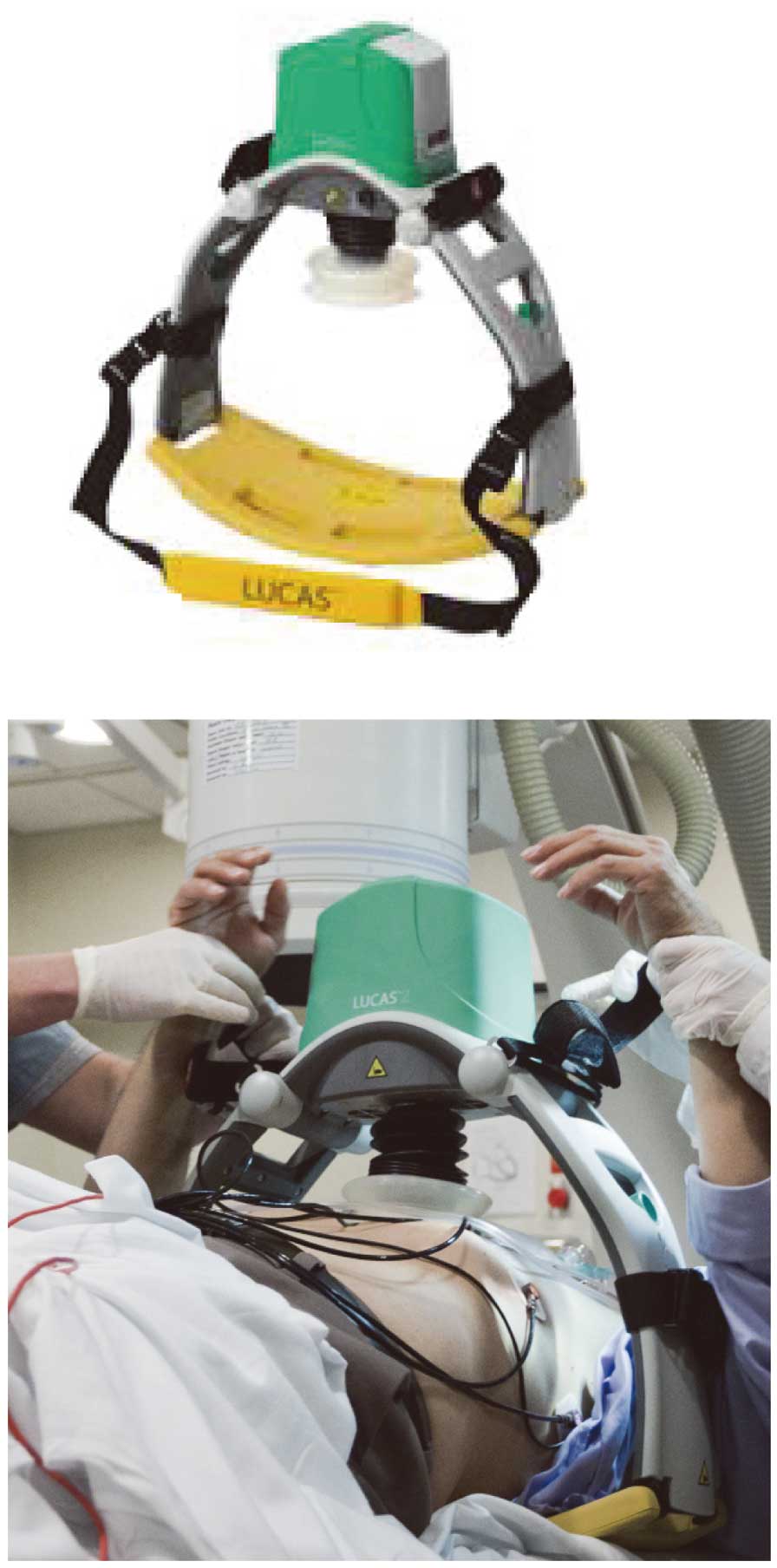
However, there are challenges with using mechanical chest compression devices in the cath lab; specifically the required time needed to place the device and begin compressions. Initially, this could take several minutes, but with practice and a regimented approach it can be done in 10–15 s.23
Another challenge is the limited radiological views obtained with a mechanical device. Both the metallic hardware in the AutoPulseTM
backboard and the column housing the piston in the LUCAS2TM
limit some standard anterior posterior angiographic views. However, the most helpful views for PCI, generally steep cranial and caudal projections, are possible with these 2 most commonly used mechanical CPR devices.
More than 13,128 patients have now been studied in randomized trials of mechanical vs. manual CPR. The 4 large-scale implementation studies, Auto Pulse Assisted Prehospital International Resuscitation (ASPIRE trial), Circulation Improving Resuscitation Care (CIRC trial), Prehospital Randomized Assessment of a Mechanical Compression Device in Cardiac Arrest (PARAMEDIC trial), and LUCAS in Cardiac Arrest (LINC trial) have not shown any significant difference in mortality and morbidity rates in patients suffering from out-of-hospital cardiac arrest (OHCA) treated with mechanical vs. manual CPR.24–27
The 2015 CPR Guidelines found that “the evidence does not demonstrate a benefit with the use of mechanical piston devices for chest compressions vs. manual chest compressions in patients with cardiac arrest.” However, they also noted that “the use of mechanical piston devices may be considered in specific settings where the delivery of high-quality manual compressions may be challenging or dangerous for the provider (eg, limited rescuers available, prolonged CPR, during hypothermic cardiac arrest, in a moving ambulance, in the angiography suite, during preparation for extracorporeal CPR [ECPR]), provided that rescuers strictly limit interruptions in CPR during deployment and removal of the devices (Class IIb, LOE C-EO)”.28
The data behind this recommendation for use in the cath lab are based on a number of case reports or cohort series. The largest experience to date was reported in 2 sequential series by Wagner et al.10,11
Together their series comprises 75 individuals treated with mechanical CPR after suffering cardiac arrest in the cath lab. The overall survival to discharge with favorable neurological function was 25% (19/75). An historical cohort group, with cardiac arrest occurring in the cath lab but treated with only manual CPR, had a survival rate of 10% (1/10).
Mechanical CPR for Refractory Cardiac Arrest
The role of mechanical chest compressions for cardiac arrest patients not responding to standard advanced cardiac life support (ACLS) efforts is being investigated. Patients refractory to standard resuscitation efforts need an alternative approach. One such alternative is using mechanical CPR devices for systemic circulatory support while transporting such patients to the hospital for more advanced therapies such as extracorporeal membrane oxygenation (ECMO) or peripheral cardiopulmonary bypass. Exactly when during the resuscitation such an approach should be considered is an important key to the success of such therapies. If caregivers wait too long, any therapy will be ineffective, but if they go too early some patients destined to be resuscitated by less invasive means will be subjected to increased morbidity.
What defines a “refractory” cardiac arrest? Is it the passage of a particular time period of unsuccessful effort or the completion of a bundle of resuscitation therapies without restoration of spontaneous circulation? Various definitions have been used, but without any consensus. Siao et al suggested that VF is refractory if no return of spontaneous circulation is achieved after more than 10 min of conventional ACLS effort.29
However, in their series this definition translated into a mean duration of resuscitation effort before ECMO was begun of 69.9±49.6 min. They also suggested that at least 3 shocks be delivered in patients with shockable rhythms before such are considered refractory. However, again that translated to a mean of 9.7±4.2 shocks before the institution of ECMO. Reynolds et al30
suggested that in OHCA a resuscitation effort between 10 and 15 min without success should be considered refractory. They noted that resuscitation duration was the primary independent variable in predicting favorable neurological survival; resuscitation efforts exceeding 15 min rarely result in favorable neurological outcomes. Belohlavek et al31
have suggested an even shorter period of failed resuscitation effort before considering other therapies. In their hyperinvasive protocol, they used 5–10 min as their cutoff point to declare efforts refractory and to move to a nonconventional ACLS approach.
Current emergency medical service (EMS) practice has morphed into a “stay and play”32
approach through numerous cycles of CPR, defibrillation, and cardiac arrest drug administrations in the field. Indeed, some systems allow the EMS personnel to declare the patient dead in the field after a certain time period has elapsed without return of spontaneous circulation (ROSC). Defining more carefully what “refractory” cardiac arrest means could allow a shift in the current paradigm towards a “scoop and run” or “load and go” approach.33
Given the persistently low long-term neurologically favorable survival rates seen today, this potential paradigm shift is becoming increasingly attractive to many EMS systems.
CPR in the Ambulance
The quality of prehospital resuscitation is thought to be a key factor in determining outcome from OHCA. High-quality CPR in adult patients with OHCA of presumed primary cardiac origin has been shown to improve rates of ROSC and survival to discharge.34
Currently, CPR is often performed manually by the EMS crew prior to arrival at the hospital. However, there are several challenges to this practice, both in relation to the safety of the EMS crew when performing CPR in a fast-moving ambulance, and the ability to generate effective blood flow through high-quality CPR with minimal interruptions.
Safety of the EMS Crew
The ambulance transport environment is inherently dangerous because of high-speed driving, risky maneuvers and possibly hazardous road conditions. This poses a significant safety risk for the EMS crew who are often standing and unrestrained in a confined space while performing CPR. It is widely recognized that unrestrained passengers face higher fatality and incapacitating injury rates than restrained passengers.35,36
Despite this fact, there is suboptimal use of standard restraints by prehospital providers during ambulance transport, particularly those located in the rear compartments.37
EMS crews often think that restraints are inconvenient, restrict movement and, in turn, inhibit patient care.
The introduction of mechanical CPR may provide a solution to the safety issue. Prehospital care providers can use restraints during mechanical CPR, while ensuring delivery of high-quality CPR to the patient.
Effective Blood Flow Generation
A number of studies have assessed the quality of manual CPR during ambulance transport in manikin models and adult patients with OHCA. Evidence from these studies suggest that during vehicle transport of a patient with OHCA the quality of resuscitation can be inadequate.38,39
For example, in a multicenter European case series of 176 adult patients with OHCA, chest compressions were given less than 50% of the time, and compressions of appropriate depth were performed less than 30% of the time.40
Potential Role of Mechanical CPR During Ambulance Transport
In principle, the use of mechanical CPR in OHCA is likely not only to reduce risk of injury and fatigue to the EMS crew, but may improve the quality of CPR, and minimize hands-off time.
Mechanical CPR has been shown to improve the quality of CPR compared with manual methods in simulated OHCA using manikin®
models.38
Mechanical CPR was noted to adhere more closely to recommended guidelines and was particularly effective in maintaining high-quality CPR during stretcher transport to the ambulance.
The benefit of mechanical CPR in OHCA, in terms of both quality of CPR and outcome, has also been demonstrated in clinical studies.39,41,42
A study conducted in Norway, which retrospectively assessed the quality of CPR in OHCA, showed an improvement in compression rate, depth and hands-off time with mechanical CPR when compared with manual CPR.39
In addition, mechanical CPR used in combination with an extrication sheet has been shown to reduce hands-off time during extrication and ambulance transport in adult OHCA of presumed cardiac origin.42
Although there has been some evidence to suggest that mechanical CPR improves the quality of resuscitation during ambulance transport, not all the data support this conclusion. In a prospective study,42
resuscitation efforts in ambulances with manual CPR and mechanical CPR were compared using time-motion analyses of digital video recordings. The results showed no significant difference in hands-off time between the 2 groups; most of the hands-off time incurred in the manual group was considered to be operator-related, as opposed to ambulance-related, factors. The authors further questioned the benefit of using mechanical CPR when ambulance transport time is short, because of the considerable time it took to deploy the mechanical device.
Though there is not uniform agreement, most data support the conclusion that the use of mechanical devices during ambulance transport will reduce the risk of injury to prehospital providers. There is also reasonable evidence to suggest that mechanical devices improve the quality of CPR. However, there are a no convincing data showing mechanical CPR during transport improves rates of survival or neurological outcome. This message seems to be largely consistent with a systematic review comparing mechanical CPR and manual CPR during OHCA and ambulance transport.43
Mechanical CPR in Refractory Cardiac Arrest Using a Hyperinvasive Approach
A newer approach to refractory cardiac arrest has been called “hyperinvasive CPR”. This involves a bundle of sequential, super-advanced care steps, generally including transitioning to mechanical CPR for transport to the hospital, early targeted temperature cooling, systemic support with either ECMO or PCAB, and early CAG/PCI. There are 2 early reports evaluating this approach.
The Prague Hyperinvasive Study
The “Prague Out-of-Hospital Study” is an ongoing, prospective, randomized study comparing prehospital standard ACLS with a hyperinvasive approach utilizing mechanical chest compressions, intra-arrest hypothermia, intrahospital extracorporeal life support, and early invasive investigation. A total of 200–400 patients are planned with a primary endpoint of 6-month survival with good neurological function. The secondary endpoint is 30-day neurological and cardiac recovery.
A methodology paper describing this in-progress study was published in 2012.32
Inclusion criteria included age between 18 and 65 years, a witnessed OHCA, and failure to establish ROSC following a minimum of 5 min of ACLS. Notably, a shockable rhythm was not a required inclusion criterion. Upon determination of eligibility, those randomized to the experimental therapy are placed on a mechanical compression device, intra-arrest cooling is begun with a nasal cooling device, and the patient is immediately transferred to a cardiac arrest center with the capacity for both coronary intervention and placement of ECMO. Mild hypothermia is continued in the post-resuscitation state. The results from their pre-intervention run-in phase have been reported.44
A total of 67 patients were evaluated: mean age was 59 years, the mean cardiac arrest time was 40 min, and 67% had VF. The admission pH was 7.06 and admission lactate was 9.9 mmol/L. The mean time to the cath lab was 64 min. Overall, 54% survived (36/67), with 75% (27/36) of survivors having good neurological function (CPC 1 or 2); 7 patients received ECMO, but none survived. The actual randomized trial is currently ongoing.
The CHEER Trial
The “CHEER” (mechanical
CPR,
Hypothermia,
ECMO and
Early
Reperfusion) trial was a single-center, prospective, observational study of selected patients with refractory cardiac arrest (both in- and out-of-hospital) that used a bundle of advanced therapies.45
Entry criteria included age 18–65 years, suspected cardiac etiology for the arrest, chest compressions begun within 10 min of collapse, initial cardiac arrest rhythm of VF, mechanical CPR available, and the patient considered a candidate for both early CAG and ECMO support. The primary endpoint was survival to hospital discharge with good neurological function. Secondary endpoints were ROSC, successful weaning from ECMO, and both ICU and hospital length of stay.
A total of 26 patients were enrolled (15 in-hospital patients, 11 out-of-hospital patients). The median age was 52 years, the median time from collapse to initiation of ECMO was 56 min, and 73% had VF. The admission pH was 6.9, and admission lactate was 10 mEq/L; 14 (54%) patients survived, including 5 of 11 (45%) with OHCA. All of the survivors had good neurological function (Table 3).
Table 3.
Outcomes in the CHEER Trial
| ROSC |
25/26 (96%) |
| Survival to DC |
14/26 (54%) |
| OHCA |
5/11 (45%) |
| Inpatient CA |
9/15 (60%) |
| Survival with favorable neurofunction (CPC 1 or 2) |
14/26 (54%) |
CA, cardiac arrest; DC, discharge; OHCA, out-of-hospital CA.
These 2 early reports suggest that mechanical CPR shows promise, as an initial therapy in an aggressive, invasive approach to refractory cardiac arrest. The importance of good circulatory support, including during transport to a facility capable of providing advanced invasive support with both ECMO and PCI is a key to long-term survival with favorable neurological function.
Conclusions
Mechanical CPR offers unique safety and efficacy advantages during the treatment of cardiac arrest in the cath lab and during transport of refractory cardiac arrest patients to the hospital and cath lab. Practice is needed to limit the time to deployment and to avoid significant interruptions to both resuscitation and emergency PCI efforts. Successful resuscitation with favorable long-term neurological function has been achieved in committed centers using mechanical CPR in these settings.
Conflict of Interest Statements
P.W., P.R., U.B.K., and A.A. have no conflicts to disclose. K.B.K., serves as a consultant and scientific advisory board member to both Zoll Medical (manufacture of the AutoPulseTM
device) and Physio-Control (manufacture of the LUCASTM
device).
References
- 1.
Moscucci M, editor. Grossman and Baim’s cardiac catheterization, angiography and intervention. 8th edn. Philadelphia: Lippincott Williams & Wilkins, 2014; 77–105.
- 2.
Kern KB, Thai HM. Cardiac arrest and resuscitation during percutaneous coronary interventions. In: Butman SM, editor. Complications of percutaneous coronary interventions. New York: Springer New York, 2005; 141–151.
- 3.
Singh M, Gersh BJ, Lennon RJ, Ting HH, Holmes DR Jr, Doyle BJ, et al. Outcomes of a system-wide protocol for elective and nonelective coronary angioplasty at sites without on-site surgery: The Mayo Clinic experience. Mayo Clin Proc 2009; 8: 501–508.
- 4.
Brennan JM, Curtis JP, Dai D, Fitzgerald S, Khandelwal AK, Spertus JA, et al. Enhanced mortality risk prediction with a focus on high-risk percutaneous coronary interventionresults from 1,208,137 procedures in the NCDR (National Cardiovascular Data Registry). J Am Coll Cardiol Interv 2013; 6: 790–799.
- 5.
Society for Cardiovascular Interventions and Angiography. SCAI QIT PCI risk assessment tool 2014 [3/10/2016]. http://scaiscientificsessions.org/PCIRiskWeb2/PCIRiskWeb2.htm (acessed April 4, 2016).
- 6.
Addala S, Kahn JK, Moccia TF, Harjai K, Pellizon G, Ochoa A, et al. Outcome of ventricular fibrillation developing during percutaneous coronary interventions in 19,497 patients without cardiogenic shock. Am J Cardiol 2005; 9: 764–765.
- 7.
Mehta RH, Harjai KJ, Grines L, Stone GW, Boura J, Cox D, et al. Sustained ventricular tachycardia or fibrillation in the cardiac catheterization laboratory among patients receiving primary percutaneous coronary intervention: Incidence, predictors, and outcomes. J Am Coll Cardiol 2004; 43: 1765–1772.
- 8.
Lavonas EJ, Drennan IR, Gabrielli A, Heffner AC, Hoyte CO, Orkin AM, et al. Part 10: Special circumstances of resuscitation: 2015 American Heart Association Guidelines update for cardiopulmonary resuscitation and emergency cardiovascular care. Circulation 2015; 132(Suppl 2): S501–S518.
- 9.
Rihal CS, Naidu SS, Givertz MM, Szeto WY, Burke JA, Kapur NK, et al. 2015 SCAI/ACC/HFSA/STS Clinical Expert Consensus Statement on the Use of Percutaneous Mechanical Circulatory Support Devices in Cardiovascular Care: Endorsed by the American Heart Assocation, the Cardiological Society of India, and Sociedad Latino Americana de Cardiologia Intervencion; Affirmation of Value by the Canadian Association of Interventional Cardiology-Association Canadienne de Cardiologie d’intervention. J Am Coll Cardiol 2015; 65: e7–e26, doi:10.1016/j.jacc.2015.02.043.
- 10.
Wagner H, Terkelsen CJ, Friberg H, Harnek J, Kern KB, Lassen JF, et al. Cardiac arrest in the catheterization laboratory: A 5-year experience of using mechanical chest compressions to facilitate PCI during prolonged resuscitation efforts. Resuscitation 2010; 81: 383–387.
- 11.
Wagner H, Hardig BM, Rundgren M, Zughaft D, Harnek J, Gotberg M, et al. Mechanical chest compressions in the coronary catheterization laboratory to facilitate coronary intervention and survival in patient requiring prolonged resuscitation efforts. Scand J Trauma Resusc Emerg Med 2016; 24: 4.
- 12.
Weisfeldt ML, Becker LB. Resuscitation after cardiac arrest: A 3-phase time-sensitive model. JAMA 2002; 288: 3035–3038.
- 13.
Webb JG, Solankhi NK, Chugh SK, Amin H, Buller CE, Ricci DR, et al. Incidence, correlates, and outcome of cardiac arrest associated with percutaneous coronary intervention. Am J Cardiol 2002; 90: 1252–1254.
- 14.
Link MS, Berkow LC, Kudenchuk PJ, Halperin HR, Hess EP, Moitra VK, et al. Part 7: Adult advanced cardiovascular life support: 2015 American Heart Association Guidelines Update for cardiopulmonary resuscitation and emergency cardiovascular care. Circulation 2015; 132(Suppl 2): S444–S464.
- 15.
Wagner H, Rundgren M, Hardig BM, Kern KB, Zughaft JD, Harnek J, et al. A structured approach for treatment of prolonged cardiac arrest cases in the coronary catheterization laboratory using mechanical chest compressions. Int J Cardiovasc Res 2013; 2: 4.
- 16.
Idris AH, Guffey D, Aufderheide TA, Brown S, Morrison LJ, Nichols P, et al. Relationship between chest compression rates and outcomes from cardiac arrest. Circulation 2012; 125: 3004–3012.
- 17.
Idris AH, Guffey D, Pepe PE, Brown S, Brooks SC, Callaway CW, et al. Chest compression rates and survival following out-of-hospital cardiac arrest. Crit Care Med 2015; 43: 840–848.
- 18.
Aufderheide TP, Pirrallo RG, Yannopoulos D, Klein JP, von Briesen C, Sparks CW, et al. Incomplete chest wall decompression: A clinical evaluation of CPR performance by EMS personnel and assessment of alternative manual chest compression-decompression techniques. Resuscitation 2005; 64: 353–362.
- 19.
Kern KB, Ewy GA, Voorhees WD, Babbs CF, Tacker WA. Myocardial perfusion pressure: A predictor of 24-hour survival during prolonged cardiac arrest in dogs. Resuscitation 1988; 16: 241–250.
- 20.
Paradis NA, Martin GB, Rivers EP, Goetting MG, Appleton TJ, Feingold M, et al. Coronary perfusion pressure and return of spontaneous circulation in human cardiopulmonary resuscitation. JAMA 1990; 263: 1106–1113.
- 21.
Halperin HR, Tsitlik JE, Guerci AD, Mellits ED, Levin HR, Shi AY, et al. Determinants of blood flow to vital organs during cardiopulmonary resuscitation in dogs. Circulation 1998; 73: 539–550.
- 22.
Vanden Hoek TL, Morrison LJ, Shuster M, Donnino M, Sinz E, Lavonas EJ, et al. Part 12: Cardiac arrest in special situations: 2010 American Heart Association guidelines for cardiopulmonary resuscitation and emergency cardiovascular care. Circulation 2010; 122(Suppl 3): S829–S861.
- 23.
Levy M, Yost D, Walker RG, Scheunemann E, Mendive SR. A quality improvement initiative to opimize use of a mechanical chest compression device within a high-performance CPR approach to out-of-hospital cardiac arrest resuscitation. Resuscitation 2015; 92: 32–37.
- 24.
Hallstrom A, Rea TD, Sayre MR, Christenson J, Anton AR, Mosesso VN, et al. Manual chest compression vs use of an automated chest compression device during resuscitation following out-of-hospital cardiac arrest: A randomized trial. JAMA 2006; 295: 2620–2628.
- 25.
Rubertssonn S, Lindgren E, Smekal D, Ostend O, Silfverstolpe J, Lichtveld RA, et al. Mechanical chest compressions and simultaneous defibrillation vs conventional cardiopulmonary resuscitation in out-of-hospital cardiac arrest: The LINC randomized trial. JAMA 2014; 311: 53–61.
- 26.
Wik L, Olsen JA, Persse D, Sterz F, Lozano M Jr, Brouwer MA, et al. Manual vs integrated automatic load-distributing band CPR with equal survival after out of hospital cardiac arrest. Resuscitation 2014; 85: 741–748.
- 27.
Perkins GD, Lall R, Quinn T, Deakin CD, Cooke MW, Horton J, et al. Mechanical versus manual chest compression for out-of-hospital cardiac arrest (PARAMEDIC): A pragmatic, cluster randomized controlled trial. Lancet 2015; 385: 947–955.
- 28.
Brooks SC, Anderson ML, Bruder E, Daya MR, Gaffney A, Otto CW, et al. Part 6: Alternative techniques and ancillary devices for cardiopulmonary resuscitation: 2015 American Heart Association Guidelines Update for cardiopulmonary resuscitation and emergency cardiovascular care. Circulation 2015; 132(Suppl 2): S436–S443.
- 29.
Siao FY, Chiu CC, Chiu CW, Chen YC, Chen YL, Hsieh YK, et al. Managing cardiac arrest with refractory ventricular fibrillation in the emergency department: Conventional cardiopulmonary resuscitation versus extracorporeal cardiopulmonary resuscitation. Resuscitation 2015; 92: 70–76.
- 30.
Reynolds JC, Frisch A, Rittenberger JC, Callaway CW. Duration of resuscitation efforts and functional outcome after out-of-hospital cardiac arrest: When should we change to novel therapies? Circulation 2013; 128: 2488–2494.
- 31.
Belohlavek J, Kucera K, Jarkovsky J, Franek O, Pokoma M, Danda J, et al. Hyperinvasive approach to out-of-hospital cardiac arrest using mechanical chest compression device, prehospital intraarrest cooling, extracorporeal life support and early invasive assessment compared to standard of care: A randomized, parallel groups comparative study proposal: “Prague OHCA Study”. J Transl Med 2012; 10: 16.
- 32.
Smith RM, Conn AK. Prehospital care - scoop and run or stay and play? Injury 2009; 40(Suppl 4): S23–S26.
- 33.
Poppe M, Weiser C, Holzer M, Sulzgruber P, Datler P, Keferbock M, et al. The incidence of “load&go” out-of-hospital cardiac arrest candidates for emergency department utilization of emergency extracorporeal life support: A one-year review. Resuscitation 2015; 91: 131–136.
- 34.
Garza AG, Gratton MC, Salomone JA, Lindholm D, McElroy J, Archer R. Improved patient survival using a modified resuscitation protocol for out-of-hospital cardiac arrest. Circulation 2009; 119: 2597–2605.
- 35.
Becker LR, Zaloshnja E, Levick N, Li G, Miller TR. Relative risk of injury and death in ambulances and other emergency vehicles. Accid Anal Prev 2003; 35: 941–948.
- 36.
Kahn CA, Pirelli RG, Kuhn EM. Characteristics of fatal ambulance crashes in the United States: An 11-year retrospective analysis. Prehosp Emerg 2001; 5: 261–269.
- 37.
Larmon B, LeGassick TF, Shriger DL. Differential front and back seat safety belt use by prehospital care providers. Am J Emerg Med 1993; 11: 595–599.
- 38.
Sunde K, Wik L, Steen PA. Quality of mechanical, manual standard and active compression-decompression CPR on the arrest site and during transport in a manikin model. Resuscitation 1997; 34: 235–242.
- 39.
Olasveengen TM, Wik L, Steen PA. Quality of cardiopulmonary resuscitation before and during transport in out-of-hospital cardiac arrest. Resuscitation 2008; 76: 185–190.
- 40.
Wik L, Kramer-Johansen J, Myklebust H, Sorebo H, Svensson L, Fellows B, et al. Quality of cardiopulmonary resuscitation during out-of-hospital cardiac arrest. JAMA 2005; 293: 299–304.
- 41.
Lyon RM, Crawford A, Crookston C, Short S, Clegg GR. The combined use of mechanical CPR and a carry sheet to maintain quality resuscitation in out-of-hospital cardiac arrest patients during extrication and transport. Resuscitation 2015; 93: 102–106.
- 42.
Wang HC, Chiang WC, Chen SY, Ke YL, Chi CL, Yang CW, et al. Video-recording and time motion analyses of manual versus mechanical CPR during ambulance transport. Resuscitation 2007; 74: 453–460.
- 43.
Ong ME, Mackey KE, Zhang ZC, Tanaka H, Ma MH, Swor R, et al. Mechanical CPR devices compared to manual CPR during out-of-hospital cardiac arrest and ambulance transport: A systematic review. Scand J Trauma Resusc Emerg Med 2012; 20: 39.
- 44.
Belohlavek B, Skalicka H, Smid O, Franek O, Pokorna M, Danda J, et al. Implementation of hyperinvasive approach to out-of hospital cardiac arrest management: Results from pre-simulation and simulation phases of the “Prague OHCA study. Resuscitation 2013; 84: S68–S69.
- 45.
Stub D, Bernard S, Pellegrino V, Smith K, Walker T, Sheldrake J, et al. Refractory cardiac arrest treated with mechanical CPR, hypothermia, ECMO, and early reperfusion (the CHEER trial). Resuscitation 2015; 86: 88–94.


