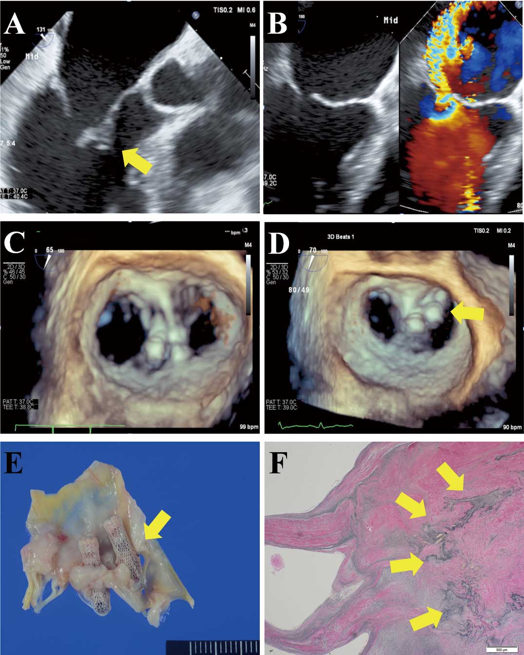
An 81-year-old man was admitted for heart failure with severe mitral regurgitation (MR). He had a history of infective endocarditis (IE;
Figure A) successfully treated 3 months earlier. No signs of infection nor vegetation were observed (Figure B). Despite surgical mitral valve replacement (MVR) recommendation, he selected MitraClip®
(Abbott Vascular, IL, USA). Two clips were carefully deployed, resulting in MR reduction to mild (Figure C,
Supplementary Movie). One week later, symptoms worsened and the medial clip was found to be detached from the posterior leaflet, resulting in severe MR (Figure D). After surgical MVR, pathology indicated a scar due to the previous IE (Figure E,F). Additional to valve tethering and annulus dilatation, we identified the cause of detachment was due to valve vulnerability and valve tension created after MitraClip deployment.
Chandrashekar et al reported a case of MR successfully treated by MitraClip 1 month after acute IE,1
although the optimal timing after IE is controversial. The present findings of pathological valve vulnerability observed even 3 months after IE suggest that there is a risk of leaflet detachment after MitraClip deployment.
Disclosures
The authors declare no conflicts of interest.
Supplementary Files
Supplementary Movie.
Transesophageal echocardiography showing reduction in mitral regurgitation to mild, after deployment of 2 clips.
Please find supplementary file(s);
http://dx.doi.org/10.1253/circj.CJ-19-0523
Reference
- 1.
Chandrashekar P, Fender EA, Al-Hijji MA, Chandrasekaran K, Rihal CS, Eleid MF, et al. Novel use of MitraClip for severe mitral regurgitation due to infective endocarditis. J Invasive Cardiol 2017; 29: E21–E22.
