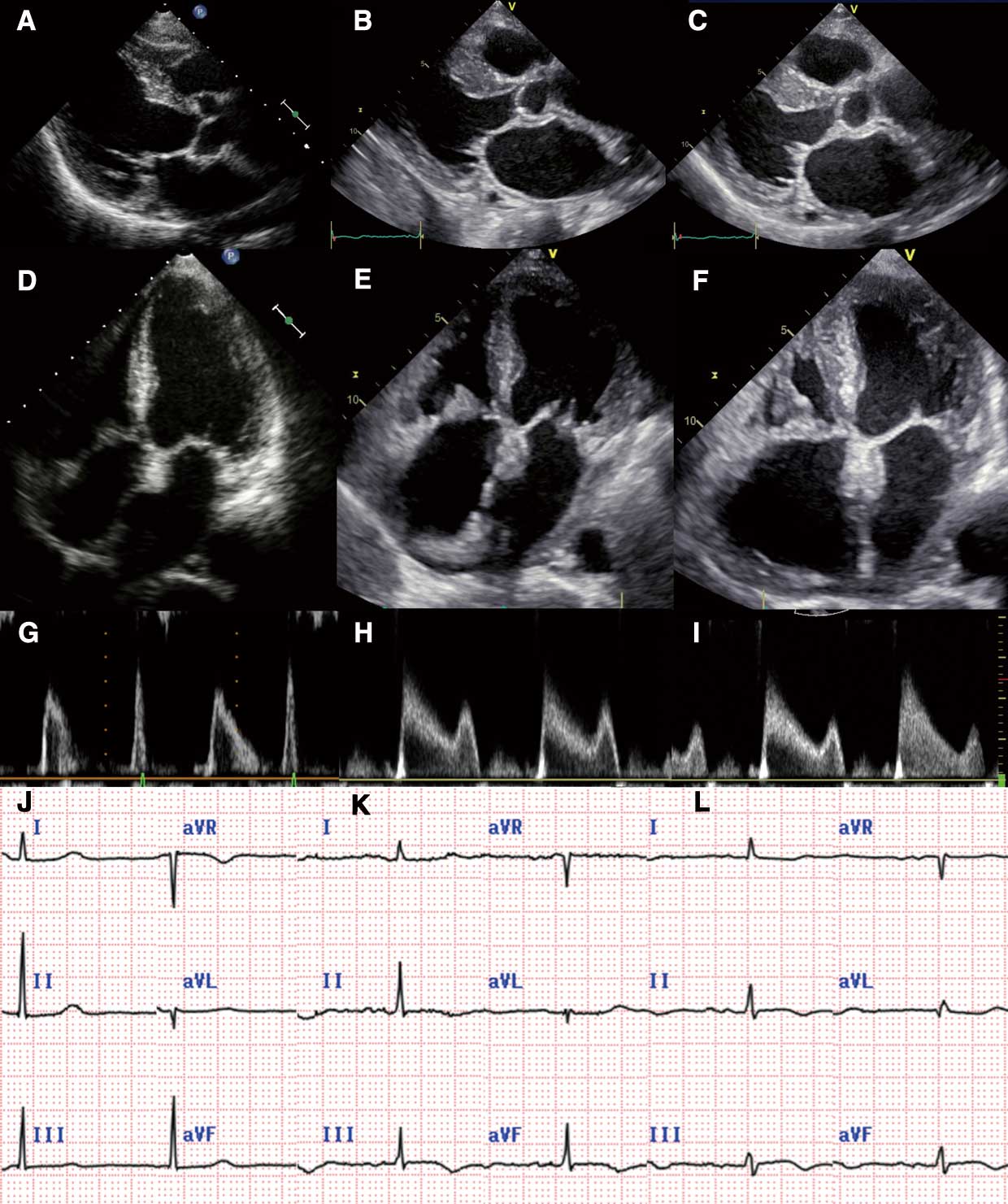
A 79-year-old woman with positive anti-centromere antibody was admitted for heart failure. As compared with images taken 2 years prior (Figure A,D), newly developed mitral and tricuspid valvular thickening (Figure E) were found and Libman-Sacks endocarditis was suspected; 3 months later, monoclonal gammopathy with Bence-Jones protein was identified. Amyloid light-chain cardiac amyloidosis (ALCA) with multiple myeloma was diagnosed by bone and cardiac biopsies (Supplementary Figure A). Review of the echocardiograms revealed left atrial wall thickening (LAWT) (Figure B). At 4 months after the ALCA diagnosis, ventricular wall thickening occurred (Figure C,F). Along with these changes, progressive decrease in both A-wave velocity and QRS voltage was observed (Figure G–L). Additionally, cardiac magnetic resonance images demonstrated late gadolinium enhancement only in the left atrium, suggesting minimal damage in the left ventricle (Supplementary Figure B). She underwent chemotherapy, but died within 1 year.
ALCA is induced by the deposition of misfolded amyloid light-chain proteins in the heart, typically presenting as valve and ventricular thickening.1
However, an ALCA diagnosis in an aged patient with suspected scleroderma is challenging because the thickened valves resemble aged degeneration or Libman-Sacks endocarditis.
LAWT is a rare feature to suggest ALCA. While its standardization is still under development, strain imaging can help identify atrial functional impairment.2
This case highlights that LAWT is crucial for the early detection of ALCA, especially when valvular or ventricular findings are indecisive.
Disclosure
Y.K. is a member of
Circulation Journal’s Editorial Team.
Supplementary Files
Please find supplementary file(s);
http://dx.doi.org/10.1253/circj.CJ-21-0485
References
- 1.
Randhawa VK, Vakamudi S, Phelan DM, Samaras CJ, McKenney JK, Hanna M, et al. Mitral and tricuspid stenosis caused by light chain cardiac amyloid deposition. ESC Heart Fail 2020; 7: 1130–1135.
- 2.
Falk RH, Alexander KM, Liao R, Dorbala S. AL (light-chain) cardiac amyloidosis: A review of diagnosis and therapy. J Am Coll Cardiol 2016; 68: 1323–1341.
