Images in Cardiovascular Medicine
Mitral Regurgitation Due to Papillary Muscle Rupture Following Elective Percutaneous Coronary Intervention for Stable Angina
2023 Volume 87 Issue 9 Pages 1251-

Details
2023 Volume 87 Issue 9 Pages 1251-
A 75-year-old man was referred for assessment of stable angina. Coronary angiography revealed severe stenosis of the distal bifurcation of left main trunk (LMT) (Figure A, Supplementary Movie 1), but the patient refused surgery. We therefore decided to perform staged percutaneous coronary intervention (PCI), which involved crossover stenting from the LMT to the left anterior descending artery. Following stenting, a high lateral branch was jailed and totally occluded (Figure B, Supplementary Movie 1), but because it was a relatively small vessel, his symptoms were self-limiting, and there was no remarkable ST-T change on ECG, we decided not to intervene on this compromised side branch. However, 5 days after the procedure, the patient presented with progressive dyspnea, and echocardiography showed mitral valve A2 prolapse due to anterolateral papillary muscle rupture (PMR) (Supplementary Movie 2). Left ventriculography revealed severe mitral regurgitation (Figure C). The patient underwent surgical mitral valve replacement. Intraoperative findings revealed complete anterolateral PMR (Figure D).
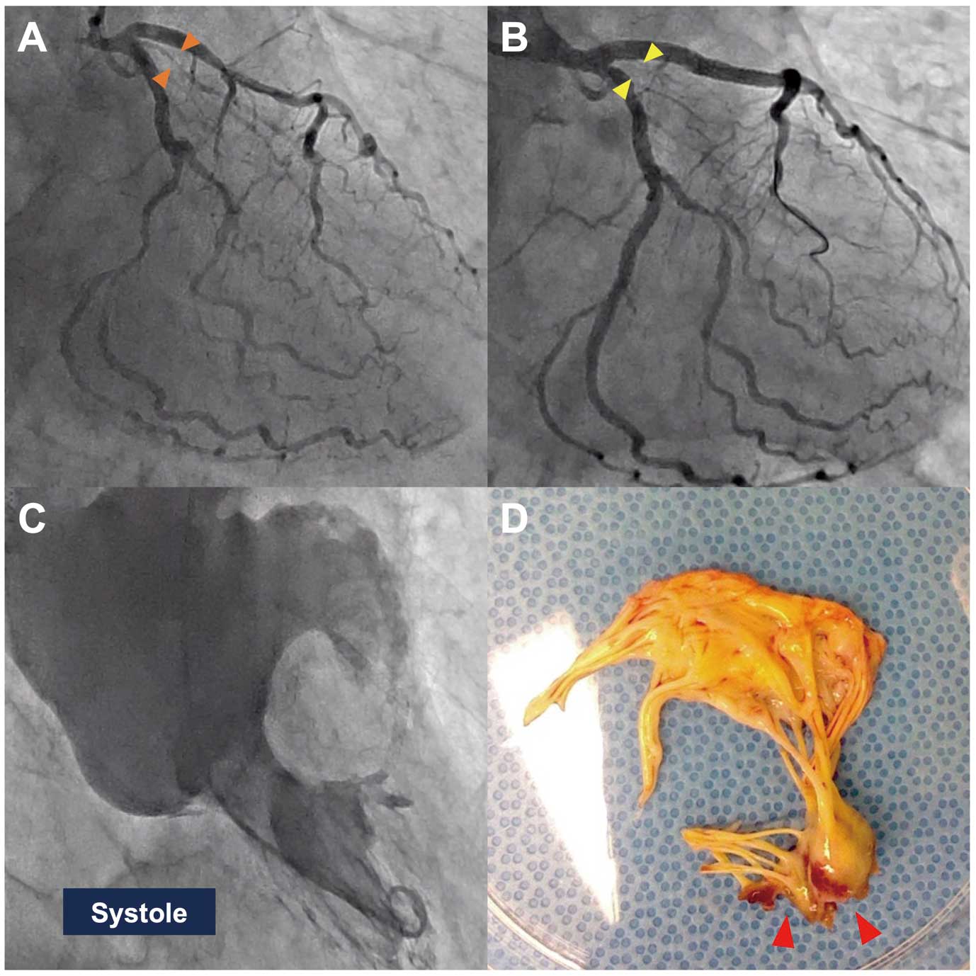
Coronary angiography (A) before PCI shows a patent high lateral branch (orange arrowheads), but after PCI (B) it is jailed and occluded (yellow arrowheads). (C) Left ventriculography shows severe mitral regurgitation. (D) Resected anterior leaflet shows rupture of the anterolateral papillary muscle (red arrowheads). PCI, percutaneous coronary intervention.
Procedural myocardial infarction due to HL occlusion followed by anterolateral PMR is a rare complication, but PCI operators should keep it in mind and carefully monitor patients following side-branch occlusion, even if relatively small.
M.I. is a member of Circulation Journal’s Editorial Team.
Supplementary Movie 1. Coronary angiography before and after PCI.
Supplementary Movie 2. Apical 3-chamber view of transthoracic echocardiography before the surgery.
Please find supplementary file(s);
https://doi.org/10.1253/circj.CJ-23-0192