A 35-year-old woman with advanced hepatocellular carcinoma was started on durvalumab and tremelimumab (STRIDE regimen). After receiving only 1 course of the STRIDE regimen, she attended hospital at week 4, just before the administration of the 2nd course, because of a fever of 39℃. She was diagnosed with septic shock and cytokine release syndrome. After admission, broad-spectrum antimicrobial and steroid pulse therapy was initiated. A few days later, cardiac enlargement and pulmonary congestion had progressed. Her serum troponin T level was 0.041 (ng/mL). Transthoracic echocardiography (TTE) revealed diffuse hypokinesis of the left ventricular myocardium (LVM) and an ejection fraction (EF) of 40%. Magnetic resonance imaging (MRI) was performed (Ingenia 3.0T, Philips Healthcare): the native T1 value of LVM was elevated to 1,399 ms (facility reference=1,237 ms; Figure A,B), and there was localized prolongation of the native T2 value (80 ms; Figure C). She was diagnosed with immune checkpoint inhibitor (ICI) myocarditis.1
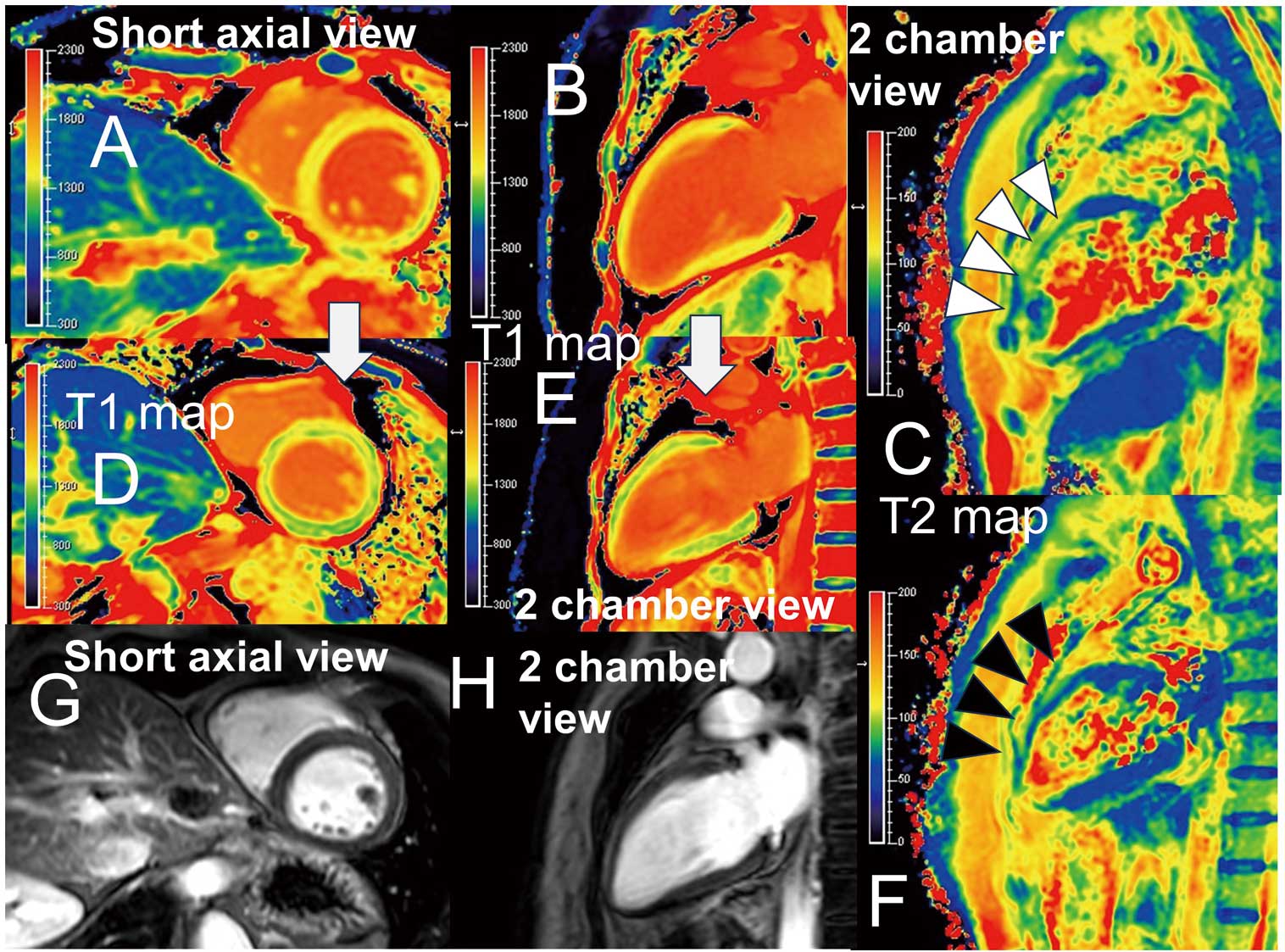
Following steroid treatment for 1 month, the EF had normalized to 60.2%, and the native T1 and T2 values of the LVM had almost normalized to 1,256 ms and 63 ms, respectively (Figure D–F), on follow-up MRI. Late gadolinium enhancement was not observed (Figure G,H).
ICI myocarditis is rare, with an incidence of 0.06% to 0.27%.1 According to our literature review, there has not been a similar imaging report revealing clearly changed images of T1 and T2 mapping pre- and post-ICI myocarditis.
Disclosure
Y.K. is a member of Circulation Journal’s Editorial Team.
