A 72-year-old woman was hospitalized with fatigue 1 week after experiencing chest pain. Her ECG revealed abnormal Q waves and ST-segment elevation in the inferior leads. Her creatine kinase level was in the normal range. Transthoracic echocardiography (TTE) revealed pericardial effusion and reduced left ventricular (LV) inferior wall motion. Enhanced computed tomography (CT) confirmed the pericardial effusion (Figure A). Coronary angiography revealed occlusion of the right coronary artery. She was in shock. We aspirated 400 mL of bloody pericardial fluid during pericardiocentesis and there was hemodynamic improvement. We concluded she had developed cardiac tamponade due to myocardial rupture following myocardial infarction.
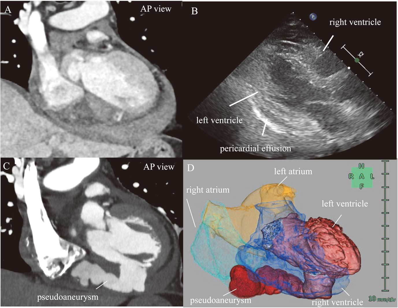
Conservative therapy included intra-aortic balloon pumping (IABP) and a β-blocker without antiplatelet agents or anticoagulants. She developed a fever on hospital day (HD) 4, resulting in IABP and drainage tube removal. TTE on HD 6 showed no pericardial effusion. However, the pericardial effusion reappeared on HD 9 (Figure B). Coronary CT angiography (CCTA) showed a myocardial defect in the LV inferior wall and a large LV pseudoaneurysm expanding along the posterior interventricular groove (Figure C,D; Supplementary Movie). The patient underwent urgent and successful repair of the pseudoaneurysm and myocardial defect. The postoperative course was uneventful.
Diagnosing myocardial infarction by TTE is often difficult.1 In this case, the pseudoaneurysm was large but unusually shaped and located in a region difficult to observe with TTE. CCTA helped diagnose an LV pseudoaneurysm.
Disclosures
Y.S. is a member of Circulation Journal’s Editorial Team. The authors declare no conflicts of interest.
Data Availability
The deidentified participant data will not be shared.
Supplementary Files
Supplementary Movie. Three-dimension reconstructed image of the cardiac CT angiography. The large LV pseudoaneurysm was shown to expand along the posterior interventricular groove.
Please find supplementary file(s);
https://doi.org/10.1253/circj.CJ-23-0906
