A 39-year-old woman with fulminant myocarditis was referred to hospital for intensive treatment. Impella CP was inserted to treat cardiogenic shock, but because of persistent hemodynamic instability and atrioventricular block, veno-arterial extracorporeal membrane oxygenation (VA-ECMO) was added to Impella (ECPELLA strategy). The rapidly progressing myocarditis induced ventricular fibrillation (VF) the day after admission, but echocardiography revealed an apparent normal heart beat even during VF (Figure A; Supplementary Movie). M-mode showed the presence of periodic left ventricular wall motion. Furthermore, the transmitral flow displayed periodic waveforms of left ventricular inflow (Figure B). Swan-Ganz catheter monitoring confirmed a significant pulmonary artery pulse pressure in response to atrial contraction (Figure C). She was in VF for 6 days, but her hemodynamics were maintained. Electrophysiological study confirmed atrioventricular dissociation, with the right atrium in sinus rhythm (Figure D) and the right ventricle in VF (Figure E). Furtheremore, simultaneous recordings revealed a pairing between the atrial electrical activity and the pulmonary artery pressure (Supplementary Figure).
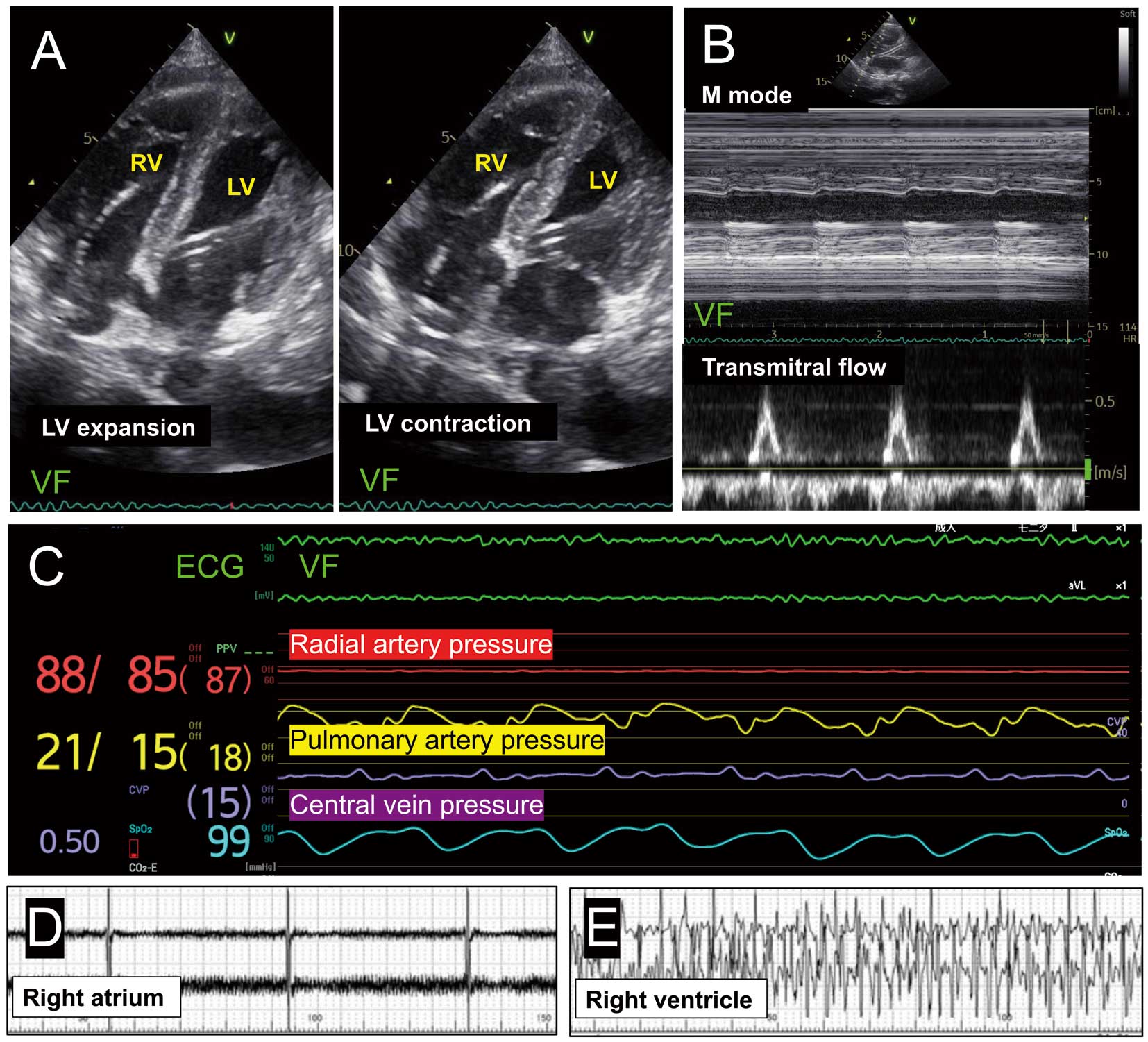
Blood flows from the left atrium (LA) into the left ventricle (LV) when the LA contracts, causing the LV to dilate. The Impella gradually pumps blood from the LV to the aorta, shrinking the LV. We hypothesize that this repetition caused the heart to appear to contract. VF was successfully treated with electrical defibrillation, and 2 days later the patient’s cardiac function improved, allowing for weaning off VA-ECMO and Impella. She was discharged after 20 days with a good clinical course.
It is important to recognize that when atrioventricular dissociation occurs during ECPELLA, the heart might appear to be beating normally, even in VF.
Supplementary Files
Supplementary Movie. Transthoracic echocardiography.
Please find supplementary file(s);
https://doi.org/10.1253/circj.CJ-23-0885
