A 42-year-old man with mild shortness of breath was referred because of a diastolic murmur at the second left sternal border and suspected aortic regurgitation. Transesophageal echocardiography did not show aortic regurgitation but revealed a ruptured right sinus of Valsalva aneurysm (SVA) protruding into the right atrium (RA), with abnormal (left-to-right) blood flow only in diastole. Aortography demonstrated abnormal blood flow into the RA during diastole only (Supplementary Movie 1). Oximetry revealed an oxygen step-up in the RA, with a pulmonary-systemic flow ratio of 2.73. Further, the SVA during diastole was identified by 4-dimensional computed tomography (Figure A, Supplementary Movie 1) and it was covered by the right coronary cusp (RCC) during systole (Figure B). A definitive repair procedure was scheduled. Intraoperatively, the ruptured SVA with an inflow diameter of 1×1 cm was confirmed to tunnel into the RA (Figure C). The inflow was completely blocked by the RCC when the valve opened (Figure D, Supplementary Movie 2). The fistulous SVA was closed directly on the RA side and closed with a Dacron patch on the aortic side. Intraoperative transesophageal echocardiography confirmed fistula closure.
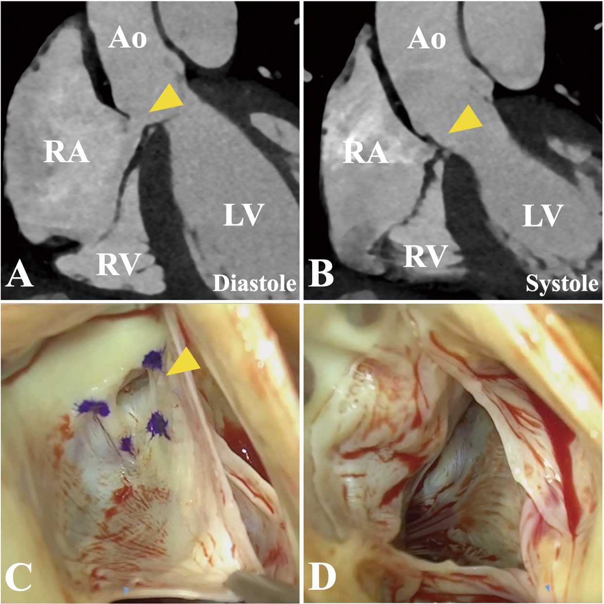
To our knowledge, this is the first report of the possible mechanism of a diastolic murmur detected in a patient with a ruptured SVA. Cardiac CT is useful for understanding anatomic alterations in the aortic root throughout a cardiac cycle by providing high-resolution 4D images.
Disclosures
The authors declare they have no conflicts of interest.
Supplementary Files
Supplementary Movie 1. Preoperative examination.
Supplementary Movie 2. Intraoperative movie and postoperative echocardiography.
Please find supplementary file(s);
https://doi.org/10.1253/circj.CJ-23-0912