Multimodal Imaging of False Lumen Thrombosis After Acute Aortic Dissection With Non-Obstructive General Angioscopy and Dual-Energy Computed Tomography
Article ID: CJ-24-0152

Details
Article ID: CJ-24-0152
False lumen thrombosis (FLT) after acute aortic dissection is critical for aortic remodeling,1 and is usually diagnosed indirectly by contrast-enhanced computed tomography (CT). We directly evaluated FLT using a multimodal approach involving intraoperative nonobstructive general angioscopy (NOGA) and dual-energy CT (DECT).
An 81-year-old man was diagnosed with type B aortic dissection (Figure A) and underwent thoracic endovascular aortic repair (TEVAR) following expansion of the false lumen (FL). Preoperative DECT with electron-density imaging (EDI) showed high attenuation in the edges of the FL (Figure B). During TEVAR, NOGA revealed a dark-red, fibrous cap on the upper edge inside the FL (Figure C, Supplementary Movie). The patient was discharged 6 days after TEVAR without complications.
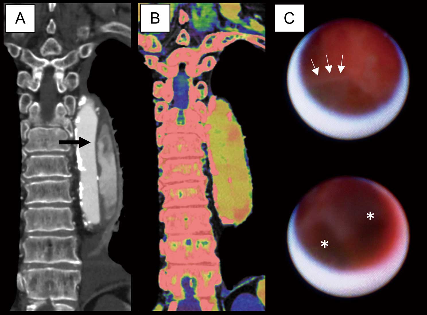
Preoperative computed tomography (CT) images and nonobstructive general angioscopy (NOGA) findings. Contrast-enhanced CT showed a filling defect area in the edges of the false lumen (FL), suggestive of thrombosis (A; arrow). DECT with electron-density imaging (EDI) revealed high attenuation in the edges of the FL (B). NOGA showed fibrous and dark-red tissue inside the edge of the FL, indicating fibrin and clot, respectively (C; arrows: fibrin; asterisks: clot. See also Supplementary Movie). The findings suggested that the high attenuation in the FL by EDI was a thrombus.
A partial FLT will affect the outcome of aortic dissection. In a previous report, DECT with EDI was used to identify an arterial thrombus,2 but application of this imaging technique in clinical settings is controversial. In this case, intraoperative NOGA directly demonstrated thrombus formation. Thus, high attenuation in the FL by EDI suggests thrombus in aortic lesions.
A multimodal approach with DECT and NOGA such as was used by us in this case may be effective for evaluating FLT without the use of contrast agent.
The authors declare no conflicts of interest or funding support.
Informed consent was obtained from the patient.
Supplementary Movie. The representative movie of non-obstructive general angioscopy during thoracic endovascular aortic repair.
Please find supplementary file(s);
https://doi.org/10.1253/circj.CJ-24-0152