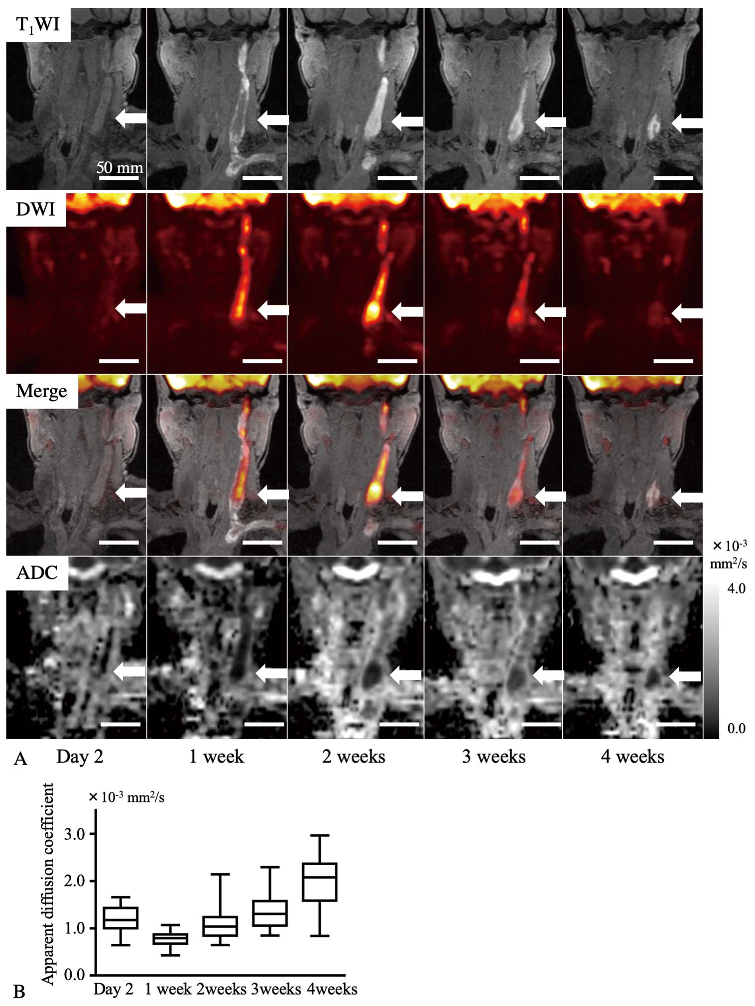
A 64-year-old woman was admitted to Koga General Hospital due to left cervical panniculitis with jugular vein thrombosis. On Day 2 after onset, T1-weighted magnetic resonance imaging (MRI) with fat suppression depicted the deep vein thrombus (DVT) as a lesion with high to iso signal intensity (SI). Diffusion weighted imaging (DWI) and a merged image of the T1-weighted image and DWI showed DVT as a high-SI lesion 1–3 weeks after onset, and as a focal high-SI lesion 4 weeks after onset (Figure A). The apparent diffusion coefficient in multiple regions of interest in the DVT was lowest 1 week after onset (Figure A
and
B). Although the patient was administered heparin and subsequently edoxaban, the DVT remained occlusive and appeared to be organized.
In a previous study, DWI showed acute (≤14 days) DVT as heterogeneous hyperintense foci, compared with non-acute (>14 days) DVT.1
These findings are compatible with those of the present case except for the findings on Day 2. The present case showed sequential signal changes of the DVT on DWI and a hyperintense signal of subacute DVT. Further studies are required to verify these findings.
Disclosures
The authors declare no conflicts of interest.
IRB Information
This study was approved by the Ethics Committee of Koga General Hospital (Reference no. 20-03).
The procedures were performed in accordance with the Declaration of Helsinki and the ethical standards of the responsible committee on human experimentation.
Reference
- 1.
Wu G, Morelli J, Xiong Y, Liu X, Li X. Diffusion weighted cardiovascular magnetic resonance imaging for discriminating acute from non-acute deep venous thrombus. J Cardiovasc Magn Reson 2019; 21: 37–45.
