Images in Cardiovascular Medicine
Left Ventricular Outflow Tract Myxoma Leading to Acute Stroke
2025 Volume 7 Issue 2 Pages 143-

Details
2025 Volume 7 Issue 2 Pages 143-
An 89-year-old woman with no significant medical history suddenly collapsed and was brought to hospital within 20 min. Initial assessments showed impaired consciousness (Glasgow Coma Score E2V1M1) and elevated blood pressure (205/90 mmHg). Brain magnetic resonance imaging revealed a hyperintense lesion in the pons on diffusion-weighted imaging, indicating an acute ischemic stroke (Figure A). Mechanical thrombectomy successfully restored blood flow in the basilar artery (Figure B–D), and histopathological examination confirmed that the embolus retrieved from the cerebral artery was a myxoma (Figure G). Subsequent echocardiography detected a 20-mm mobile mass in the left ventricular outflow tract (LVOT), leading to emergency surgical resection (Figure E,F). The mass was pedunculated, gelatinous, and had histological features identical to the cerebral embolus, confirming it as a myxoma (Figure H).
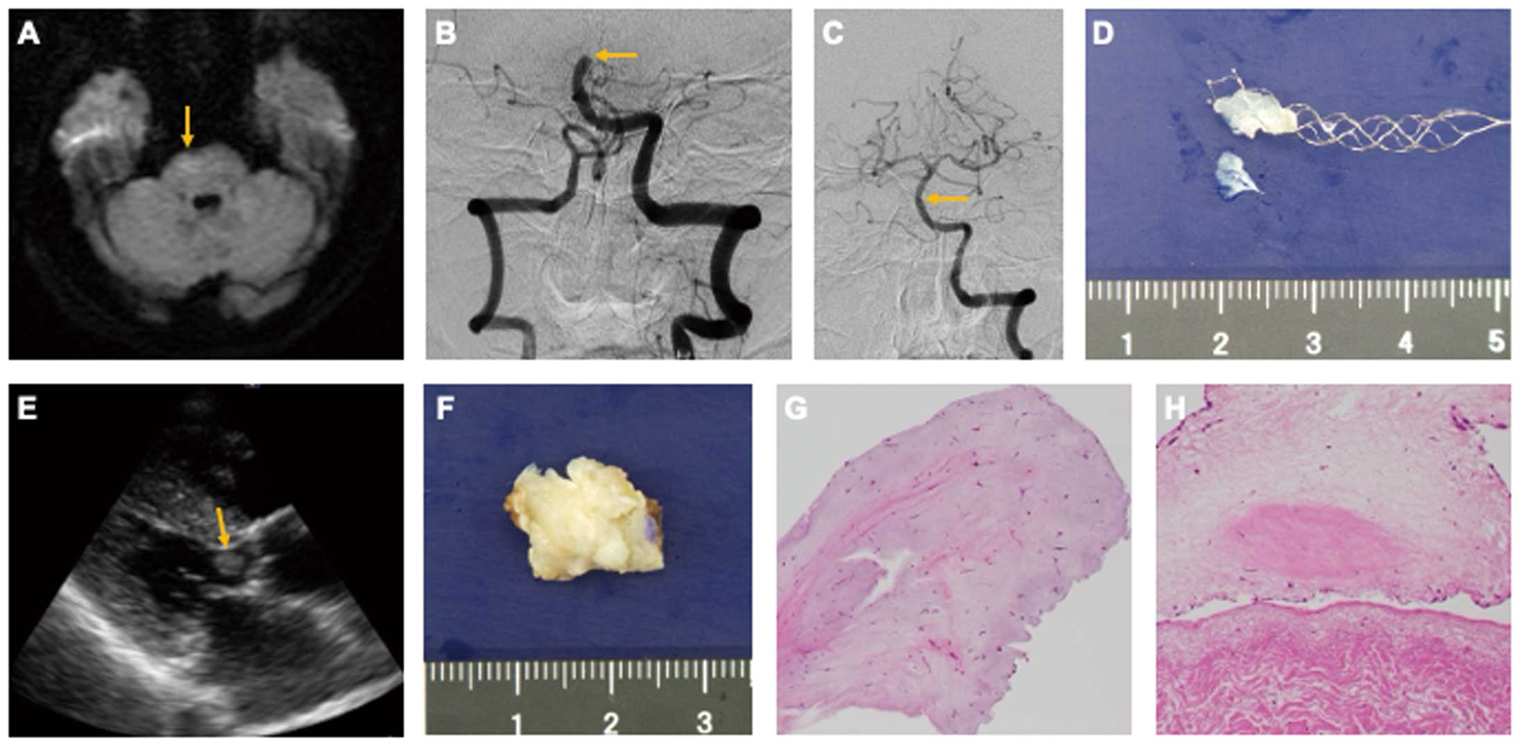
Axial diffusion-weighted imaging shows a hyperintense lesion (arrow) in the pons (A). Digital subtraction angiography shows a mid-basilar artery occlusion (arrow); mechanical thrombectomy successfully restored blood flow (arrows) (B,C). The embolus retrieved by stent retriever: a soft, white, tumorous mass (D). Parasternal long-axis echocardiographic image of a 20-mm mobile mass (arrow) within the left ventricular outflow tract (LVOT) (E). Surgically excised LVOT mass is yellowish-white with an irregular surface (F). Pathological examination of both the embolus (G) and the LVOT tumor (H) revealed scattered stellate or spindle-shaped cells within a mucopolysaccharide-rich matrix, confirming the diagnosis of myxoma.
This case illustrates an extremely rare presentation of LVOT myxoma diagnosed after an embolic stroke, highlighting the importance of thorough evaluation in stroke patients. It aligns with previous reports suggesting that atypical tumor locations outside the left atrium and irregular tumor morphology increase the risk of embolization.1