Bicuspid Aortic Valve Stenosis With Membranous Interventricular Septal Aneurysm Treated Using Transcatheter Aortic Valve Replacement
Article ID: CR-19-0092

Details
Article ID: CR-19-0092
A 90-year-old woman presented with symptomatic severe aortic stenosis (AS). Echocardiography showed a sac-like aneurysm of the membranous ventricular septum (AMS; Figure A). Computed tomography (CT) showed a type 1 bicuspid valve due to right-left coronary cusp fusion and an AMS just below the right coronary cusp (Figure B,C). Although the annular perimeter was measured as 93 mm, the practical perimeter was estimated at 80 mm, excluding the aneurysmal area (Figure D). The inter-commissural distance was 26 mm (Figure E). Because determining the appropriate valve size was difficult, transcatheter aortic valve replacement (TAVR) using a self-expandable valve was planned. A 29-mm CoreValve Evolut R (Medtronic, MN, USA) was successfully implanted. Post-procedural echocardiography showed a mild paravalvular leakage unrelated to the aneurysm (Figure F,G; Supplementary Movie), and CT showed a partially covered AMS with no thrombus (Figure H–J).
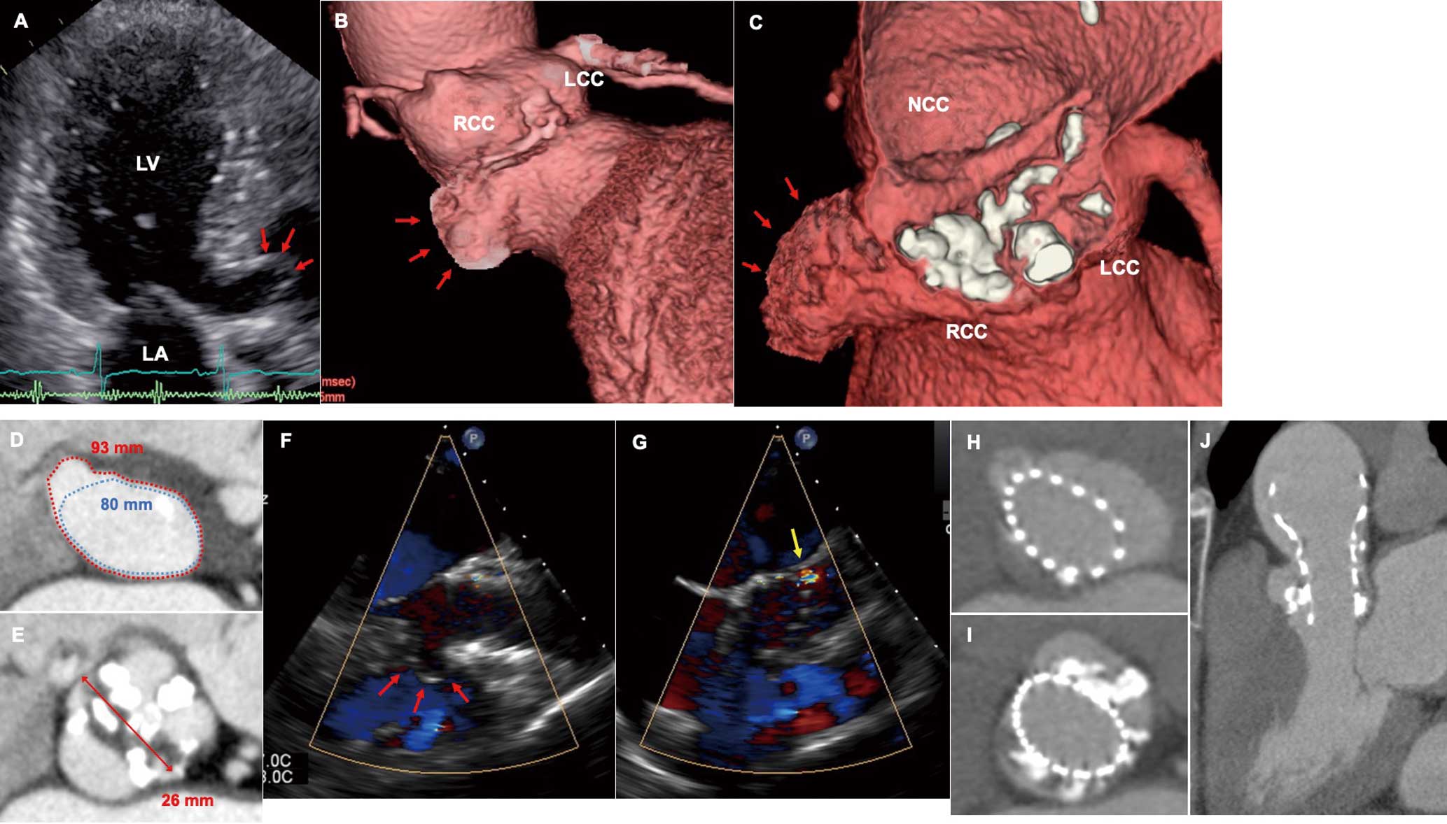
(A) Echocardiography showing an aneurysm of the membranous ventricular septum (AMS; arrows). LA, left atrium; LV, left ventricle. (B,C) Computed tomography (CT) showing a bicuspid valve and AMS (arrows). LCC, left coronary cusp; NCC, non-coronary cusp; RCC, right coronary cusp. (D) The annular perimeters were 93 and 80 mm with (red dotted line) and without (blue dotted line) the aneurysm, respectively. (E) Pre-procedural CT showing the supra-annulus level. (F,G) Post-procedural echocardiography showing a mild paravalvular leakage (yellow arrow) unrelated to the aneurysm (red arrows; F, long-axis view; G, short-axis view). (H–J) Post-procedural CT showing the (H) annulus and (I) supra-annulus levels, and (J) valve position.
AMS is a rare congenital disorder caused by spontaneous closure of a pre-existing ventricular septal defect or other etiological factors.1 This is the first reported case of TAVR in a patient with bicuspid AS and AMS. Because determining the optimal valve size is challenging, a repositionable and retrievable valve might be safer.
The authors declare no conflicts of interest.
Supplementary Movie. Post-procedural echocardiography showing a mild paravalvular leakage unrelated to the aneurysm.
Please find supplementary file(s);
http://dx.doi.org/10.1253/circrep.CR-19-0092