A 22-year-old male with no past medical history presented with a 1-day history of chest pain, which commenced 1 day after the third dose of the mRNA-1273 (Moderna) coronavirus disease 2019 (COVID-19) vaccine. He had received 2 prior doses of the mRNA-1273 COVID-19 vaccine. An electrocardiogram demonstrated widespread ST-segment elevation (Figure A), whereas echocardiography findings were normal (Figure B,C). The creatinine kinase (CK) concentration was 63 U/L, CK-MB was 1.9 ng/mL, troponin I was 0.35 ng/mL, and B-type natriuretic peptide was 6.9 pg/mL. COVID-19 antigen quantification was negative. Myocarditis was diagnosed after excluding vasospastic angina and stress-induced cardiomyopathy based on symptoms, repeated echocardiograms, cardiogenic enzyme courses, and electrocardiography results. Because there were no laboratory abnormalities, suggesting that the cause was viral, allergic, autoimmune, collagen disease, or sarcoidosis, it was concluded that the cause was the COVID-19 vaccine. Concentrations of CK (253 U/L), CK-MB (21.0 ng/mL), and troponin I (3.65 ng/mL) peaked 3 days after vaccination, and cardiac magnetic resonance imaging (cMRI) showed no myocardial edema or late gadolinium enhancement (Figure D–G). The patient was carefully monitored; electrocardiogram findings improved (Figure H) and echocardiography findings remained normal.
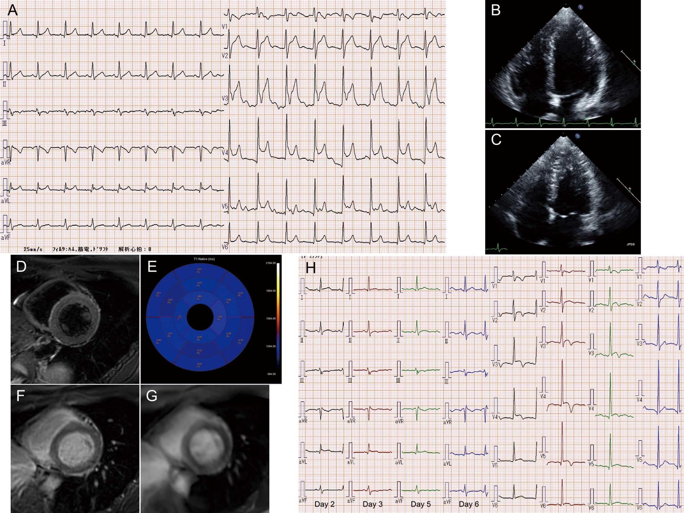
This patient developed myocarditis after the third mRNA-1273 COVID-19 vaccine, with distinct electrocardiographic changes and mild elevation of CK, CK-MB, and troponin I. The occurrence of first-episode myocarditis after a third dose of the mRNA-1273 COVID-19 vaccine may be difficult to detect due to the 75–95% diagnostic accuracy of cMRI.1
Disclosures
K.T. is a member of Circulation Reports’ Editorial Team. The remaining authors declare no conflicts of interest.
IRB Information
This study was approved by Shimane University Faculty of Medicine (No. 6229).
