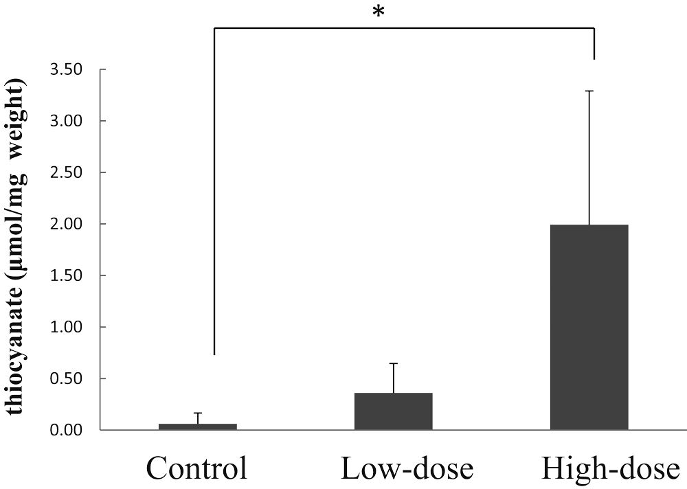
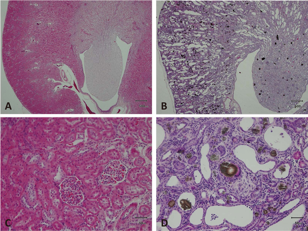
Food Safety
Online ISSN : 2187-8404
ISSN-L : 2187-8404

この記事には本公開記事があります。本公開記事を参照してください。
引用する場合も本公開記事を引用してください。

The Acute Encephalopathy Induced by Intake of Sugihiratake Mushroom in the Patients with Renal Damage Might Be Associated with the Intoxication of Cyanide and Thiocyanate
Hiroshi Akiyama Hideki MatsuokaTakanori OkuyamaKyohei HigashiToshihiko ToidaHiroyuki KomatsuYoshiko Sugita-KonishiSatomi KoboriYukio KodamaMidori YoshidaHitoshi Endou
著者情報
ジャーナル フリー HTML 早期公開

論文ID: 2014036

この記事には本公開記事があります。
詳細
Abstract

A novel type of encephalopathy associated with the ingestion of Sugihiratake mushroom (Pleurocybella porrigens) occurred in patients with chronic renal failure treated on hemodialysis in fall, 2004 in Japan. To clarify the mechanism of encephalopathy onset, we, for the first time, purified the cyanogen glycoside fraction (CG) from Sugihiratake mushroom using reversed phase high-performance liquid chromatography and hydrophilic interaction chromatography. Furthermore, we investigated single dose toxicity of the CG in an adenine-induced rat model of chronic renal damage (CRD). Pathological examination of kidneys indicates the development of CRD. Oral administration of the CG induces the accumulation of thiocyanate in the hemolyzed blood and brain in CRD rats, although no morphological changes were found in the brain. No further enhancement of kidney damage is observed after the oral administration of the CG in CRD rats. This is the first experimental report to suggest that acute encephalopathy, induced by Sugihiratake mushroom intake in the patients with chronic renal failure, is associated with intoxication of cyanide and thiocyanate, presumably produced metabolically produced after the ingestion of Sugihiratake mushroom.

Figures
Fig. 1.

Cyanide (%) content of fractions obtained from hydrophilic interaction chromatography.

The vertical axis represents the ratio of cyanide content detected in each fraction to total cyanide content detected in the sample before the injection to HPLC.

Fig. 2.

Temporal changes of cyanide levels in the hemolyzed blood in an adenine-induced rat model of CRD.

Control represents the control group administered distilled water alone, Low-dose represents the low-dose group administered 360 mg cyanogen glycoside fraction (CG)/kg body weight (corresponding to 0.31 mg sodium cyanide/kg body weight), and High-dose represents the high-dose group administered 2840 mg CG/kg body weight (corresponding to 2.44 mg sodium cyanide/kg body weight). Bars represent means ± standard deviation (3 rats/group).

The bar represents the change of cyanide level in the hemolyzed blood from 0 h to 8 h after administration of CG

The bar represents the change of cyanide level in the hemolyzed blood from 0 h to 24 h after administration of CG

Fig. 3.

Temporal changes of thiocyanate levels in the hemolyzed blood in an adenine-induced rat model of CRD.

Control represents the control group administered distilled water alone, Low-dose represents the low-dose group administered 360 mg CG/kg body weight (corresponding to 0.31 mg sodium cyanide/kg body weight), and High-dose represents the high-dose group administered 2840 mg CG/kg body weight (corresponding to 2.44 mg sodium cyanide/kg body weight). Bars represent means ± standard deviation (3 rats/group). Asterisks indicate significant differences from control values (**p < 0.01).

The bar represents the change of thiocyanate level in the hemolyzed blood from 0 h to 8 h after administration of CG

The bar represents the change of thiocyanate level in the hemolyzed blood from 0 h to 24 h after administration of CG

Fig. 4.

Thiocyanate content in the brains of adenine-induced CRD rats.

Control represents the control group administered distilled water only, Low-dose represents the low-dose group administered 360 mg CG/kg body weight (corresponding to 0.31 mg sodium cyanide/kg body weight), and High-dose represents the high-dose group administered 2840 mg CG/kg body weight (corresponding to 2.44 mg sodium cyanide/kg body weight). Bars represent means ± standard deviation (3 rats/group). Asterisks indicate significant differences from control values (*p < 0.05).

The bar represents brain thiocyanate levels after administration of CG

Fig. 5.

Macroscopy of the normal kidney (left), and Adenine represents the adenine-induced crystal nephropathy observed in CRD rats (right).

Fig. 6.

Histopathology of the cerebrum cortex and cerebrum caudal nucleus of CRD rats after administration of Sugihiratake extract.

Control represents the control group administered distilled water alone, and High-dose represents the high-dose group administered 2840 mg CG/kg body weight (corresponding to 2.44 mg sodium cyanide/kg body weight. Microscopically no treatment related abnormalities were observed in neurons and glial cells. x100. Hematoxylin-eosin staining.

Fig. 7.

Histopathological images of the kidneys of CRD rats.

A-D, Histopathological changes in the kideny. A and C, Normal structure obtained from a normal adult rat kidney. B and D, Adenine-induced chronic renal damage. The normal structure was destroyed, and brown to black dots, adenine crystals, were diffusely distributed throughout the kidney at low magnification (x10). C and D, Higher magnification (x100) of A and B. C. Normal structure. D, Various sizes of crystals are accumulated in the dilated lumen of renal tubules or deposited in the tubules. Inflammatory cells infiltrate the interstitium. Hematoxylin-eosin staining.

Tables
Table 1.The body weight changes
IndividualNo.Animal*No.Quarantine and acclimation periodsAdenine-component feed feeding period
---- 0.75% ----------- 0.5% --------
Day15918152228
11302298334355362281270270259
2exc.308355381384267---
31203295344366368268282285282
41103288317332334257220219220
51301304337368378270282285256
61303297343366375321303298282
71101315354385388293258260262
81201306348368378301279279261
91102303340361362275271270273
101202305345363369266268264254
Mean302342365370280270270261
S.E.23556886

Unit: g.

No.2: died on day 8 of adenine-component feed feeding period (266 g body weight).

* Identification numbers after allocation to treatment groups (exc.: excluded from this study).

Quarantine period: 5 days.

Acclimation period: 9 days (with quarantine period).

Table 2.Pathological findings in rats with adenine-induced renal failure after oral administration of Sugihiratake extract
Test substanceControlSugihiratake extract
360 mg/kg2840 mg/kg
Animal No.110111021103120112021203130113021303
Heart
      Myocardium
            Focal calcification001100110
      Coronary artery
            Cellular infiltration, mononuclear cell101100110
            Calcification003300321
      Arcus aorta
            Tunica media, calcification203310321
            Tunica intima, cellular infiltration, neutrophil /
mononuclear cell
100100000
Lung
      Alveolar septa, calcification003000200
      Bronchial smooth muscle, calcification002000100
Kidney
      Artery
            Calcification102210221
      Renal tubule
            2,8-dihydroxyadenine (DHA) crystals333333333
            Tubular dilatation333333333
            Tubular basophilic change333333333
            Cellular infiltration, neutrophil222222222
            Giant cells111111111
      Interstitium
            Cellular infiltration, neutrophil /
mononuclear cell
111111111
            Fibrosis222222222

0: No change, 1: Slight, 2: Moderate, 3: Marked.

No significant changes were detected: Spleen, Liver.

Table 3.Pathological findings in rats with adenine-induced renal failure after oral administration of Sugihiratake extract
Test substanceControlSugihiratake extract
360 mg/kg2840 mg/kg
FindingsAnimal No.110111021103120112021203130113021303
Kidney
    Enlargement, pale yellow+++++++++
Thymus
    Small size+++++++++
Arcus aorte
    Grayish white--+------
Coronary artery
    Grayish white--+------
Glandular stomach
    Grayish white--+---+--
Colon
    Grayish white--+---+--

-: No change, +: Change.

References
 
© 2015 Food Safety Commission, Cabinet Office, Government of Japan
feedback
Top