原著論文
Identification of Flavonoid Pigments and Coloration Mechanisms in the Bluish-purple Flowers of Platycodon grandiflorus (Jacq.) A. DC. (Campanulaceae)
2023 年 92 巻 3 号 p. 342-353

詳細
2023 年 92 巻 3 号 p. 342-353
We investigated bluish-purple Platycodon grandiflorus flowers for yet unidentified flavonoid-related compounds and their flower coloration mechanisms. We identified a new polyacylated anthocyanin, delphinidin 3-O-[6-O-(α-rhamnopyranosyl)-β-glucopyranoside]-7-O-[6-O-(4-O-(6-O-(4-O-(6-O-(4-O-(β-glucopyranosyl)-trans-caffeoyl)-β-glucopyranosyl)-trans-caffeoyl)-β-glucopyranosyl)-trans-caffeoyl)-β-glucopyranoside] (3) as the minor anthocyanin component along with four known anthocyanins, four known flavones, and chlorogenic acid. The major anthocyanin of the bluish-purple P. grandiflorus flowers is platyconin. While platyconin has two caffeic acids linked in series via glucose molecules at the 7-position of delphinidin, anthocyanin 3 has three caffeic acids linked in series via glucose molecules at the 7-position of delphinidin. To investigate the effects of the number of aromatic acyl groups in polyacylated anthocyanin on bluing and stability, the color and stability of anthocyanin 3 in diluted aqueous solution (5 × 10⁻⁵ M) at a weak acidic condition (pH 5.7, the same pH as the petal sap) were compared with those of platyconin. Results showed that even if the three caffeic acids were bound in series at the 7-position of delphinidin, there was no further bluing or stabilizing effects of anthocyanin 3 compared to platyconin, but a tendency toward weakening color intensity was observed. Investigation of the change in anthocyanin content at the flower developmental stages showed that delphinidin 3-rutinoside-7-glucoside, which is the deacylated form of platyconin, first accumulated up to sufficient amounts at the bud stage, followed by an increase in platyconin content. At the bud stage, the petals were not purple or violet, except for veins, despite the presence of a good amount of delphinidin 3-rutinoside-7-glucoside, indicating the delphinidin 3-rutinoside-7-glucoside was present in some colorless state.
Platycodon grandiflorus (Jacq.) A. DC. (Campanulaceae), commonly called the balloon flower, is the only species of the genus Platycodon and is natively distributed in Northeast Asia, including Japan, China, and Korea. The plant has been used as a traditional medicinal plant in these regions for long and numerous compounds have been identified from this plant (Zhang et al., 2015). Platycodon grandiflorus is used as an ornamental plant due to its beautiful flowers (The Royal Horticultural Society, 2016). Platycodon grandiflorus produces purple-blue flowers with darker purple-blue veins. The major pigment of the bluish-purple flowers is an anthocyanin named platyconin. Saito et al. isolated platyconin in 1971, and the complete structure of platyconin was determined as delphinidin 3-O-[6-O-(α-rhamnopyranosyl)-β-glucopyranoside]-7-O-[6-O-(4-O-(6-O-(4-O-(β-glucopyranosyl)-trans-caffeoyl)-β-glucopyranosyl)-trans-caffeoyl)-β-glucopyranoside] by Goto et al. in 1983. Anthocyanins that have two or more aromatic acyl groups in their structure as in platyconin are called polyacylated anthocyanins. Platyconin is known as the first isolated polyacylated anthocyanin and several polyacylated anthocyanins have been isolated from violet to blue flowers of various plants to date (Honda and Saito, 2002; Yoshida et al., 2009). Polyacylation of anthocyanins gets anthocyanins to develop a bluer color (Goto and Kondo, 1991; Honda and Saito, 2002; Yoshida et al., 2009). In addition, in weakly acidic or neutral aqueous solutions, polyacylated anthocyanins maintain a stable color, whereas non-polyacylated anthocyanins rapidly decolorize into colorless forms due to hydration (Goto and Kondo, 1991; Honda and Saito, 2002). The bluer coloration and the color stability are proposed to be produced by stacking interactions between the aromatic acyl moieties and the anthocyanidin chromophore. (Dangles and Fenger, 2018; Goto and Kondo, 1991; Honda and Saito, 2002; Yoshida et al., 2009). In addition, the number of aromatic acyl groups in polyacylated anthocyanin has been reported to affect the bluing effect and stability (Honda and Saito, 2002).
In this study, we investigated bluish-purple P. grandiflorus flowers for previously unreported flavonoid compounds including anthocyanins, and to further understand their flower color expression mechanisms. First, we conducted analysis and identification of flavonoid compounds. The structures of compounds were determined by chromatographic analyses (TLC and HPLC) and spectroscopic analyses (UV-vis, FABMS, and 1H and 13NMR). We identified a new polyacylated anthocyanin in which three caffeic acids were linked in series via glucose molecules at the 7-position; this is longer than that of platyconin by one glucosyl-caffeic acid. Thus, the color and stability of this anthocyanin were compared with those of platyconin in a buffer solution with the same pH as the petal sap to determine the effect of the length of the polyacylated side chain (the effect of the number of aromatic acyl groups) on color and stability. Next, we investigated the change in anthocyanin content in the flower development stages to obtain insights into the biosynthesis of the polyacylated anthocyanins in P. grandiflorus.
TLC was performed on cellulose-coated plastic sheets (Merck KGaA, Darmstadt, Germany) using the following mobile phases: For anthocyanins, BAW (n-BuOH/HOAc/H2O, 4:1:2, v/v/v), BuHCl (n-BuOH/2N HCl, 1:1, v/v, upper layer), AHW (HOAc/HCl/H2O, 15:3:82, v/v/v) and 1% HCl were used. For sugars, BAW, EAA (EtOAc/HOAc/H2O, 3:1:1, v/v/v), ETN (EtOH/NH4OH/H2O, 16:1:3, v/v/v), and EFW (EtOAc/HCOOH/H2O, 5:2:1, v/v/v) were used with aniline hydrogen phthalate spray reagent to detect spots. For anthocyanidins, Forestal (HOAc/HCl/H2O, 30:3:10, v/v/v) was used (Harborne, 1984).
Analytical HPLC was performed on an LC 20A system (Shimadzu Corporation, Kyoto, Japan), using a Spherisorb ODS2 column (4.6 × 250 mm; Waters Corporation, Milford, MA, USA) at 40°C with a flow rate of 1 mL·min−1 and monitoring was done with a photodiode array detector. The eluant was applied as a linear gradient elution for 40 min from 20 to 85% solvent B (1.5% H3PO4, 20% HOAc, 25% MeCN in H2O, v/v/v/v) in solvent A (1.5% H3PO4 in H2O, v/v).
As authentic samples, delphinidin prepared from Vanda (Tatsuzawa et al., 2004), p-coumaric acid (Tokyo Chemical Industry Co., Ltd., Tokyo, Japan), caffeic acid (Sigma Chemical Company, St. Louis, MO, USA), glucose (Nacalai Tesque, Inc., Kyoto, Japan), and rhamnose (FUJIFILM Wako Pure Chemical Corporation, Osaka, Japan) were used.
UV-vis spectra of purified anthocyanins in 0.1% HCl-MeOH were recorded on a UV-vis spectrophotometer (MPS-2450; Shimadzu Corporation, from 200 to 700 nm). UV-vis spectra of purified flavones were recorded on MPS-2450 in MeOH, MeOH+NaOMe, MeOH+AlCl3, MeOH+AlCl3+HCl, MeOH+NaOAc, and MeOH+NaOAc+H3BO3 (from 200 to 500 nm).
High resolution FAB mass spectrometry (HR-FABMS) was obtained in the positive ion mode using glycerol as a matrix with a mass spectrometer (JMS-700; JEOL Ltd., Tokyo, Japan).
NMR spectra were recorded on a JNM-AL400 (JEOL Ltd.) at 400 MHz for 1H spectra and 100 MHz for 13C spectra using CD3OD-CF3COOD (9:1) for anthocyanins, and dimethyl sulfoxide (DMSO)-d6 for flavones, as solvents. Chemical shifts were reported on the δ-scale from tetramethylsilane as the internal standard and coupling constants (J) were denoted in Hz.
Plant materialsSeeds of P. grandiflorus ‘Samidare’ were purchased from Takii & Co., Ltd. (Kyoto, Japan). The seeds were sown in December 2018, and the plants were grown in a greenhouse and an experimental farm at Iwate University. Fresh petals of the bluish-purple flowers were collected. Some of them were dried overnight at 45°C and kept at −20°C until use.
Flower color measurementThe flower color was recorded by comparing it directly with the Royal Horticultural Society Colour Chart (5th edition, RHS CC). The CIELAB chromaticity values were recorded on a CM-700d Spectro Color Meter (Konica Minolta, Inc., Tokyo, Japan). Ten flowers were measured and the average ± SE was obtained. The absorption spectrum of the flower color was directly measured for an intact petal sandwiched in a pierced (5 mm) black holder with a spectrophotometer (MPS-2450; Shimadzu Corporation).
Measurement of fresh petal sap pHPetals were mashed and the pH of the sap was measured using a compact pH meter (B-212; Horiba, Ltd., Kyoto, Japan). The average ± SE from the repetitions was obtained.
Analysis of flavonoids in flowersDried petals (10 mg) or intact petals (200 mg) were immersed in MAW (MeOH/HOAc/H2O, 4:1:5, v/v/v, 500 μL or 4 mL, respectively) and the extracts were analyzed by HPLC. Anthocyanins were detected at 530 nm and the other compounds were detected at 350 nm.
Isolation and purification of compoundsThe dried petals of bluish-purple flowers (ca. 400 g) were immersed in 10% HOAc-H2O (v/v, 5 L) at room temperature for 24 h. The extract was loaded onto a Diaion HP-20 (Nippon Rensui Co., Tokyo, Japan) column (60 × 300 mm). Absorbed pigments were washed with 5% HOAc-H2O (v/v, 20 L) and eluted with 5% HOAc-MeOH (v/v, 1 L). After concentration, pigments were separated with paper chromatography (PC) using BAW. The separated anthocyanins were further separated by SephadexTM LH-20 (GE Healthcare UK Ltd., Buckinghamshire, England) column chromatography using 5% HOAc-H2O. They were then further purified with preparative HPLC (prep. HPLC). Prep. HPLC was performed on an LC 10A system (Shimadzu Corporation) using a μ-Bondasphere column (19 × 150 mm, Waters Corporation) at 40°C with a solution flow rate of 4 mL·min−1 monitored at 530 nm. The solvent was subjected to isocratic elution for 15 min with 40% solvent B in solvent A. Each fraction was transferred to a Diaion HP-20 column. Anthocyanins were eluted with 5% HOAc-MeOH, concentrated, had excess Et2O added, and were then dried. The purified anthocyanins 2 (ca. 416 mg), 3 (ca. 35 mg), and 4 (ca. 48 mg) were obtained as dark violet powders. The crude compounds other than anthocyanins separated by PC were purified by SephadexTM LH-20 column chromatography. The purified compound 5 (ca. 12 mg) was obtained as a pale brown powder and compounds 6 (ca. 21 mg), 7 (ca. 6 mg), 8 (ca. 23 mg), and 9 (ca. 24 mg) were obtained as pale yellow powders.
Acid hydrolysis of anthocyanins 2–4 (detection of anthocyanidins, hydroxycinnamic acids, and sugars)Acid hydrolysis of 2–4 (ca. 0.5 mg each) was performed with 2N HCl (1 mL) at 100°C for 2 h. The hydrolysates were analyzed by TLC and HPLC. The Rf-values in TLC and retention time (tR) in HPLC (detected at 530 nm for anthocyanidins and at 315 nm for hydroxycinnamic acids) were compared with those of authentic anthocyanidins (delphinidin, Rf-value Forestal 0.22; tR 23.5 min), hydroxycinnamic acids (trans-p-coumaric acid, tR 16.4 min and trans-caffeic acid, tR 11.6 min), sugars (glucose, Rf-value BAW 0.32, EAA 0.59, ETN 0.46, EFW 0.59 and rhamnose, Rf-value BAW 0.44, EAA 0.75, ETN 0.61, EFW 0.79).
Alkaline hydrolysis of anthocyanins 2–4 (detection of deacylated anthocyanin and glucosyl-hydroxycinnamic acids)Each of the purified anthocyanins 2–4 (ca. 1 mg each), “Leschenaultia blue anthocyanin 1” (ca. 1 mg) (Saito et al., 2007), and “Laeliocattleya anthocyanin 1” (ca. 1 mg) (Tatsuzawa et al., 1994) were dissolved separately in 2 N NaOH (100 μL) in a degassed syringe and left for 15 min. The solutions were then acidified with 2 N HCl (110 μL). The alkaline hydrolysates were analyzed immediately by HPLC (detected at 530 nm for deacylated anthocyanins and at 315 nm for glucosyl-hydroxycinnamic acids). Alkaline hydrolysates from “Leschenaultia blue anthocyanin 1” and “Laeliocattleya anthocyanin 1” were used as comparative standards for 4-O-glucosyl-caffeic acid (HPLC tR 8.6 min) and 4-O-glucosyl-p-coumaric acid (HPLC tR 8.8 min), respectively. In order to identify the deacylated anthocyanin of anthocyanins 2–4, the deacylated anthocyanin (ca. 40 mg) was purified from an alkaline hydrolysate made from a crude mixture of 2–4 in the process of purification (ca. 1 g).
Structural determination of compoundsThe structural determination of compounds was performed by chromatographic (TLC and HPLC) and spectroscopic (UV-vis, NMR, HR-FABMS) techniques. NMR spectra of 1H NMR, 13C NMR, HH-COSY, NOESY, CH-COSY (or HMQC), and HMBC were measured.
Measurement of color and stability of anthocyanin 3 and comparisons with platyconin.Anthocyanin 3 and platyconin solutions were prepared to a concentration of 5 × 10⁻⁵ M in 0.1 M acetate buffer (pH 5.7). The absorption spectrum (wavelength range of 400–800 nm) of the solution was measured using a UV-2450 spectrophotometer (Shimadzu Corporation). For the color stability measurement, the solutions were placed in the dark at room temperature (around 23°C), and the absorption spectrum measurements were repeated every day. The color stability was expressed as the ratio of the absorbance at λmax, normalizing the measured values using absorbance immediately after dissolution as 100%.
Measurement of changes in anthocyanin content in various flower developmental stagesAs the flowering stage designations, green buds were designated as stage 0, green buds with bluer colored veins as stage 1, bluish-purple colored buds as stage 2, open flowers with unopened anthers as stage 3, open flowers with opened anthers as stage 4, and withered flowers as stage 5 (Fig. 5). The pH of petal sap at each stage was measured and the content of anthocyanins at each stage was examined by HPLC. The quantification was done based on the peak areas. Calibration curves were created using the purified delphinidin 3-rutinoside-7-glucoside and platyconin.
The opened flower color of the bluish-purple P. grandiflorus was close to Violet-blue 93B based on the RHS CC. The CIELAB chromaticity values were L* (51.05 ± 0.65), a* (14.54 ± 0.60), and b* (−24.69 ± 0.58). The light absorption spectrum of petals exhibited three λmax at 625, 573, and 541 nm with one shoulder at 504 nm (using a spectral measurement range from 400–750 nm). The pH value of fresh petal sap was 5.7 ± 0.0 (average ± SE, n = 10).
HPLC analysis of the extract from the dried petals showed several anthocyanin peaks at 530 nm (Fig. 1), with a preponderance of peak 2. Three peaks 2–4 were purified and isolated in this study. In addition, five of the peaks observed at 350 nm (5–9, Fig. 1) were isolated. The percentages of the anthocyanin 1–4 contents to the total HPLC peak areas at 530 nm were 1, 3.5 ± 0.7%; 2, 61.0 ± 2.7%; 3, 3.1 ± 0.3%, and 4, 7.9 ± 1.1%, and those of the compound 5–9 contents to the total HPLC peak areas at 350 nm were 5, 4.7 ± 0.7%; 6, 12.3 ± 1.2%; 7, 2.7 ± 0.5%; 8, 23.3 ± 1.7%, and 9, 10.6 ± 1.1% (average ± SE, n = 10).
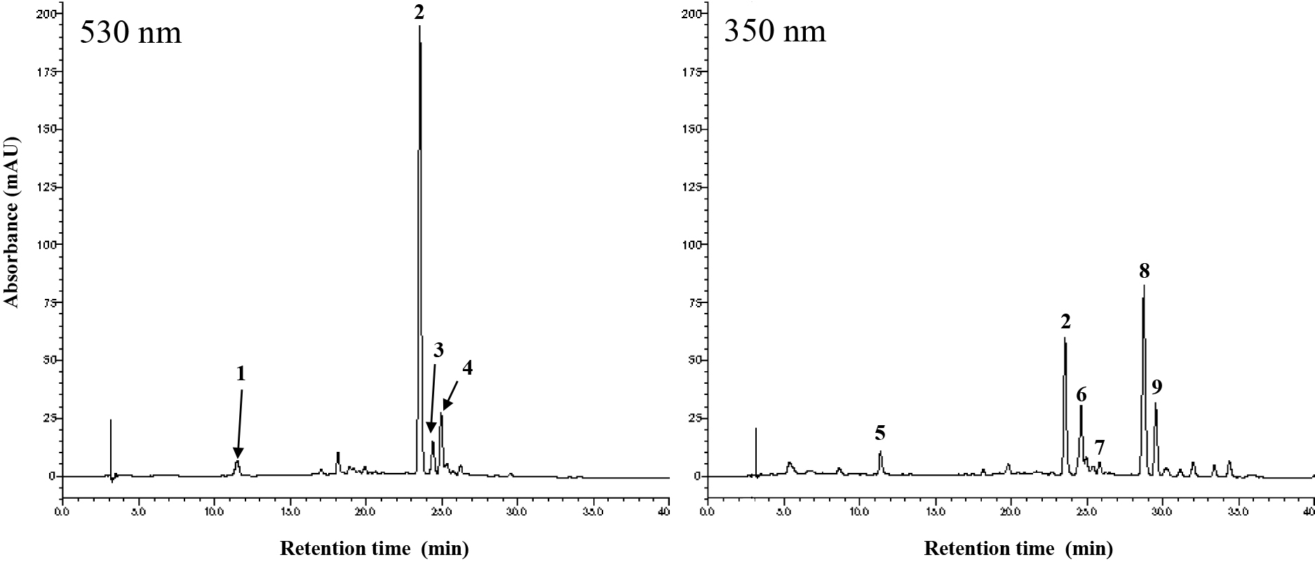
HPLC chromatograms of an extract from bluish-purple petals of P. grandiflorus.
Compounds 2–9 were isolated from ca. 400 g of dried petals through several chromatographic separations. Acid hydrolysis of the purified anthocyanins 2–4 yielded delphinidin, glucose, rhamnose, and caffeic acid. In addition, p-coumaric acid was detected in the hydrolysate of 4. From the alkaline hydrolysate of anthocyanins 2–4, 4-glucosyl-caffeic acid was detected. In addition, 4-glucosyl-p-coumaric acid was detected in the alkaline hydrolysate of anthocyanin 4. A deacylated anthocyanin was observed from the alkaline hydrolysates from 2–4, which was identified as delphinidin 3-rutinoside-7-glucoside (section 3.1). The retention time of peak 1 observed in the HPLC analysis of the petal extract (Fig. 1) corresponded to that of delphinidin 3-rutinoside-7-glucoside (HPLC tR 11.4 min). Kondo et al. (2021) previously reported the presence of delphinidin 3-rutinoside-7-glucoside in bluish-purple P. grandiflorus flowers. In this study, we identified peak 1 to be delphinidin 3-rutinoside-7-glucoside. A FABMS molecular ion [M+H]+ of compound 5 was detected at 355 m/z. Compound 5 was identified to be chlorogenic acid by direct comparison with the commercial standard by TLC and HPLC analyses. The chromatographic properties of compound 5 are shown in section 3.3. Chlorogenic acid has already been reported for P. grandiflorus (Mazol et al., 2004).
The structures of anthocyanins 2–4 and compounds 6–9 were elucidated in detail by HR-FABMS spectra and NMR spectra. Anthocyanin 3 was a previously unreported anthocyanin. Peak 4 was revealed to contain two anthocyanins with similar structure, which were designated as 4A and 4B. The determination of anthocyanins 3, 4A and 4B are detailed in below sections 1 and 2. The chromatographic and spectroscopic properties of compounds 2, and 6–9 are shown in sections 3.2), and 3.4)–3.7). Signal assignments for 1H and 13C NMR spectra of 2 are shown in Table 1 and Figure S1. Anthocyanin 2 was identified as delphinidin 3-O-[6-O-(α-rhamnopyranosyl)-β-glucopyranoside]-7-O-[6-O-(4-O-(6-O-(4-O-(β-glucopyranosyl)-trans-caffeoyl)-β-glucopyranosyl)-trans-caffeoyl)-β-glucopyranoside] (Fig. 2), which is a platyconin. Compounds 6–9 were identified as luteolin 7-glucoside (6), luteolin 7-O-[2-O-(α-rhamnosyl)-β-glucoside] (luteolin 7-neohesperidoside, 7), apigenin 7-glucoside (8), and apigenin 7-O-[2-O-(α-rhamnosyl)-β-glucoside] (apigenin 7-neohesperidoside, 9). These are known flavones (Harborne and Baxter, 1999). Flavones 6 and 8 have often been reported in P. grandiflorus (Inada et al., 1992; Jang et al., 2010; Jeong et al., 2010; Mazol et al., 2004). Although flavones 7 and 9 have been reported in traditional Chinese medicines including P. grandiflorus as one of the ingredients (Huang et al., 2007; Wang et al., 2022), to our knowledge, these have not yet been reported in P. grandiflorus itself (SciFindern; December 24, 2022).
1H and 13C NMR spectroscopic dataz of polyacylated anthocyanins from the bluish-purple flowers of P. grandiflorus. Coupling constants are shown in parentheses (J in Hz).

Continued

Polyacylated anthocyanins from the bluish-purple flowers of P. grandiflorus. Observed NOEs are indicated by solid arrows. Observed HMBCs are indicated by dotted arrows.
The purified anthocyanin 3 was a dark violet powder. The FABMS molecular ion peak [M]+ of anthocyanin 3 was observed at 1745 m/z, indicating that anthocyanin 3 is composed of delphinidin with five molecules of glucose, one molecule of rhamnose, and three molecules of caffeic acid. The elemental components of 3 were confirmed to be C78H89O45 by HR-FABMS ([M]+: found 1745.4701, calc. for C78H89O45: 1745.4676). Signal assignments for 1H and 13C NMR spectra of 3 are shown in Table 1. The 1H NMR spectrum of 3 was similar to that of 2, except for the additional signals of Glc E and caffeic acid III moieties (Table 1; Fig. S2). The proton signals of H-6a and b of Glc D in 3 (δ 3.97 and 4.60) were shifted to a lower field compared to those of Glc D in 2 (δ 3.78 and 3.85), indicating that the OH-6 of Glc D in 3 is acylated by the additional caffeic acid III. NOE between H-1 of Glc E and H-5' of caffeic acid III was observed (Fig. 2), supporting glycosylation of the OH-4' of caffeic acid III by Glc E. Based on the coupling constants, the five glucose molecules were assumed to be in the β-pyranose forms and the rhamnose to be in the α-pyranose form (Table 1). Therefore, anthocyanin 3 was determined to be delphinidin 3-O-[6-O-(α-rhamnopyranosyl)-β-glucopyranoside]-7-O-[6-O-(4-O-(6-O-(4-O-(6-O-(4-O-(β-glucopyranosyl)-trans-caffeoyl)-β-glucopyranosyl)-trans-caffeoyl)-β-glucopyranosyl)-trans-caffeoyl)-β-glucopyranoside] (Fig. 2), which was the first reported anthocyanin (SciFindern; December 24, 2022). This polyacyl group at the 7-position is longer than that of platyconin by one 4-glucosyl-caffeic acid. Other spectroscopic and chromatographic properties about anthocyanin 3 are as follows: HPLC tR (min): 24.4; UV-vis (in 0.1% HCl-MeOH): λmax 551, 318sh (= shoulder), 284 nm, E440/Emax (%) = 18, Eacyl/Emax (%) = 143, AlCl3 shift +; TLC (Rf-values): BAW 0.11, BuHCl 0.01, AHW 0.35, 1% HCl 0.10.
2. Anthocyanin 4 (mixture of 4A and 4B)The isolated anthocyanin 4 was a dark violet powder. The FABMS molecular ion peak [M]+ of 4 was observed clearly as one peak at 1405 m/z, indicating that anthocyanin 4 is composed of delphinidin with four molecules of glucose, one molecule each of rhamnose, caffeic acid, and p-coumaric acid. The elemental components of anthocyanin 4 were confirmed to be C63H73O36 by HR-FABMS ([M]+: found 1405.3864, calc. for C63H73O36: 1405.3882). However, the 1H NMR signal data of the purified 4 were complex and more signals than expected were detected, as shown in Fig. S3. Considering the result of HR-FABMS data and acid and alkaline hydrolysates, 1H NMR signal data should have reflected the 12 aromatic proton signals derived from delphinidin, one caffeic acid, and one p-coumaric acid moiety. However, 24 aromatic proton signals were detected between 6.00 and 8.60 ppm (Fig. S3). These indicated that the 12 aromatic proton signals appeared twice with the same integral ratio. Moreover, from the signals detected in the region between δ 4.50–5.40, where anomeric protons of sugars should be detected, it appeared that two kinds of acylated delphinidin glycosides were mixed in the purified 4 sample. We focused on the H-4 of delphinidin proton signal which appears as a typical singlet signal with the lowest chemical shift. In the 1H NMR spectral data of anthocyanin 4, two singlet signals with the same integral value were observed at δ 8.57 and 8.50 (Fig. S3). Therefore, the lowest signal at δ 8.57 was assigned as H-4 of delphinidin of anthocyanin designated as 4A, and the next signal at δ 8.50 was assigned as H-4 of delphinidin of anthocyanin designated as 4B (Fig. S3). The assigned 1H NMR signal data of 4A and 4B were similar to that of anthocyanin 2, except for the signals of caffeic acid II and I in anthocyanin 2 (Table 1), which were replaced by signals of p-coumaric acid II and I in anthocyanins 4A and 4B, respectively (Fig. 2; Table 1). Four sets of two pairs of doublet resonances were assigned to be the olefinic proton signals derived from the caffeic acid and p-coumaric acid moieties in 4A and 4B. The coupling constants (J = 16.1 Hz each) indicated that the acids were in the trans configuration form. Five pairs of anomeric protons derived from five sugars each in 4A and 4B were assigned as shown in Table 1 and Figure S3. Based on the coupling constants, the eight glucose molecules were assumed to be in the β-pyranose forms and the two rhamnoses to be in the α-pyranose form. The positions of the attachment of sugars and acyl groups were determined based on NOESY experiments. NOEs were observed as shown in Figure 2. Thus, anthocyanins 4A and 4B were determined to be delphinidin 3-O-[6-O-(α-rhamnopyranosyl)-β-glucopyranoside]-7-O-[6-O-(4-O-(6-O-(4-O-(β-glucopyranosyl)-trans-p-coumaroyl)-β-glucopyranosyl)-trans-caffeoyl)-β-glucopyranoside] and delphinidin 3-O-[6-O-(α-rhamnopyranosyl)-β-glucopyranoside]-7-O-[6-O-(4-O-(6-O-(4-O-(β-glucopyranosyl)-trans-caffeoyl)-β-glucopyranosyl)-trans-p-coumaroyl)-β-glucopyranoside], respectively (Fig. 2). Anthocyanins 4A and 4B were reported by Kondo et al. (2021). Other spectroscopic and chromatographic properties of anthocyanin 4 (mixture of 4A and 4B) were as follows: HPLC tR (min): 24.9; UV-vis (in 0.1% HCl-MeOH): λmax 547, 308sh, 284 nm, E440/Emax (%) = 18, Eacyl/Emax (%) = 144, AlCl3 shift +; TLC (Rf-values): BAW 0.16, BuHCl 0.01, AHW 0.30, 1%HCl 0.07.
3. The chromatographic and spectroscopic properties of compounds 1, 2, and 5–9 1) Delphinidin 3-rutinoside-7-glucoside (1, Deacylated anthocyanin of 2–4)Dark violet powder; HPLC tR (min): 11.4; HR-FABMS [M]+: found 773.2140 (calc. for C33H41O21: 773.2140); 1H NMR δ Delphinidin: 8.84 (s, H-4), 6.88 (d, J = 2.0 Hz, H-6), 7.21 (d, J = 2.0 Hz, H-8), 7.80 (s, H-2',6'), Glucose A: 5.40 (d, J = 7.8 Hz, H-1), 3.79 (t, J = 9.0 Hz, H-2), 3.65 (t, J = 9.3 Hz, H-3), 3.46 (t, J = 9.2 Hz, H-4), 3.67 (m, H-5), 3.60 (m, H-6a), 4.10 (brd, J = 10.8 Hz, H-6b), Glucose B: 5.24 (d, J = 7.6 Hz, H-1), 3.62 (t, J = 9.3 Hz, H-2), 3.59 (m, H-3), 3.44 (t, J = 9.3 Hz, H-4), 3.76 (m, H-5), 3.77 (m, H-6a), 4.02 (dd, J = 1.9, 12.0 Hz, H-6b), Rhamnose: 4.68 (brs, H-1), 3.83 (dd, J = 1.2, 3.2 Hz, H-2), 3.66 (dd, J = 3.2, 9.4 Hz, H-3), 3.34 (t, J = 9.4 Hz, H-4), 3.60 (m, H-5), 1.17 (d, J = 6.4 Hz, CH3); 13C NMR δ Delphinidin: 165.5 (C-2), 147.1 (C-3), 134.0 (C-4), 158.4 (C-5), 104.2 (C-6), 167.4 (C-7), 96.2 (C-8), 156.6 (C-9), 113.8 (C-10), 119.9 (C-1'), 113.5 (C-2',6'), 147.7 (C-3',5'), 146.3 (C-4'), Glucose A: 103.2 (C-1), 74.7 (C-2), 78.8 (C-3), 71.4 (C-4), 78.0 (C-5), 67.9 (C-6), Glucose B: 101.9 (C-1), 74.7 (C-2), 78.0 (C-3), 71.5 (C-4), 77.6 (C-5), 62.8 (C-6), Rhamnose: 102.3 (C-1), 72.0 (C-2), 72.6 (C-3), 74.0 (C-4), 69.9 (C-5), 18.0 (CH3); NOEs: between H-1 of glucose A and H-4 of delphinidin, between H-1 of glucose B and H-6 of delphinidin, and between H-1 of glucose B and H-8 of delphinidin; HMBC correlations: between H-1 of glucose A and C-3 of delphinidin and between H-1 of rhamnose and C-6 of glucose A.
2) Anthocyanin 2 (Platyconin)Dark violet powder; HPLC tR (min): 23.5; HR-FABMS [M]+: found 1421.3808 (calc. for C63H73O37: 1421.3831); UV-vis (in 0.1% HCl-MeOH): λmax 550, 318sh, 284 nm, E440/Emax (%) = 17, Eacyl/Emax (%) = 102, AlCl3 shift +; TLC (Rf-values): BAW 0.14, BuHCl 0.01, AHW 0.33, 1% HCl 0.08; For 1H and 13C NMR spectra, see Table 1 and Figure 2 and S1.
3) Compound 5 (Chlorogenic acid)Pale brown powder; HPLC tR (min): 11.4; FABMS [M+H]+: 355 m/z; TLC (Rf-values): BAW 0.75, BuHCl 0.77, AHW 0.67, 1%HCl 0.43, under UV: Blue, under UV + NH3: Bright Blue.
4) Flavone 6 (Luteolin 7-glucoside)Pale yellow powder; HPLC tR (min): 24.6; HR-FABMS [M+H]+: found 449.1075 (calc. for C21H21O11: 449.1084); TLC (Rf-values): BAW 0.51, BuHCl 0.44, AHW 0.06, 1%HCl 0.01; 1H NMR δ Luteolin: 6.56 (s, H-3), 6.46 (d, J = 2.0 Hz, H-6), 6.75 (d, J = 2.0 Hz, H-8), 7.37 (s, H-2'), 6.89 (d, J = 8.6 Hz, H-5'), 7.38 (dd, J = 2.2, 8.6 Hz, H-6'), Glucose: 5.06 (d, J = 7.3 Hz, H-1), 3.51 (m, H-2), 3.52 (t, J = 9.0 Hz, H-3), 3.42 (t, J = 9.3 Hz, H-4), 3.54 (m, H-5), 3.73 (dd, J = 5.9, 12.0 Hz, H-6a), 3.94 (dd, J = 2.0, 12.0 Hz, H-6b); 13C NMR δ Luteolin: 166.8 (C-2), 104.2 (C-3), 184.0 (C-4), 162.9 (C-5), 101.1 (C-6), 164.8 (C-7), 96.0 (C-8), 158.9 (C-9), 107.1 (C-10), 123.5 (C-1'), 114.3 (C-2'), 147.1 (C-3'), 151.2 (C-4'), 116.8 (C-5'), 120.5 (C-6'), Glucose: 101.6 (C-1), 74.7 (C-2), 77.8 (C-3), 71.3 (C-4), 78.4 (C-5), 62.5 (C-6); NOEs: between H-1 of glucose and H-6 of luteolin and between H-1 of glucose and H-8 of luteolin.
5) Flavone 7 (Luteolin 7-neohesperidoside)Pale yellow powder; HPLC tR (min): 25.8; HR-FABMS [M+H]+: found 595.1656 (calc. for C27H31O15: 595.1663); TLC (Rf-values): BAW 0.59, BuHCl 0.58, AHW 0.19, 1%HCl 0.01; 1H NMR δ Luteolin: 6.59 (s, H-3), 6.45 (d, J = 2.2 Hz, H-6), 6.75 (d, J = 2.2 Hz, H-8), 7.39 (s, H-2'), 6.90 (d, J = 8.6 Hz, H-5'), 7.41 (dd, J = 2.2, 8.6 Hz, H-6'), Glucose: 5.19 (d, J = 7.6 Hz, H-1), 3.70 (t, J = 8.3 Hz, H-2), 3.63 (t, J = 8.8 Hz, H-3), 3.41 (t, J = 9.7 Hz, H-4), 3.54 (ddd, J = 2.2, 5.9, 9.7 Hz, H-5), 3.71 (dd, J = 5.8, 12.0 Hz, H-6a), 3.92 (dd, J = 2.0, 12.0 Hz, H-6b), Rhamnose: 5.29 (d, J = 1.7 Hz, H-1), 3.95 (brd, J = 3.4 Hz, H-2), 3.61 (dd, J = 3.4, 9.7 Hz, H-3), 3.41 (t, J = 9.7 Hz, H-4), 3.95 (dd, J = 6.2, 9.7 Hz, H-5), 1.33 (d, J = 6.3 Hz, CH3); 13C NMR δ Luteolin: 166.9 (C-2), 104.2 (C-3), 184.1 (C-4), 163.0 (C-5), 101.0 (C-6), 164.4 (C-7), 96.0 (C-8), 159.0 (C-9), 107.1 (C-10), 123.5 (C-1'), 114.3 (C-2'), 147.1 (C-3'), 151.3 (C-4'), 116.8 (C-5'), 120.6 (C-6'), Glucose: 99.8 (C-1), 79.1 (C-2), 79.1 (C-3), 71.4 (C-4), 78.3 (C-5), 62.5 (C-6), Rhamnose: 102.6 (C-1), 72.3 (C-2), 72.3 (C-3), 74.0 (C-4), 70.1 (C-5), 18.3 (CH3); NOEs: between H-1 of glucose and H-6 of luteolin, between H-1 of glucose and H-8 of luteolin, and between H-1 of rhamnose and H-2 of glucose; HMBC correlations: between H-1 of glucose and C-7 of luteolin, between H-1 of rhamnose and C-2 of glucose, and between H-2 of glucose and C-1 of rhamnose.
6) Flavone 8 (Apigenin 7-glucoside)Pale yellow powder; HPLC tR (min): 28.7; HR-FABMS [M+H]+: found 433.1137 (calc. for C21H21O10: 433.1135); TLC (Rf-values): BAW 0.71, BuHCl 0.74, AHW 0.16, 1%HCl 0.01; 1H NMR δ Apigenin: 6.63 (s, H-3), 6.47 (d, J = 2.0 Hz, H-6), 6.78 (d, J = 2.0 Hz, H-8), 7.86 (d, J = 8.8 Hz, H-2',6'), 6.92 (d, J = 8.8 Hz, H-3',5'), Glucose: 5.06 (d, J = 7.0 Hz, H-1), 3.49 (t, J = 9.5 Hz, H-2), 3.50 (m, H-3), 3.41 (t, J = 9.3 Hz, H-4), 3.55 (m, H-5), 3.72 (dd, J = 5.9, 12.0 Hz, H-6a), 3.94 (dd, J = 1.7, 12.0 Hz, H-6b); 13C NMR δ Apigenin: 166.7 (C-2), 104.1 (C-3), 184.1 (C-4), 162.9 (C-5), 101.2 (C-6), 164.8 (C-7), 96.1 (C-8), 158.9 (C-9), 107.1 (C-10), 123.0 (C-1'), 129.6 (C-2',6'), 117.0 (C-3',5'), 162.9 (C-4'), Glucose: 101.6 (C-1), 74.7 (C-2), 77.8 (C-3), 71.3 (C-4), 78.4 (C-5), 62.5 (C-6); NOEs: between H-1 of glucose and H-6 of apigenin and between H-1 of glucose and H-8 of apigenin.
7) Flavone 9 (Apigenin 7-neohesperidoside)Pale yellow powder; HPLC tR (min): 29.5; HR-FABMS [M+H]+: found 579.1720 (calc. for C27H31O14: 579.1714); TLC (Rf-values): BAW 0.78, BuHCl 0.78, AHW 0.36, 1%HCl 0.04; 1H NMR δ Apigenin: 6.62 (s, H-3), 6.42 (d, J = 2.0 Hz, H-6), 6.72 (d, J = 2.0 Hz, H-8), 7.84 (d, J = 8.8 Hz, H-2',6'), 6.90 (d, J = 8.8 Hz, H-3',5'), Glucose: 5.18 (d, J = 7.6 Hz, H-1), 3.72 (m, H-2), 3.64 (t, J = 8.8 Hz, H-3), 3.41 (t, J = 9.3 Hz, H-4), 3.54 (m, H-5), 3.70 (m, H-6a), 3.93 (m, H-6b), Rhamnose: 5.29 (d, J = 2.0 Hz, H-1), 3.96 (dd, J = 2.0, 3.2 Hz, H-2), 3.63 (dd, J = 3.2, 9.5 Hz, H-3), 3.41 (t, J = 9.5 Hz, H-4), 3.94 (m, H-5), 1.33 (d, J = 6.3 Hz, CH3); 13C NMR δ Apigenin: 166.7 (C-2), 104.2 (C-3), 184.0 (C-4), 162.9 (C-5), 101.0 (C-6), 164.4 (C-7), 96.0 (C-8), 158.9 (C-9), 107.1 (C-10), 123.0 (C-1'), 129.7 (C-2',6'), 117.1 (C-3',5'), 162.9 (C-4'), Glucose: 99.8 (C-1), 79.1 (C-2), 79.1 (C-3), 71.4 (C-4), 78.3 (C-5), 62.5 (C-6), Rhamnose: 102.6 (C-1), 72.3 (C-2), 72.3 (C-3), 74.0 (C-4), 70.1 (C-5), 18.3 (CH3); NOEs: between H-1 of glucose and H-6 of apigenin, between H-1 of glucose and H-8 of apigenin, and between H-1 of rhamnose and H-2 of glucose; HMBC correlations: between H-1 of glucose and C-7 of apigenin, between H-1 of rhamnose and C-2 of glucose, and between H-2 of glucose and C-1 of rhamnose.
Color and stability of anthocyanin 3It has been reported that the number of aromatic acyl groups in polyacylated anthocyanin affect the bluing effect and stability (Honda and Saito, 2002). Thus, the color and the color stability of anthocyanin 3, which has three caffeic acids linked in series via glucose molecules at the 7-position of delphinidin (Fig. 2), were compared with those of platyconin (2), which has two caffeic acids linked in series via glucose molecules at the 7-position of delphinidin (Fig. 2). Anthocyanin 3 and platyconin solutions were prepared at a concentration of 5 × 10⁻⁵ M in 0.1 M acetate buffer at pH 5.7, which was the same pH as that of the opened flower petal sap. Visually, the hues of both solutions were violet and similar to each other, but the platyconin solution was darker in color than that of the anthocyanin 3 solution (Fig. 3A). The shapes of absorption spectra of the anthocyanin 3 and platyconin solutions were similar to each other in a wavelength range of 400–800 nm with three absorption peaks at 536, 572, 618 nm and 538, 571, 618 nm, respectively (Fig. 3B). The ratios of the absorbances at 536 and 618 nm to the absorbance at 572 nm in anthocyanin 3 were 0.76 and 0.73, and the ratios of the absorbances at 538 and 618 nm to the absorbance at 571 nm in platyconin were 0.79 and 0.71. We assumed that the similarity of the hues of the two solutions was due to the similarity in the shape of the two absorption spectra, but the absorbance in the spectrum of 3 was lower than that of platyconin (Fig. 3B), corresponding to visual color intensity. To investigate color stability, the solutions were placed in the dark at room temperature (about 23°C). Both solutions gradually faded, but the color remained for more than two weeks in both (Fig. 4). From these results, it appeared that due to polyacylation at the 7-position, even if the three caffeic acids were linearly bonded, there was no further bluing or stabilizing effect compared to the case in which two caffeic acids were linearly bonded. Instead, we observed a tendency towards weakening of the color intensity (hypochromic shift). Anthocyanin 3 was a minor pigment that was present only at a very small amount (Fig. 1), so we cannot preclude the possibility that contaminants that could not be removed during purification remained in 3, resulting in the low absorbance of the anthocyanin 3 solution. However, Kondo et al. (2021) demonstrated the color and stability of five polyacylated anthocyanins with two aromatic acyl residues at the 7-position of delphinidin at a concentration of 50 μM at pH 6.0. These five diacylated anthocyanins were isolated from bluish-purple P. grandiflorus flowers, including platyconin, 4A and 4B. The four anthocyanins other than platyconin were the minor pigments. Between the five diacylated anthocyanin solutions, the platyconin solution also showed the highest absorbance at the same concentration (Kondo et al., 2021). Our work and the study by Kondo et al. (2021) used a cultivar of P. grandiflorus as plant material, while Saito et al. (1971) used flowers collected from plants grown naturally in a habitat in Japan and their study showed that platyconin was the single major pigment in the wild P. grandiflorus plants. Platyconin could be selected as the single major pigment among the polyacylated anthocyanins that P. grandiflorus can biosynthesize because of its ability to produce high color intensity most efficiently, in addition to offering color stability.

Colors and absorption spectra of anthocyanin 3 and platyconin solutions (5 × 10⁻⁵ M in 0.1 M acetate buffer at pH 5.7). (A) Photograph of anthocyanin 3 (left) and platyconin (right) solutions. (B) Absorption spectra of anthocyanin 3 and platyconin solutions (optical path length, 10 mm).

Color stability of anthocyanin 3 and platyconin solutions (5 × 10⁻⁵ M in 0.1 M acetate buffer at pH 5.7). Solutions were placed in the dark at room temperature and the ratio of the absorbance at λmax was plotted with measured values normalized to absorbance immediately after dissolution as 100%.
We investigated the anthocyanin content in the petals at six flower developmental stages (stage 0–5) shown in Figure 5 by HPLC analysis. Concurring with the results of Saito et al. (1971), our results showed that platyconin (2) was the major anthocyanin pigment of bluish-purple P. grandiflorus flowers in the open flower stages (Fig. 1 and stages 3 and 4 in Fig. 5; Table 2). However, in the buds in stage 1, delphinidin 3-rutinoside-7-glucoside (1), which is the deacylated form of platyconin, was observed as the major anthocyanin (Table 2). In Figures 5 and S4, stage 1 represents buds with green petals and blue-colored veins. The amount of delphinidin 3-rutinoside-7-glucoside in stage 1 was comparable to the platyconin concentration in stages 3 and 4 even though the petals were not purple or violet, except in the vines. Platyconin was at trace levels in stage 1 (Table 2). In the bluish-purple colored buds in stage 2 (Fig. 5), platyconin became the major anthocyanin component. Delphinidin 3-rutinoside-7-glucoside was also detected in stage 2, but at a decreased level (Table 2). When flowers opened, delphinidin 3-rutinoside-7-glucoside further decreased to trace levels. These results indicate that delphinidin 3-rutinoside-7-glucoside is accumulated first up to a sufficient amount in the early bud stage, and then the major pigment platyconin is produced through polyacylation of delphinidin 3-rutinoside-7-glucoside and the flowers turn bluish-purple with increases in platyconin levels. The HPLC peaks of anthocyanins 3 and 4 were observed from stage 2 onwards as minor peaks. Kondo et al. (2021) suggested that the biosynthetic route of acylation may not be strictly controlled and that anthocyanins 4A and 4B may be biosynthesized under such not-strict control conditions. Similarly, the minor anthocyanin 3 may also be biosynthesized under such not-strict control in the process of platyconin synthesis. The flowers wilted about a week after flower opening (stage 5 in Fig. 5). In the petals at stage 5, 59% of platyconin content in the petals in stage 4 was detected (Table 2). The pH of the petal sap was 6.0 in stage 1 and decreased as the flowering progressed (Table 2).
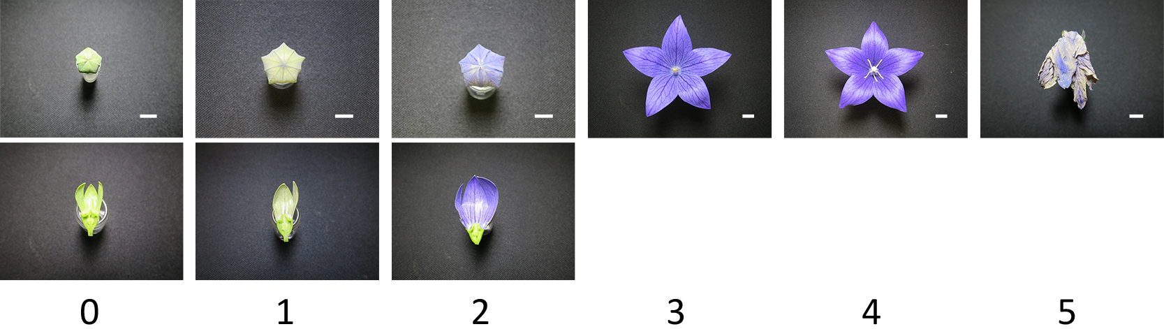
Flower developmental stages. Stage 0, green bud; Stage 1, green bud with blue-colored veins; Stage 2, bluish-purple colored bud; Stage 3, open flower with unopened anthers; Stage 4, open flower with opened anthers; Stage 5, withered flower. Bar = 10 mm. The lower photos of stage 0, 1, and 2 show the adaxial side of the petal.

Delphinidin 3-rutinoside-7-glucoside and platyconin content and the pH of the petal saps in flower developmental stages of P. grandiflorus.
The petals in stage 1 were not purple or violet except in the veins despite the fact that a good amount of delphinidin 3-rutinoside-7-glucoside had accumulated. When the petals of stage 1 were dried at 45°C, a dull blue-black color that was thought to be derived from delphinidin 3-rutinoside-7-glucoside appeared (Fig. S4). Anthocyanins can change to colored forms and colorless forms by various processes such as acid-base, water addition-elimination, tautomerization, and isomerization (Dangles and Fenger, 2018; Pina, 2014). If delphinidin 3-rutinoside-7-glucoside exists in colored forms, its quinonoidal forms (neutral quinonoid base and anionic quinonoid base) will increase under around pH 6.0 and produce a purple or violet color. Another possibility is that delphinidin 3-rutinoside-7-glucoside is present in some colorless state in the petals at stage 1 rather than in colored forms. When flowers opened, delphinidin 3-rutinoside-7-glucoside was only slightly detected and platyconin was dominant, suggesting that delphinidin 3-rutinoside-7-glucoside is efficiently modified to platyconin without any wastage. Delphinidin 3-rutinoside-7-glucoside may be present bound to the modification enzyme for subsequent modification without being released. An example of white flower color despite the presence of delphinidin 3-rutinoside-7-glucoside was reported in some white flowers of delphinium (Hashimoto et al., 2000). Delphinium is also a plant that biosynthesizes the 7-position polyacylated anthocyanins. Anthocyanins are well-known as “flower dyes”. Cases in which anthocyanins are in an invisible or hard-to-see state are interesting. Whether these examples in flower colors also involve other anthocyanins or if they are specific to delphinidin 3-rutinoside-7-glucoside is an interesting subject for further study.