抄録
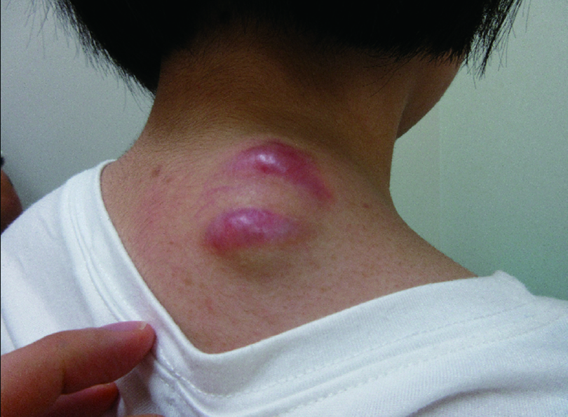
The patient was a 16-year-old girl. She noticed a gradual increase in the size of a skin mass on the right posterior neck and observed redness 1 month prior to visiting our department. The mass was 4 cm in size, dome-shaped, and on the right side of the neck. Ultrasound images showed a well-defined oval heterogeneous isoechoic mass. The interior was rich in blood flow with some coarse calcifications. Magnetic resonance imaging showed uniform hypointensity on T1-weighted images and variable hypointensity with hyperintensity in some superficial layers on T2-weighted images. On the basis of these findings, a solitary fibrous tumor was suspected. The tumor was resected at the proximal margin under general anesthesia. The mass was erythematous, overlying and partially adherent to the fascia. The mass was diagnosed as a calcified epithelioma.
Solitary fibrous tumors are histologically fibroblastic mesenchymal neoplasms, which include hemangiopericytomas. Generally, these tumors arise within the thoracic cavity, but few cases have been reported in the skin and subcutaneous soft tissue. We encountered a calcified epithelioma that we suspected to be a solitary fibrous tumor on imaging. Because similar cases are rare, we report this case along with a literature review.