International Journal of Surgical Wound Care
Online ISSN : 2435-2128
4 巻, 1 号
選択された号の論文の6件中1~6を表示しています
Original Article
  • Masaki Fujioka, Kiyoko Fukui, Kentaro Yoshino, Marie Idemitsu
    2023 年 4 巻 1 号 p. 1-5
    発行日: 2023年
    公開日: 2023/03/01
    ジャーナル フリー
    Introduction: In general, exposed bone wounds are covered with flaps. If the wound is small, skin grafting with perifascial areolar tissue can be performed for resurfacing; however, it is not suitable for large bone-exposed wounds exceeding 5 cm in length.
    Methods: Six patients underwent skin grafting for bone-exposed wounds at our medical center between 2014 and 2020. In all cases, a suitable local flap donor site could not be found around the wound, and a suitable donor vessel to anastomose the free flap was difficult to find. After a sequestrectomy, slit artificial dermis was applied to the wounds, and basic fibroblast growth factor was sprayed. Ointment-impregnated gauze was applied to the wounds. After confirming the creation of a suitable wound bed, skin grafting was performed.
    Results: In all cases, after skin grafting, the exposed bone wounds were closed. Four cases were due to extensive burns, one was due to malignant tumor resection, and one was due to exposed bone after injury. For burn injuries, suitable skin flap donors were difficult to find. Three cases of bone exposure involved the skull, and the rest involved the anterior surface of the tibia. Four cases required split-thickness skin grafts (including two mesh skin grafts), and two required full-thickness skin grafts. Covering a wound after skin grafting took 3 weeks to 3 months (median, 1 month), which is much longer than the time taken when using a skin flap.
    Conclusion: Even for exposed bone wounds with poor blood circulation, a good wound bed can be created by the combination of artificial dermis and bFGF, and wound closure can be achieved by skin grafting. Bone-exposed wounds should be closed early using a skin flap. However, when the patient’s general condition is poor or when temporary closure is difficult due to the unavailability of a suitable donor, wound closure by skin grafting is a simple and useful treatment option, although it takes a long time.
    Case 2. Skin grafts for exposed skull wounds due to burn injury. Fullsize Image
    (a) The patient had deep dermal burns > 45% of the total body surface area (TBSA) to the head, neck, anterior chest, and both upper extremities. The photograph was taken 1 and a half months after the injury, showing an 18 × 12 cm skull-exposed wound on the back of the head. (b) First surgery was performed 1 month after the injury. After sequestrectomy of the necrotic skull cortex, the artificial dermis was applied. (c) Second surgery was performed 1 and half months after the injury. The initial bone-exposed wound was a mixture of granulationdeveloped and bone-exposed parts. (d) After re-sequestrectomy of the part of the skull cortex not covered with granulation, the artificial dermis was applied again. A patch graft was applied to the part where the granulation tissue was formed. (e) Photograph taken 3 months after the injury. The exposed skull surface is mostly covered with granulation. (f) View of the third resurfacing surgery 3 months after the injury, showing the mesh graft applied on the new granulation. (g) Photograph taken at 6 months post-injury showing the exposed skull completely resurfaced with skin grafts.
Case Reports
  • A Rare Tumor Case Report
    Miho Baba, Yushi Suzuki, Junji Takano
    2023 年 4 巻 1 号 p. 6-11
    発行日: 2023年
    公開日: 2023/03/01
    ジャーナル フリー
    Solitary fibrous tumors (SFTs) are caused by the proliferation of mesenchymal cells. Facial SFTs are rare. Resection scar (of a subcutaneous tumor in the buccal area) is distinctive if it is located at the center of the cheek. To avoid this complication, we resected the lesion using a preauricular incision to approach the superficial musculoaponeurotic system, an approach similar to that used during rhytidectomy (face-lift). A 42-year-old woman underwent tumor resection using this approach. Eight months after, no tumor recurrence was observed. Additionally, no scarring or facial nerve paralysis was observed. Immunostaining of the tumor cells revealed CD34-positive, Bcl-2-positive, STAT6-positive, and some SMA-positive cells, and SFT was diagnosed. This case falls into the low-risk SFT group. Therefore, careful follow-up is necessary for SFTs because they have a high risk of recurrence and local metastasis.
    Operative findings. Fullsize Image
    (a), (b) A tumor found at the central right cheek. (c) Dissection above the SMAS and approach to the tumor. (d) Complete resection of the tumor could be achieved. (e), (f) Operation had good cosmetic outcomes. SMAS: superficial musculo-aponeurotic system.
  • Takayuki Hirao
    2023 年 4 巻 1 号 p. 12-16
    発行日: 2023年
    公開日: 2023/03/01
    ジャーナル フリー
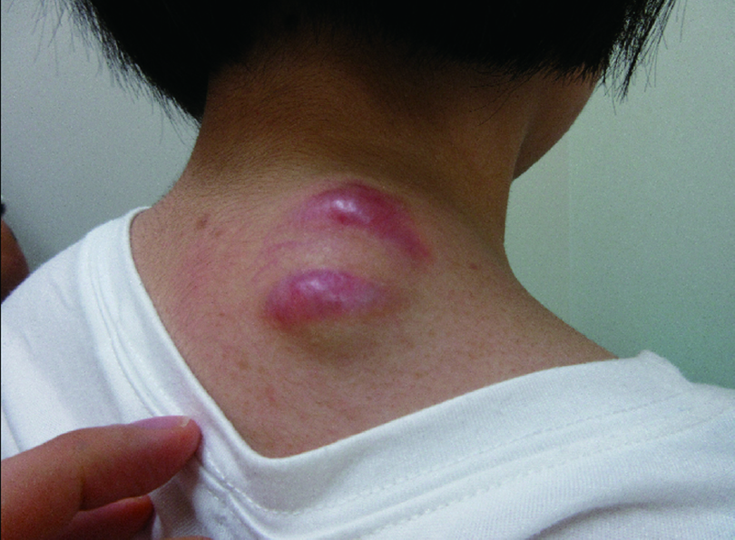
    The patient was a 16-year-old girl. She noticed a gradual increase in the size of a skin mass on the right posterior neck and observed redness 1 month prior to visiting our department. The mass was 4 cm in size, dome-shaped, and on the right side of the neck. Ultrasound images showed a well-defined oval heterogeneous isoechoic mass. The interior was rich in blood flow with some coarse calcifications. Magnetic resonance imaging showed uniform hypointensity on T1-weighted images and variable hypointensity with hyperintensity in some superficial layers on T2-weighted images. On the basis of these findings, a solitary fibrous tumor was suspected. The tumor was resected at the proximal margin under general anesthesia. The mass was erythematous, overlying and partially adherent to the fascia. The mass was diagnosed as a calcified epithelioma.
    Solitary fibrous tumors are histologically fibroblastic mesenchymal neoplasms, which include hemangiopericytomas. Generally, these tumors arise within the thoracic cavity, but few cases have been reported in the skin and subcutaneous soft tissue. We encountered a calcified epithelioma that we suspected to be a solitary fibrous tumor on imaging. Because similar cases are rare, we report this case along with a literature review.
    Picture of the tumor at the first visit to the hospital. Fullsize Image
    A 4-cm sized, dome-shaped mass is seen on the right posterior neck, accompanied by thinning of the skin and erythema.
  • Masaki Fujioka, Kiyoko Fukui, Kentaro Yoshino, Miho Noguchi
    2023 年 4 巻 1 号 p. 17-21
    発行日: 2023年
    公開日: 2023/03/01
    ジャーナル フリー
    Gustilo-Anderson grade IIIB severe open fracture of the olecranon with skin defect require early wound covering to treat the fracture, but wound covering with simple topical flaps or skin grafts is often difficult due to surrounding skin damage and bone exposure. Consequently, it requires some kind of flap reconstruction. We presented 2 cases of treatment for exposed olecranon degloving trauma (Gustilo-Anderson grade IIIB), and discussed the selection of flaps for these complex injuries, based on the points of reflection on the complications. When injured by this type of high-energy trauma, the surgeon should be aware of the following two points. Firstly, the arterial perforator branch to the skin around the wound is torn by shear stress, and secondly, delayed arterial embolism can occur.
    Therefore, a flap should be obtained outside the zone of injury. Therefore, for forearm injuries, the flap should be raised from the upper arm, and for upper arm injuries, the flap should be raised from the forearm. A free flap is the choice for extensive injuries from the upper arm to the forearm.
    Case 1. Fullsize Image
    (a) Left elbow skin loss with exposed fractured bone is visualized. The upper arm was primarily exfoliated. (b) The wound defect was covered with a posterior interosseous flap. (c) Two days after reconstructive surgery, the posterior interosseous flap developed ischenia and necrosis. (d) Image of the flap 3 months after the second reconstructive surgery.
  • A Case Report with Literature Review
    Tomoya Kawabata, Mika Ikeda, Haruka Matsuzoe, Shungo Oka, Hiroaki Oham ...
    2023 年 4 巻 1 号 p. 22-28
    発行日: 2023年
    公開日: 2023/03/01
    ジャーナル フリー
    When pleural empyema is treated with open-window thoracostomy, only 35% of cases are successfully closed, requiring an average of 4.5 months of treatment until wound closure. Conventional treatments such as daily gauze replacement often result in insufficient re-expansion of the collapsed lung, and surgical procedures such as thoracoplasty, muscle flap transposition, and omentoplasty are often necessary. We report a case of acute pleural empyema secondary to coronavirus disease 2019 treated with negative-pressure wound therapy (NPWT). After a month of conservative treatment following open-window thoracostomy, NPWT was performed for three weeks, and the thoracostoma was completely closed with the expanded lung. Approximately one week later, epithelialization of the exposed pleura was completed, and the patient was discharged. Six months after healing, there was no recurrence of pleural empyema. In this case, NPWT resulted in good pulmonary expansion and likely shortened the healing time. Although NPWT is contraindicated in wounds with exposed organs, the use of a contact layer and appropriate negative-pressure settings ensured safe treatment in our patient. We report the results of this study, along with a review of the available literature.
    Three-dimensional reconstruction of the computed tomography images showing each lung lobe. Fullsize Image
    (a), (b) Day of the open window thoracostomy. Atelectasis is indicated by orange color. (c), (d) Postoperative day 10. (e), (f) Postoperative day 27 (4 days before initiating negative-pressure wound therapy [NPWT]). (g), (h) Postoperative day 45 (14 days after NPWT initiation).
Brief Communication
  • Hideharu Nakamura, Takaya Makiguchi, Yasuko Hasegawa, Yukie Yamatsu, K ...
    2023 年 4 巻 1 号 p. 29-33
    発行日: 2023年
    公開日: 2023/03/01
    ジャーナル フリー
    Inguinal lymphorrhea is a complication of surgery, such as lymphadenectomy and revascularization. The inguinal region repeatedly flexes and extends, and lymphorrhea is often intractable. Persistence of this condition for a long period can lead to impaired wound healing and infection, which reduces patient’s quality of life and is a concern from the perspective of medical costs. Conservative treatment is often the first choice for managing lymphorrhea, but surgery is considered if the condition becomes intractable. In recent years, negative pressure wound therapy (NPWT) has been used as a conservative treatment, but there are few reports on the use of this method for inguinal lymphorrhea due to problems with management of air leaks and contamination. We used NPWT and NPWT with instillation and dwelling in four cases of inguinal lymphorrhea and obtained improved wound healing with the formation of granulation tissue in the early phase. Outcomes of these cases suggest that NPWT may be a useful treatment method for inguinal lymphorrhea.
    Case 1. Fullsize Image
    (a) Preoperative findings. (b) Wound debridement under general anesthesia. (c) After the start of NPWTi-d. Hydrocolloid wound dressing was attached to the movable part of the hip joint (black arrow). (d) At 10 months after treatment, there was no recurrence. NPWTi-d: NPWT with instillation and dwelling
feedback
Top