Regular Article
A note on the occurrence of Zoelucasa sablensis Nicholls, 2012 (Cercozoa, Incertae Sedis): Morphology and environment, recorded from the east coast of Scotland, North Sea
2023 年 55 巻 p. 1-5
詳細
2023 年 55 巻 p. 1-5
Zoelucasa sablensis is a small (under 22 µm) pyriform heterotrophic flagellate recorded from nearshore marine environments from the northern hemisphere, which have only been recorded in limited numbers. The paucity of further material, including the current lack of southern hemisphere examples, may be due to the small size of the Zoelucasa and the relative smaller numbers of investigation into relevant environments in the southern hemisphere. The current paper illustrates and discusses the occurrence of Z. sablensis from the east coast of Scotland, North Sea; confirming the siliceous nature of the test, reinterpreting the microstructure of the scales and extending the known environmental range to brackish water conditions.
Zoelucasa sablensis Nicholls, 2012 is a rare monospecific marine protist, with a limited record of occurrence (Nicholls, 2012; Prokina et al., 2017; 2018). Z. sablensis is thought to represent a heterotrophic flagellate, from the phylum Cercozoa, possibly belonging to Class Imbricatea, but classified as Incertae Sedis (Nicholls, 2012). Originally recorded from near-shore benthic environments from the Pacific and Atlantic coasts of Canada (Nicholls, 2012), its only other apparent record appears to be from the Black Sea on the coast of Crimea near Sevastupol (Prokina et al., 2017; 2018). Here we describe examples of Z. sablensis from the east coast of Scotland, North Sea, from littoral beach sands and brackish stream water, as well as discussing aspects of the round scales that cover the surface of the test.
Two spatially related samples were collected from Pease Bay (The Borders, east coast of Scotland), 55°55′51.7″N and 2°19′59.4″W (Fig. 1). A water sample from a stream, Cockburnspath Burn, on the beach at Pease Bay, and a medium grain size, quartz dominated, moist beach sand containing water menisci (Fig. 2) collected from the top 100 mm of sand, between low- and high-tide marks, approximately 30 m south of Cockburnspath Burn. Testate amoebae and other micro-fauna were separated from the sand by stirring in water and decanting the supernatant liquid, and both samples were vacuum filtered and processed using the technique described in Buckman & Krivtsov (2022). Collected material examined by scanning electron microscopy (SEM), using a Quanta 650 FEG SEM equipped with an Oxford Instruments energy-dispersive X-ray (EDX) system. Salinity values of both samples were measured using a HACH Pocket Pro+ Multi 2, conductivity monitor, with results in ppt (‰).

A) Green pin marks burn sample, B) yellow pin marks beach sample. HWM=high water mark, LWM=low water mark, asterisk=major river entrance. Arrow in inset marks approximate position of Pease Bay.
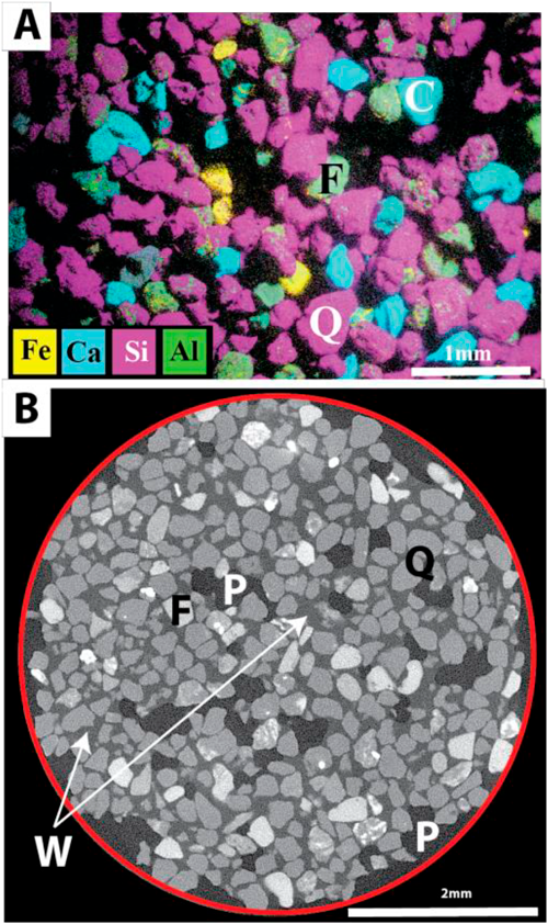
A) Scanning electron microscopy energy dispersive X-ray (SEM-EDX) map of the beach sand. Medium grain size (250–500 µm). Predominantly quartz. Grains (Q)—magenta, carbonate particles (C)—blue, and feldspar (F)—green. B) X-ray tomography (XRT) slice through the beach sand. Key as in (A) with the addition of pore (P) and water (W). Note that water forms a meniscus between grains, as viewed in the sands moist collected state—leaving ample environment for microorganisms including testate amoebae.

A), C) Secondary electron (SE) and backscattered electron (BSE) images of the same area, illustrating overall test morphology, arrangement of plates, and plate surface ornamentation. B) SE enlargement of one of the scales in (A), showing granular texture. D) and E) BSE images, with terminal distal plate (arrowed), and surface plate ornament. Note in D) linear depression structure on surface of plate (dotted lines). F) SE image of partially disarticulated specimen, illustrating the lightly cemented nature of individual plates. G) SE image of test with potential remnants of flagella (arrowed). H) BSE image, illustrating pair of protruding aperture scales. A)–D) and H) from beach sand, E)–G) from burn water flowing on beach.

A), B) from beach burn water A) whole test, with white box illustrating the area in (B), which shows granular sub-micron texture and smooth margin. C) Enlargement of original transmission electron microscopy (TEM) illustration from Nicholls (2012), showing meshwork of electron dense material and smoother marginal rim. D) BSE image of individual from beach deposits, illustrating the brittle nature of the siliceous scales, and granular texture of scales.
Four individuals from the Cockburnspath Burn (water salinity of 1‰), and four from the beach sand (water salinity of 21‰), collected during October 2019. Zoelucasa sablensis is not particularly common, with testate amoebae dominating, such as Trinema in the stream waters, and Psammonobiotus and Micramphora in the beach sands.
DescriptionSmall, pyriform tests, 8 to 12 µm in length by 8 to 5 µm wide, with a 2 to 3 µm aperture. Surface covered by thin (< 100 nm), overlapping, 3 to 5 µm circular curved scales (Figs. 3 and 4). Surface of scales have a micro-indented (granular) external surface, and typically a smooth rim (Figs. 3A, B, H and 4A, B, D). Scales loosely held together, with test often observed to be falling apart (Fig. 3F, G). Scales brittle and easily fractured (Fig. 4D). One possible partial flagella observed (Fig. 3G). EDX analysis indicates that scales are composed of silicon and oxygen (Fig. 5), with an approximate ratio of 1 : 2.

A), B) for the whole test of Zoelucasa sablensis, indicating concentration of silicon and oxygen respectively, and C) spectrum analysis for part of a plate, indicating siliceous composition (SiO2).
In terms of general morphology and scale shape, the current material compares well with previous illustrations of Zoelucasa sablensis by Nicholls (2012) and Prokina et al. (2017). As can be seen from Table 1, the Scottish material compares favourably with all three metrics recorded by Prokina et al. (2017), and both works are lower in value to measurements made by Nicholls (2012); the latter being up to twice the size of subsequent discoveries of the taxa, with the exception of scale diameter which are closer in size. The size differences may represent local geographic, environmental factors, or even be grounds for a new species. However, given the current small numbers, size differences are here not currently considered to be of significance in terms of species diagnosis. Both Nicholls (2012) and Prokina et al. (2017) noted the occurrence of a pair of flagellae, one short and one long. Examination of the Scottish material lacked any clear flagellae, with the possible occurrence in a single case of one flagellum (Fig. 3G).
| Length (µm) | Width (µm) | Scale diameter (µm) | |
|---|---|---|---|
| Nicholls (2012) | 14–22 | 9–13 | 3–6 |
| Prokina et al. (2017) | 8–10 | 5–7 | 3–4 |
| Herein | 8–12 | 5–8 | 3–5 |
This genus and species was originally described from the west and east coast of Canada, with a global marine distribution expected within the northern hemisphere (Nicholls, 2012). Subsequently, noted and illustrated from the Black Sea of Crimea (Prokina et al., 2017; 2018). A widespread geographical distribution within the northern hemisphere oceans is further supported by the current occurrence on the east coast of Scotland, from the North Sea. It is noteworthy that to date no examples of Zoelucasa have been recorded from the southern hemisphere. This may in part be due to an apparent bias of study of supralittoral, littoral and nearshore marine environments to areas within Europe, and in particular the Black Sea, Altantic coast of Europe and Baltic Sea (e.g., Golemansky 1974; 1983; 1992). In addition, the small size of Zoelucasa, and particularly examples illustrated by Prokina et al. (2017) and herein, may help explain the small number of citations on the occurrence of Zoelucasa. Further investigation of suitable environments worldwide may well expand the current geographical range and abundance of the genus. However, it is noteworthy that investigation of other littoral environments from around the UK by the current author have not solicited further occurrences of the genus.
Scale composition, morphology and ornamentNicholls (2012) deduced that the scales of Z. sablensis were likely to be siliceous in nature, but could not provide definitive evidence, other than their insolubility within sulphuric and nitric acid. Analysis of the current material by EDX analysis conclusively illustrates that these scales are siliceous (Fig. 5), and therefore similar to many testate amoebae that construct siliceous scales (idiosomes). The current work also confirms the delicate nature of the scales and their curved “shield-like” morphology. Nicholls (2012) described Z. sablensis scales as having a smooth unornamented exterior, and illustrated an internal meshwork of electron dense material, with a marginal plain margin (Fig. 4C). SEM imaging of the current material at 5 kV clearly indicate the marginal rim (Figs. 3A, B and 4A, B), but unlike Nicholls (2012) illustrates a fine grained granular and pitted exterior surface to the scales (Figs. 3 and 4B, D). These observations suggest that the scales have a microgranular texture, similar to that apparent within the major scales of Trinema (Fig. 6: 5 kV), which can also have a plain marginal zone. This brings into question the model of Nicholls (2012) of a proximal and distal surface sandwiching an electron dense meshwork—replacing the model with a simpler microgranular and microporous secreted siliceous scale structure, comprising just the one layer, with a more homogeneous plain outer rim.

Illustrating microporosity/microgranular texture at centre of round scales and plain margin, similar to scales of Zoelucasa.
The occurrence of Zoelucasa sablensis from material with recorded salinities of 1 and 21‰ respectively, implies a much broader salinity range than that of 31 to 33‰ recorded by Nicholls (2012). The known range of salinity tolerance for Z. sablensis can now be tentatively extended from 1–33‰, and with more assurance to that of 21–33‰, or 18–33‰ taking into account the work of Prokina et al. (2018).
The original description of this species recorded sampling from nearshore beach sand and water (Nicholls, 2012), and that for Prokina et al. (2018) is from supralittoral Miocene clays. The current samples were obtained from littoral medium grained siliciclastic dominated beach sands (between low- and high-tide levels) and stream water on the beach (Cockburnspath Burn). Therefore, both the current work and that of Prokina et al. (2017; 2018) support an increase in known environmental occurrence from supralittoral, through littoral to nearshore beach environments. Given their small size and occurrence within moist beach sand, Zoelucasa sablensis from the current study can be interpreted as representing at least in part a psammobiotic mode-of-life, although a motile phase swimming within water as noted by Nicholls (2012) cannot be excluded, and is worthy of future research.
The current work further confirms the more widespread occurrence of Zoelucasa sablensis, with new records from the east coast of Scotland, North Sea; with all records of the species still confined to northern hemisphere oceans. The siliceous composition of the scales that form the test of Z. sablensis have been confirmed beyond doubt through SEM based EDX analysis. Examination of scale morphology and ornamentation indicates that the model proposed by Nicholls (2012) is too complex, and are better explained as a simpler microgranular and microporous single layered siliceous scales. Environment of habitation is extended from 18–33‰ (supralittoral to near-shore benthic) to that of a slightly brackish (1‰) flowing stream water habitat, with further support for a psammobiotic mode-of-life within medium grained siliciclastic dominated beach sands in the littoral zone, as well as a free-swimming mode.
Special thanks to the Centre for Environmental Scanning Electron Microscopy (CESEM), and the X-ray tomography facilities at the Institute of GeoEnergy Engineering, Heriot-Watt University for access to SEM and XRT.