Abstract
Factors influencing on in vitro evaluation of UV protecting ability of sunscreens were analyzed. It was found that any factors making the sunscreen layer spatially inhomogeneous, such as directional viscous fingering during the sunscreen application, dewetting of applied sunscreen layer, and the surface roughness of the standard PMMA plate, alter the UV transmittance. New application procedure and new type of flat hydrophilic plate were developed for inhibiting the generation of spatial inhomogeneity in applied sunscreen layer. The method created by the combination of these newly developed technologies succeeded in providing reliable and reproducible in vitro evaluation of UV protecting ability.
1 Introduction
For the evaluation of UV protecting ability of sunscreens such as sun protection factor (SPF)1) and UVA protection factor (UVA-PF)2), in vivo methods that require UV light irradiation on human’s back have been employed. Concerns for the establishment of in vitro evaluation method of UV protecting ability of sunscreens have been increased, since it gives results more quickly, is less expensive and is more ethical3),4),5),6),7),8),9),10). Attempts have been made to develop reliable and reproducible in vitro evaluation method of UV protection ability11),12),13),14),15),16). However, in 2010, Rohr et al. pointed out the list of problems for the developed in vitro evaluation method and explained that further work is necessary for the establishment of satisfactory method17). Although ISO method of in vitro UVA-PF evaluation was approved in 201218), no worldwide standard in vitro method has been established for the determination of SPF up to date. In addition, some countries, such as Japan, Korea, and Taiwan, do not admit indicating in vitro UVA-PF value for selling sunscreens, since there are many problems in terms of its repeatability and reliability.
In the in vitro UVA-PF evaluation based on the ISO method18) and the determination of critical wavelength for “Broad Spectrum” approval based on US FDA rule19), recommended amount of sunscreen samples are applied on a cosmetic standard PMMA UV evaluation plate to measure the UV transmission of the applied sample sunscreen layer using SPF analyzer. However, spontaneous formation of spatially periodic pattern is usually observed during the application of viscous liquid on a solid substrate. Ilya Prigogine was awarded the 1977 Nobel Prize in Chemistry by his achievement on the development of the concept of dissipative structure, i.e. self-organization in far-from equilibrium systems due to the growth of fluctuations20),21),22). Application of viscous liquid usually makes the system in far-from-equilibrium condition, since mechanical energy is constantly supply to the applicator to move it to apply the liquid. Fingering instability called directional viscous fingering is thus usually generated as the result of the growth of morphological fluctuation in interface to generate spatially periodic stripe pattern23),24),25),26),27),28),29),30). Although we began to study the directional viscous fingering during the application of pseudo-sunscreen sample for the fabrication of water-repellant surface31),32),33), further studies on directional viscous fingering were carried out for analyzing its influence on the in vitro evaluation of UV protecting ability34),35),36), since one can simply understand that spatial inhomogeneity in applied sunscreen layer alters the UV absorbance. In addition, we have studied dewetting as another phenomenon that makes thin liquid film spatially inhomogeneous. Dewetting is rupture of thin liquid film when spreading coefficient change the sign from positive to negative. The concept of spinodal dewetting explain how thin liquid film becomes unstable to morphological fluctuation in far-from-equilibrium condition to lead to spatially periodic dewetting structure37),38),39),40),41),42),43),44),45),46),47). Although we began to study the dewetting for the fabrication of water-repellant surface31),33),48) similar to the viscous fingering, its influence on the in vitro evaluation of UV protecting ability was also analyzed49),50).
It was anticipated that not only these far-from-equilibrium phenomena spontaneously generated in applied sunscreen layers but also any factors to make the applied layer spatially inhomogeneous vary the value of in vitro UV protecting ability. The roughened cosmetic standard PMMA UV evaluation plates should be used for the in vitro evaluation of UVA-PF by ISO 2444318) and critical wavelength for the Broad Spectrum approval by US FDA’s Final Sunscreen Rule19). We have studied the influence of surface roughness of the cosmetic standard PMMA UV evaluation plate on the value of in vitro UV protecting ability51), since it surely affects the spatial inhomogeneity in applied sunscreen layer.
In addition to those studies to verify the problems on in vitro evaluation method of UV protecting ability of sunscreens34),35),36),48),49),50),51), attempts were made to establish reliable and reproducible in vitro evaluation method of UV protecting ability of sunscreens52). New type of plate was developed by coating the super-hydrophilic plate by polysaccharide. Here, we have encountered another type of far-from-equilibrium phenomenon. In the process of drying the aqueous solution of polysaccharide on the plate, special attention had to be paid for inhibiting the generation of Marangoni convection to lead to the spatial inhomogeneity in the resulting polysaccharide layer53). New method utilizing this new plate was proposed, and the validity of the new method was verified by ring test.
2 Generation of Viscous Fingering during the Sunscreen Application and Its Influence on the in vitro Evaluation of UV Protecting Ability
Fingering instability is one example of spontaneous pattern formation in far-from-equilibrium systems20),21),22). Fingering patterns are generated in moving interface of viscous liquid as the results of the growth of morphological fluctuations, and this phenomenon is called viscous fingering54),55),56),57). Stripe patterns are spontaneously formed when a thin liquid film is produced by passing it through a small gap23),24),25),26),27) or when two pieces of adhesive tapes that are stuck together are peeled28),29),30), and these pattern formations are called directional viscous fingering. Since the growth rate of the morphological fluctuation is a function of its wavelength, the stripe patterns formed by this process are usually spatially periodic, and the characteristic length of the spatial periodicity corresponds to the wavelength of the fastest growing fluctuation23). We have found that the stripe pattern was spontaneously formed by the directional viscous fingering during the application of viscous fluid composed of ingredients of sunscreens on a glass plate and analyzed the influence of application condition on the geometry of the surface and its influence on the water-repellant property31),32),33).
When the viscous fingering is generated during the application of sample sunscreen, thickness of sunscreen layer becomes spatially inhomogeneous, and it is represented as a function of the position, h(x), where x is the position (Fig. 1a). In this case, intensity of the UV light passed through the sunscreen layer, I, and the corresponding net UV absorbance, A, are represented as
(1)
(2)
in which, I0, l, ε, and c are intensity of incident light, characteristic length of the spatial periodicity, absorption coefficient, and concentration of UV absorber, respectively. Value of A of the patterned sunscreen layer possessing square, triangular, and trigonometric curve cross section (Figs. 1b-1d) is theoretically calculated as
(3)
(4)
(5)
in which d0 and 2d are the average thickness and the distance between the top or bottom, respectively. Calculations were carried out by setting α=0.15 μm–1 and d0=20 μm. Significant decrease in UV absorbance was observed as increasing the distance between the top and bottom of the applied sunscreen layer in each case (Fig. 2).

Experimental analyses were carried out to verify the validity of the theoretical prediction on the relation between the geometry of applied sunscreen layer and the UV absorbance. We have succeeded in finding the procedure to form flat applied sunscreen layer34),35). Using four-side applicator for the sample application was found to inhibit the spontaneous formation of spatially periodic stripe pattern in sunscreen layer. Clear solution type pseudo-sunscreen sample was prepared and used for the evaluation of influence of pure geometric factor of the applied sample layer on the UV absorbance34),36). UV absorber liquid was prepared by mixing UVA absorber, diethylamino hydroxybenzoyl hexyl benzoate (DHHB) and butyl methoxydibenzoylmethane (BMDM), UVB absorber, 2-ethylhexyl methoxycinnamate (EHMC), and cosmetic oil, 2-ethylhexyl ester of oligomer of hydroxystearic acid (EH-O-HAS), to make the weight ratio at DHHB : BMDM : EHMC : EH-O-HAS=0.220 : 0.214 : 0.470 : 0.096, and it was dissolved in iso-propanol solution of acrylsilicone resin to prepare pseudo-sunscreen solution sample. Weight ratio of the pseudo-sunscreen solution sample was UV absorber liquid : acrylsilicone resin : iso-propanol=0.030 : 0.485 : 0.485. Linear motor coater was used to control the application velocity, and the pseudo-sunscreen sample solution was applied on a quartz plate using block applicator and four-sided applicator. Stripe pattern was formed on the applied layer when the block applicator was used, while flat applied layer was formed when the four-sided applicator was used34),36) (Fig. 3). UV absorbance of these applied sample layer was measured using SPF analyzer. Although the average thickness of both applied pseudo-sunscreen layer was almost identical with each other at 11.8 and 11.5 μm for the patterned and flat layer, respectively, relatively large difference was observed in their UV absorbance34),36) (Fig. 4). To verify that the difference in UV absorbance of the pseudo-sunscreen layers was exclusively due to their morphologies, simulations for predicting the value of A at each wavelength was carried out. The charts of surface profile measurement were pixelized to determine the thickness of the layer at each position. Thickness of the layer at N positions, hn (n:1–N), were determined from the chart obtained by the surface profile measurement. The local UV transmittance, In (n:1–N), were then calculated to predict the net UV absorbance, A, using the equation
(6)
in which AFlat is the UV absorbance of the flat pseudo-sunscreen layer having the thickness of h0. The results of the simulation correlated well to the experimental results34),36) (Fig. 4).

In 2011, US FDA launched a new rule using the term “Broad Spectrum” for labeling the sunscreens. Experimental procedure for the measurement of in vitro UVA-PF18) is required for obtaining “Broad Spectrum” approval19). Sample sunscreen is applied on the roughened cosmetic standard PMMA UV evaluation plate, and the UV absorbance of the sunscreen layer is measured at each wavelength from 290 to 400 nm. Critical wavelength, λC, is calculated using equation
(7)
Thus, the critical wavelength is identified as the wavelength at which the integral of the spectral absorbance from 290 nm reaches 90 percent of the integral over the UV spectrum from 290 nm to 400 nm. The sunscreens possessing critical wavelength of 370 nm or greater are classified in “Broad Spectrum” protection sunscreens, since they can be regarded as the sunscreens exhibiting UV protecting ability in broad range of UV wavelength. However, UV absorbance measured by the SPF analyzer is a series of discrete data evaluated at each wavelength of the integer. Thus, the integral equation (7) should be replaced by the summation formula
(8)
The value of ∑λλ=290A (λ) / ∑400λ=290 A (λ)) was then calculated for the UV absorbance data in Fig. 4 to find the wavelength at which the value was the closest to 0.9. It was observed that degree of declining UV absorbance, A, by the generation of spatial inhomogeneity in the applied pseudo-sunscreen layer is larger at the wavelength showing large value of AFlat (Fig. 4), and the validity of this dependence of degree of declining A on the value of AFlat was mathematically verified in our study36). The value of λC of the flat pseudo-sunscreen layer, i.e. the intrinsic critical wavelength of the pseudo-sunscreen, was determined to be 369 nm, while that of the patterned pseudo-sunscreen layer was 370 nm (Fig. 5). It is the morphology of the pseudo-sunscreen layer that leads to the discrepancy between the critical wavelength, since it was also calculated to be 370 nm by the simulation using equation (6) for determining A at each wavelength. Thus, it can be noted that the pseudo-sunscreen that is not essentially approved to be “Broad Spectrum” was approved to be “Broad Spectrum” by the spontaneous pattern formation by the viscous fingering during its application36).

3 Influence of the Surface Roughness of Cosmetic Standard PMMA UV Evaluation Plate on the in vitro Evaluation of UV Protecting Ability
Not only the stripe pattern of applied sunscreen layer generated by viscous fingering during the application, but also any factor that makes the sunscreen layer spatially inhomogeneous should alter the UV protecting ability. For the in vitro evaluation of UVA-PF by ISO 2444318) and critical wavelength for the “Broad Spectrum” approval by US FDA’s Final Sunscreen Rule19), it is required to apply sample sunscreens on the roughened cosmetic standard PMMA UV evaluation plate. The surface roughness of the plate should make the applied sunscreen layer spatially inhomogeneous to alter the UV protecting ability. In addition, some groups have suggested the relation between the surface roughness of the cosmetic standard PMMA UV evaluation plates and the low reproducibility of the in vitro evaluations of UV protection ability58),59). However, no detailed regulation was established for the surface roughness of the standard plate. This situation motivated us to study the influence of surface roughness of the cosmetic standard PMMA UV evaluation plate on the in vitro evaluation of UV protecting ability. Three types of cosmetic standard PMMA UV evaluation plates employed in our study were Helioplates HD6 (Sa=6 μm) (HelioScreen), HD6, and ISO Plates ISO2 and ISO5 (Ra=2 and 5 μm) (Solar Light Company, Inc.). All plates are square of one side of 5 cm. Surface roughness of three types of plates were analyzed, and its influence on the in vitro evaluation of UV protecting ability of sunscreens was studied51).
Acrylsilicone resin was dissolved in a mixed solvent of iso-propanol and decamethylcyclopentasiloxane, and the solution thus obtained was slowly dropwised on HD6, ISO2 and ISO5 plate. The plates were broken into 3 pieces after the completion of the solvent evaporation, and the broken sample plate was put on a SEM sample holder to observe the cross section. Distribution of silicone atom in the cross section was evaluated by SEM/EDS mapping images to analyze the infiltration behavior of acrylsilicone resin solution into the inside of these plates. SEM/EDS mapping images of the cross section represent the situation of acrylsilicone resin deposited on each cosmetic standard PMMA UV evaluation plate (Fig. 6). Yellow part in EDS mapping image is the region consisting of high density of Si atom, and they indicates the regions in which acrylsilicone resin solution soaked, since no Si atom is incorporated in PMMA molecules. It was found that these plates possess small cracks in addition to the surface roughness51).

To investigate the influence of surface roughness of the cosmetic standard PMMA UV evaluation plates on the in vitro evaluation of UV protecting ability, clear solution type pseudo-sunscreen samples were dropwised on these plates, and UV absorbance was measured using SPF analyzer after the completion of the evaporation of the solvent. Three types of pseudo-sunscreen solutions, 1, 2, and 3, were prepared by dissolving UV absorber liquid having the composition of DHHB : BMDM : EHMC : EH-O-HAS=0.220 : 0.214 : 0.470 : 0.096 into hexane solution of acrylsilicone resin. Content of acrylsilicone resin was the only difference between three types of pseudo-sunscreen solution samples (Table 1). Although all samples 1, 2, and 3 contained identical amount of UV absorbers, their UV absorbance was significantly different with each other for all cosmetic standard roughened PMMA evaluation plates (Fig. 7). Sample 3 did not contain acrylsilicone resin, and the UV absorbers were more likely to localize at the vicinity of the bottom of the hollows of the roughened surface. The transmitted UV intensity was thus not decreased at the rest of the part of the plate to scarcely increase net UV absorbance. On the other hand, samples 1 and 2 contained acrylsilicone resin, and their deposition fill the hollows of the roughened surface. The UV absorbers were thus more likely to extend on relatively larger part of the roughened surface of the plate. The degree of spatial extension of the UV absorbers increased as increasing the amount of acrylsilicone resin. Sample 1 containing the largest amount of acrylsilicone resin exhibited the highest net UV absorbance for all cosmetic standard roughened PMMA evaluation plates. All the cosmetic standard PMMA UV evaluation plates without the deposition of pseudo-sunscreen samples exhibited a nonzero value of UV absorbance51). However, UV absorption by PMMA was scarce. In these cases, decline in UV intensity upon passing through the plates was mostly due to diffuse reflection at the roughened surface. Relatively large value of A at higher wavelength when sample 3 was deposited was thus mostly due to the diffuse reflection, since UV absorbers used in this study does not show significant UV absorption at these higher wavelength. The value of A at the higher wavelength became closed to zero when sample 1 or 2 was deposited on all cosmetic standard PMMA UV evaluation plate, since filling the hollows of the roughened surface by the sample contained acrylsilicone resin decreased diffuse reflection. In all cases, thanks to the surface roughness of the cosmetic standard PMMA UV evaluation plate, the critical wavelength, λC, was equal to or larger than 370 nm, and astonishingly large λC was obtained when sample 3 was employed. The results of this study suggest the inappropriateness of employing surface roughened plate as the substrate for the in vitro evaluation of UV protecting ability of sunscreens. If the surface roughness is to correlate the in vitro value with the corresponding in vivo value, the surface structure should be a precise mimic of human skin and determined by standards and regulations.
Table 1
Pseudo-sunscreen solutions prepared by dissolving acrylsilicone resin and UV absorber liquid containing DHHB, BMDM, EHMC, and EH-O-HAS51). (The final, published version of this article is available at https://doi.org/10.5650/jos.ess18207. Copyright©2019 Japan Oil Chemists’ Society, Tokyo, Japan.)

4 Preparation of New Hydrophilic Plate for Hydrophilic Sunscreens
Another problem for the cosmetic standard PMMA UV evaluation plate is its wettability to the hydrophilic sunscreen samples such as O/W emulsion sunscreens. Although hydrophilic sunscreens have become popular from their smooth and light texture60),61),62), they cannot wet the hydrophobic PMMA surface. Dewetting, a rupture of thin liquid film on a solid substrate to generate holes, occurs when the spreading coefficient, S=ΓSG– (ΓSL+ΓLG), in which ΓSG, ΓSL, and ΓLG are interfacial tension between solid-gas, solid-liquid, and liquid-gas interface, respectively, changes its sign from positive to negative37),38),39),40),41),42),43),44),45),46),47). Hydrophilic sunscreens usually possess higher ΓSL for their interface to the hydrophobic PMMA surface in addition to its higher ΓLG to make the sign of spreading coefficient negative.
We have developed two types of hydrophilic substrate, super-hydrophilic plate (SHP plate)49),50) and hydroxyalkyl cellulose coated plate (HCC plate)52),53). Contact angle of water on a quartz plate was in a range between 70 to 80 deg, and SHP plate was prepared by Corona-discharge treatment on a flat square quartz plate using Laboratory Corona Treater (BD-20AC, Electro-Technic Products). This treatment succeeded in making the surface super-hydrophilic to have a contact angle of water at 0 deg. The contact angle of water, however, exponentially recovered to the initial value, and its half-life was around five days49),50). Therefore, attempts were made to prepare new type of hydrophilic substrate. Aqueous solution of hydroxyalkyl cellulose was spread on SHP plate to let water to be evaporated to prepare HCC plate. However, Marangoni convection is usually generated during the evaporation process of volatile solvent from the solution spread on a solid substrate to lead to the spatial non-uniformity such as Marangoni contraction63) and tears of wine64). In the case of evaporating water from the aqueous solution of hydroxyalkyl cellulose spread on the SHP plate, various drying patterns were found to be generated (Fig. 8)52). Thus, special care had to be taken for the preparation of flat and uniform film of hydroxyalkyl cellulose53). Surface roughness of HCC plate was found to be less than μm level except for some tiny cracks and bumps (Fig. 9)52). The contact angle of sessile drop of water on HCC plate was determined to be 44±2°, and the water composing the sessile drop gradually osmosed into the hydroxyalkyl cellulose film. Contrary to the super-hydrophilic plate whose half-life of the super-hydrophilicity is around five days49),50), the contact angle of sessile drop of water on the surface of HCC plate scarcely varied during six months of storage at room temperature (25°C) and in a refrigerator52).
Six sunscreen samples whose in vivo SPF have already been evaluated (Table 2) were employed for the in vitro SPF measurement, and they were applied on SHP plate, HCC plate, as well as flat PMMA plate (PMMA plate) using four-sided applicator to inhibit the spontaneous pattern formation by viscous fingering during their applications52). UV protecting ability of each six sample was evaluated by measuring the thickness and UV absorbance of each applied layer. UV absorbance of 10 μm thickness sunscreen layer and in vitro SPF was calculated using these values for each sample. Evaluation of the UV protecting ability was carried out for four time for each sample and each substrate, and the average was calculated. It was found that PMMA plate was not wetted by sample A and C (Fig. 10) and this character was well accordance with the fact that these samples do not wet the surface of the roughened cosmetic standard PMMA UV evaluation plate. On the other hand, no dewetting was observed in the layer of these samples applied on SHP and HCC plates. These samples exhibited much lower UV absorbance and in vitro SPF when they were applied on PMMA plate (Fig. 11). Both SHP and HCC plates were thus found to be appropriate for the in vitro SPF evaluation of these hydrophilic sunscreens52),53).

Table 2
Six types of sunscreen samples employed for the in vitro SPF evaluation52). (The final, published version of this article is available at https://doi.org/10.5650/jos.ess21266. Copyright©2022 Japan Oil Chemists’ Society, Tokyo, Japan.)

5 Development of New in vitro Evaluation Method of UV Protecting Ability of Sunscreens and Its Validity
It is very important for the standard test method that anyone can follow the instruction to carry out the experiments at anyplace to obtain similar results. In addition, time and labor for the experimenters are expected to be relieved. Twelve times experiments are required for each sample if four times measurements using three types of substrates are required to verify the validity of the test. We thus considered to establish a simplified procedure for the new in vitro SPF evaluation method including following three steps, i) selecting the most appropriate substrate for evaluating in vitro SPF from the three substrates, i.e. SHP, HCC, PMMA plate, by finding the substrate that recorded the highest in vitro SPF, ii) repeating three more times identical experiments using the most appropriate substrate, iii) deleting the highest value of in vitro SPF among the four times measurements and calculating average and standard deviation52). In order to verify the validity of this simplified in vitro SPF evaluation method, ring tests were carried out by three researchers at different laboratory in three organizations.
The values shown with color in Table 3 indicates the highest in vitro SPF evaluated using SHP, HCC, PMMA plate in the first step by three researchers. Number of choosing HCC plate among six samples was four, three, and five by Researcher 1, 2, and 3, respectively. Although Researcher 2, and 3 chose PMMA plate for sample B and D, respectively, no one chose it for sample A and C that did not wet the surface of PMMA plate. Coefficient of variation, i.e. the quotient of standard deviation by average, of in vitro SPF obtained by the test by three researchers was less than 0.35 in five out of six sunscreen samples (Table 4)52). However, linear relationship was not observed between in vivo SPF and in vitro SPF52). The samples exhibiting in vivo SPF ranging from 10 to 30 showed lower in vitro SPF values, while samples exhibiting in vivo SPF values higher than 50 showed higher in vitro SPF values. The value of in vitro SPF calculated by the prescribed equation increases proportional to the reciprocal of UV transmittance18), while there is no such physiological evidence that the in vivo SPF linearly increases as increasing the reciprocal of UV transmittance. This fact may thus be one candidate for generating the deviation from linear relation between in vivo SPF and in vitro SPF.
Table 3
First step of ring test for selecting the most appropriate substrate among the three substrates by three experimenters. Colored bold character number indicates the highest value of in vitro SPF for each sample. Additional three times experiments were conducted using the selected substrate52). (The final, published version of this article is available at https://doi.org/10.5650/jos.ess21266. Copyright©2022 Japan Oil Chemists’ Society, Tokyo, Japan.)
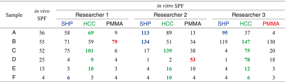
Table 4
Average (AVG), standard deviation (STD), and coefficient of variation (CV) of in vitro SPF of six sunscreen samples evaluated by three experimenters at different laboratory. Since the data of AVG and STD are the value rounding off one decimal place, CV does not exactly consistent with the value of quotient of STD divided by AVG52). (The final, published version of this article is available at https://doi.org/10.5650/jos.ess21266. Copyright©2022 Japan Oil Chemists’ Society, Tokyo, Japan.)

6 Conclusion
Concerns have been increased for the establishment of in vitro evaluation method of UV protecting ability of sunscreens, since it gives results more quickly, is less expensive and is more ethical. However, no worldwide standard in vitro method has been established, since there are many problems in terms of its reproducibility and reliability. This review article summarizes our recent studies for clarifying the problems on in vitro evaluation method of UV protecting ability of sunscreens and development of new evaluation method overcoming these problems (Fig. 12).
We have analyzed the factors that influence in vitro evaluation of UV protecting ability of sunscreens. For the in vitro evaluation of UV protecting ability, sample sunscreens are applied on a substrate to measure the UV transmission of the applied layer. It was clarified that any factors making the sunscreen layer spatially inhomogeneous alter the UV transmittance to lead to the lack of reproducibility and reliability.
Two far-from-equilibrium phenomena, directional viscous fingering during sample application on a substrate and spinodal dewetting generated in the applied layer, were determined as the factors to make the sunscreen layer spatially inhomogeneous. The former forms spatially periodic stripe patterns and the latter forms holes in the applied sunscreen layer. Not only theses far-from-equilibrium pattern formation but also the surface roughness of cosmetic standard PMMA UV evaluation plate was also determined as the factors to make the sunscreen layer spatially inhomogeneous. Generation of spatial inhomogeneity in the applied sunscreen layer lowers the UV protecting ability, since the reduction of UV absorption by the generation of the thinner part is larger than the increment of that by the generation of the thicker part. In addition, UV absorbance was found to be largely declined by the generation of spatial inhomogeneity at the wavelength showing large UV absorbance for the flat applied layer. Thus, the sunscreen that is not essentially approved to be “Broad Spectrum” could be approved to be “Broad Spectrum” by the generation of spatial inhomogeneity in the applied layer.
To overcome these clarified problems, application procedure to inhibit the generation of directional viscous fingering during the application of sample sunscreens and two types of hydrophilic substrates to prevent dewetting on the applied sunscreen layer were developed as well as proposing employing flat plate instead of using surface roughened plate as a substrate for the sample sunscreen application. Using four-side applicator for the sample application was found to inhibit the generation of directional viscous fingering. Super-hydrophilic plate was prepared by Corona-discharge treatment on a flat square quartz plate, and this treatment succeeded in making the surface to have a contact angle of water at 0 deg. In addition, hydroxyalkyl cellulose coated plate was prepared by spreading its aqueous solution on the super-hydrophilic plate followed by water evaporation with special care to prevent generation of Marangoni convection.
New in vitro evaluation method of UV protecting ability of sunscreens was proposed by combining all these techniques developed based on the scientific considerations. In addition, simplified evaluation method was also proposed to reduce time and labor of the experimenters. Ring tests were carried out by three researchers at different laboratory in three organizations to verify the validity of this simplified in vitro SPF evaluation method. Six sunscreen samples whose in vivo SPF have already been evaluated were employed for the ring test. Coefficient of variation of in vitro SPF obtained by the test by three researchers was less than 0.35 in five out of six sunscreen samples. However, no linear relations were observed for the in vivo and in vitro SPF values. The value of in vitro SPF calculated by the prescribed equation increases proportional to the reciprocal of UV transmittance. But, there is no such physiological evidence that the in vivo SPF linearly increases as increasing the reciprocal of UV transmittance. This fact may be one candidate for generating the deviation from linear relation between in vivo and in vitro SPF.
Acknowledgements
This work was supported in part by Cosmetology Research Grants from The Cosmetology Research Foundation and the fund from Keio Leading-edge Laboratory of Science and Technology.
Conflicts of Interest
All authors declare that there is no conflict of interest.
References
- 1) International Standards Organization, Cosmetics -Sun protection test methods -in vivo determination of the sun protection factor (SPF), ISO 24444 (2010).
- 2) International Standards Organization, Cosmetics -Sun protection test methods -in vivo determination of sunscreen UVA protection, ISO 24442 (2011).
- 3) Stokes, R.P.; Diffey, B.L. in vitro assay of high-SPF sunscreens. J. Soc. Cosmet. Chem. 48, 289-295 (1997).
- 4) Diffey, B.L.; Tanner, P.R.; Matts, P.J.; Nash, J.F. in vitro assessment of the broad-spectrum ultraviolet protection of sunscreen products. J. Am. Acad. Dermatol.43, 1024-1035 (2000).
- 5) Gers-Barlag, H.; Klette, E.; Bimczok, R.; Springob, C.; Finkel, P.; Rudolph, T.; Gonzenbach, H.U.; Schneider, P.H.; Kockott, D.; Heinrich, U.; Tronnier, H.; Bernklau, R.; Johncock, W.; Langner, R.; Driller, H.J.; Westenfelder, H. in vitro testing to assess the UVA protection performance of sun care products. Int. J. Cosmetic Sci.23, 3-14 (2001).
- 6) Heinrich, U.; Tronnier, H.; Kockott, D.; Kuckuk, R.; Heise, H.M. Comparison of sun protection factors determined by an in vivo and different in vitro methodologies: A study with 58 different commercially available sunscreen products. Int. J. Cosmetic Sci.26, 79-89 (2004).
- 7) Ferguson, J.; Brown, M.W.; Hubbard, A.W.; Shaw, M.I. Determination of sun protection factors. Correlation between in vivo human studies and in vitro skin cast method. Int. J. Cosmetic Sci. 10, 117-129 (1988).
- 8) Diffey, B.L.; Robson, J. A new substrate to measure sunscreen protection factors throughout the ultraviolet spectrum. J. Soc. Cosmet. Chem.40, 127-133 (1989).
- 9) Kelley, K.A.; Laskar, P.A.; Ewing G.D.; Dromgoole, S.H.; Lichtin, J.L.; Sakr, A. in vitro sun protection factor evaluation of sunscreen products. J. Soc. Cosmet. Chem.44, 139-151 (1993).
- 10) Soruce, S.R.; Hewitt, J.P. in vitro SPF, methodology and correlation with in vivo data. Euro Cosmet.6, 14-20 (1995).
- 11) Department of Health, Education and Welfare, Food and Drug Administration, Sunscreen drug products for over-the-counter human drugs: proposed safety, effective and labeling conditions. Federal Register 43/166, 38206-38269 (1978).
- 12) Deutsches Institut für Normung: Experimentelle dermatologische Bewertung des Erythemschutzes von externen Sonnenschutzmitteln für die menschliche Haut. DIN 67501. Berlin, DIN, 1984.
- 13) COLIPA, Sun protection factor test method. COLIPA Publication. Ref 94/289. October 1994.
- 14) COLIPA, JCIA, CTFA SA, International sun protection factor (SPF) test method. COLIPA, 2006.
- 15) Sunscreen products - evaluation and classification. Joint Australian/New Zealand Standard AS/NZS 2604, 1998.
- 16) COLIPA Guideline, Method for the in vitro determination of UVA protection provided by sunscreen products (2007).
- 17) Rohr, M.; Klette, E.; Ruppert, S.; Bimzcok, R.; Klebon, B.; Heinrich, U.; Tronnier, H.; Johncock, W.; Peters, S.; Pflücker, F.; Rudolph, T.; Flösser-Müller, H.; Jenni, K.; Kockott, D.; Lademann, J.; Herzog, B.; Bielfeldt, S.; Mendrok-Edinger, C.; Hanay, C.; Zastrow. L. in vitro sun protection factor: Still a challenge with no final answer. Skin Pharmacol. Physiol.23, 201-212 (2010).
- 18) International Standards Organization, Cosmetics -Sun protection test methods- Determination of sunscreen UVA photoprotection in vitro. ISO 24443 (2012).
- 19) Department of Health and Human Services, Food and Drug Administration, Sunscreen drug products for over-the-counter human use. Federal Register 76/117, 35620-35665 (2011).
- 20) Nicolis, G.; Prigogine. I. Self-organization in non-equilibrium systems, John Wiley & Sons, New York (1977).
- 21) Kondepudi, D.K.; Prigogine, I. Modern thermodynamics -from heat engines to dissipative structure, John Wiley & Sons, New York (1998).
- 22) Epstein, I.R.; Pojman, J.A. An introduction to nonlinear chemical dynamics -oscillations, waves, patterns, and chaos. Oxford University Press, New York (1998).
- 23) Pearson, J.R.A. The instability of uniform viscous flow under rollers and spreaders. J. Fluid Mech.7, 481-500 (1960).
- 24) Taylor, G.I. Cavitation of a viscous fluid in narrow passages. J. Fluid Mech.16, 595-619 (1963).
- 25) Pitts, E.; Greiller, J. The flow of thin liquid films between rollers. J. Fluid Mech.11, 33-50 (1961).
- 26) Rabaud, M.; Michalland, S.; Couder, Y. Dynamical regimes of directional viscous fingering -spatiotemporal chaos and wave propagation. Phys. Rev. Lett.64, 184-187 (1990).
- 27) Hakim, V.; Rabaud, M.; Thome, H.; Couder, Y. Directional growth in viscous fingering. in New Trends in Nonlinear Dynamics and Pattern-Forming Phenomena (Coullet, P.; Huerre, P. eds.), Springer, New York, vol 237, pp 327-337 (1991).
- 28) McEwan, A.D.; Taylor, G.I. Peeling of a flexible strip attached by a viscous adhesive. J. Fluid Mech.26, 1-15 (1966).
- 29) Yamazaki, Y.; Toda, A. Dynamical-morphological property of adhesive tape in peeling. J. Phys. Soc. Jpn. 71, 1618-1621 (2002).
- 30) Yamazaki, Y.; Toda, A. Stability of tunnel structure and relationship between peel load and spatiotemporal pattern by deformed adhesive during peeling. J. Phys. Soc. Jpn.73, 2342-2346 (2004).
- 31) Kuroda, A.; Ishihara, T.; Takeshige, H.; Asakura, K. Fabrication of spatially periodic double roughness structures by directional viscous fingering and spinodal dewetting for water-repellent surfaces. J. Phys. Chem. B112, 1163-1169 (2008).
- 32) Joly, P.; Kuroda, A.; Asakura K. Preparation of highly water-repellent surface by spontaneous formation of double scale roughness pattern. J. Oleo Sci.59, 89-94 (2010).
- 33) Asakura, K.; Kuroda, A.; Takeshige H.; Ishihara, T. Jpn. Pat. 5283819.
- 34) Fujikake, K.; Tago, S.; Plasson, R.; Nakazawa, R.; Okano, K.; Maezawa, D.; Mukawa, T.; Kuroda, A.; Asakura, K. Problems on in vitro SPF measurements brought by viscous fingering generated during sunscreens applications. Skin Pharmacol. Physiol.27, 254-262 (2014).
- 35) Asakura, K.; Kuroda, A. Jpn. Pat. 5825654.
- 36) Wakabayashi, M.; Okano, K.; Mukawa, T.; Maezawa, D.; Masaki, H.; Kuroda, A.; Asakura, K. Problems on the evaluation of the critical wavelength of sunscreens for “Broad Spectrum” approval brought by viscous fingering during sunscreen application. Photochem. Photobiol.92, 637-643 (2016).
- 37) Ulman, A. Introduction to thin organic films, Academic Press, New York (1991).
- 38) Vrij, A. Possible mechanism for the spontaneous rupture of thin, free liquid films. Faraday Soc.42, 23-33 (1966).
- 39) Ruckenstein, E.; Jain, R.K. Spontaneous rupture of thin liquid films, J. Chem. Soc. Faraday Trans. 270, 132-147 (1974).
- 40) Brochard-Wyart, F.; Daillant, J. Drying of solids wetted by thin liquid films. Can. J. Phys.68, 1084-1088 (1990).
- 41) Williams, M.B.; Davis, S.H. Nonlinear theory of film rupture. J. Colloid Interface Sci. 90, 220-228 (1991).
- 42) Sharma, A. Relationship of thin film stability and morphology to macroscopic parameters of wetting in the apolar and polar systems. Langmuir9, 861-869 (1993).
- 43) Sharma, A. Equilibrium contact angles and film thicknesses in the apolar and polar systems: Role of intermolecular interactions in coexistence of drops with thin films, Langmuir9, 3580-3586 (1993).
- 44) Sharma, A.; Jameel, A.T. Nonlinear stability, rupture, and morphological phase separation of thin fluid films on apolar and polar substrates. J. Colloid Interface Sci.161, 190-208 (1993).
- 45) Xie, R.; Karim, A.; Douglas, J.F.; Han, C.C.; Weiss, R.A. Spinodal dewetting of thin polymer films. Phys. Rev. Lett.81, 1251-1254 (1998).
- 46) Sharma, A.; Khanna, R. Pattern formation in unstable thin liquid films. Phys. Rev. Lett.81, 3463-3466 (1998).
- 47) Reiter, G.; Khanna, R.; Sharma, A. Self-destruction and dewetting of thin polymer films: The role of interfacial tensions. J. Phys.: Condens. Matter15, S331-S336 (2003).
- 48) Kuroda, A.; Takeshige, H.; Asakura, K. Control of spatial periodicities of dewetting structures of oleo-liquid films. J. Oleo Sci.55, 277-282 (2006).
- 49) Asakura, K.; Kuroda, A. Hydrophilicity of the substrate surface under the applied sunscreen layer changes in vitro UV protection efficacies. IFSCC Magazine21, 53-57 (2018).
- 50) Asakura, K.; Kuroda, A. WO2018/047707.
- 51) Kuroda, A.; Sakai, K.; Yahagi, S.; Mukawa, T.; Sato, N.; Nakamura, N.; Maezawa, D.; Masaki, H.; Banno, T.; Asakura, K. Surface structures of cosmetic standard poly methyl methacrylate UV evaluation plates and their influence on the in vitro evaluation of UV protection abilities of cosmetic sunscreens. J. Oleo Sci.68, 175-182 (2019).
- 52) Ikawa, K.; Aizawa, A.; Banno, T.; Fujishiro, M.; Yahagi, S.; Kuroda, A.; Asakura, K. Proposal of new in vitro SPF evaluation method being available for hydrophilic sunscreen samples. J. Oleo Sci.71, 321-331 (2022).
- 53) Kuroda, A. WO2021/020432.
- 54) Hele-Shaw, H. The flow of water. Nature58, 34-36 (1898).
- 55) Saffman P.G.; Taylor, G. The penetration of a fluid into a porous medium or Hele-shaw cell containing a more viscous liquid. Proc. R. Soc. Lon. Ser.-A245, 312-329 (1958).
- 56) Paterson, L. Radial fingering in a Hele Shaw cell. J. Fluid Mech.113, 513-529 (1981).
- 57) Casademunt, J. Viscous fingering as a paradigm of interfacial pattern formation: Recent results and new challenges. Chaos14, 809-824 (2004).
- 58) Ferrero, L.; Pissavini, M.; Dehais, A.; Marguerie, S.; Zastrow, L. Importance of substrate roughness for in vitro sun protection assessment. IFSCC Magazine9, 1-13 (2006).
- 59) Fageon, L.; Moyal, D.; Coutet, J.; Can-dau, D. Importance of sunscreen products spreading protocol and substrate roughness for in vitro sun protection factor assessment. Int. J. Cosmetic Sci.31, 405-417 (2009).
- 60) Osterwalder, U.; Sohn, M.; Herzog, B. Global state of sunscreens. Photodermatol. Photoimmunol. Photomed.30, 62-80 (2014).
- 61) Binks, B.P.; Fletcher, P.D.I.; Johnson, A.J.; Marinopoulos, I.; Crowther, J.M.; Thompson, M.A. Evaporation of particle-stabilized emulsion sunscreen films. ACS Appl. Mater. Interfaces.8, 21201-21213 (2016).
- 62) Otto, A.; Plessis, J.; Wiechers, W.J. Formulation effects of topical emulsions on transdermal and dermal delivery. Int. J. Cosmet. Sci.31, 1-19 (2009).
- 63) Karpitschka, S.; Liebig, F.; Riegler, H. Marangoni contraction of evaporating sessile droplets of binary mixtures. Langmuir 33, 4682-4687 (2017).
- 64) Fournier, J.B.; Cazabat, A.M. Tears of wine. Europhys. Lett.20, 517-522 (1992).


;%0A%09%09%09newWindow.document.open();%0A%09%09%09newWindow.document.write('<img src=%22./Graphics/73_ess23258_t1.jpg%22>');%0A%09%09%09newWindow.document.close();%0A%09%09)


;%0A%09%09%09newWindow.document.open();%0A%09%09%09newWindow.document.write('<img src=%22./Graphics/73_ess23258_t2.jpg%22>');%0A%09%09%09newWindow.document.close();%0A%09%09)


;%0A%09%09%09newWindow.document.open();%0A%09%09%09newWindow.document.write('<img src=%22./Graphics/73_ess23258_t3.jpg%22>');%0A%09%09%09newWindow.document.close();%0A%09%09)
;%0A%09%09%09newWindow.document.open();%0A%09%09%09newWindow.document.write('<img src=%22./Graphics/73_ess23258_t4.jpg%22>');%0A%09%09%09newWindow.document.close();%0A%09%09)
