Introduction
Quality of life and the health of the planet are major issues in the modern era. The use of chemicals is ubiquitous in modern day life, and the increasing reliance on pesticides in today’s agriculture poses a real threat to both the environment and to food safety.1) Given such issues, the detection of these highly toxic compounds is crucial for human health. One toxic agrochemical worthy of attention is paraquat, 1,1-dimethyl-4,4-bipyridyl dichloride (PQ), which sees widespread use given its high herbicidal efficacy and useful weed control properties.2) Unfortunately, PQ possesses very high solubility in water, and this means that residual paraquat, particularly after overuse, can readily enter the environment and impact on fish and other organisms. It can also affect human health via drinking water and can lead to food contamination. Indeed, the extremely toxic nature of PQ means that it is potentially lethal to both animals and humans.3) What makes matters worse, is that there is no effective antidote for PQ, which consequently may result in a high mortality rate. A number of paraquat-targeting sensors have appeared in the literature in recent years, with the sensing devices varying from molecular organics to nanomaterials.4–10) However, these methods typically require sophisticated instrumentation, long response times, cumbersome sample preparations, or skilled data analysts, all of which severely limits their application in field testing.
Cucurbit[n]urils (n=5–8, 10, 13–15) are herein abbreviated as Q[n]s and consist of n glycoluril units linked by 2n methylene bridges and possess hydrophobic cavities and polar carbonyl oxygens at the portal (Fig. 1).11–16) In previous studies, it has been shown that cucurbiturils are capable of binding to a range of guests in aqueous solution, including organic molecules, metal ions, amino acids, peptides as well as some pesticides.17–29) The isoquinoline alkaloid palmatine hydrochloride (PAL) due to its structural peculiarities does not exhibit intrinsic fluorescence in aqueous environments. By contrast, following the interaction of PAL with Q[7], the resulting host–guest complex emits a moderately intense fluorescence.30,31) With this in mind, we note the widespread use of fluorescence sensing for the detection of various substances, where the attraction is the simplicity, operability, and sensitivity of the method.32–44) Examples include Li et al., who have utilized a fluorescent probe constructed using Q[7] and PAL in order to detect ethambutol.45) Also, Yang et al. reported a fluorescent probe constructed using Q[7] and PAL for the detection of L-cystine.46) In the field of cucurbit[n]urils, many fluorescent probes have appeared in the literature constructed from PAL/Q[n] for the detection of a variety of small molecules,47–51) however few seem to be available for the detection of pesticides. Based on this, we decided to explore if the PAL probe could be encapsulated within the cavity of Q[7] and thereby form an inclusion complex and also if Q[7]-PAL can subsequently be employed for pesticide detection.
In this study, the binding ability of Q[7] to PAL was investigated by employing a variety of techniques, including 1H NMR, UV-vis and fluorescence spectroscopies, and isothermal titration calorimetry (ITC). These studies revealed the formation of a 1 : 1 host–guest inclusion complex that exhibited moderate fluorescence. Importantly, there was significant quenching of the fluorescence intensity of the inclusion complex on addition of paraquat (PQ) (Fig. 1) to the host-guest (1 : 1) inclusion complex Q[7]@PAL. By contrast, on addition of 12 other pesticides (Fig. S1) namely Metalaxyl, Tricyclazole, Pyroquilon, Dodine, Pyrimethanil, Dinotefuran, Thiamethoxam, Pymetrozine, Acetamiprid, Metoxazon, Ethiofencarb, Hymexazol to the same inclusion complex, there were no significant changes in the fluorescence. These results suggested that this system can be utilized for the selective detection and determination of PQ in an aqueous environment. Moreover, the method is characterized by a fast response, high sensitivity and a readable signal output. We note that the inclusion of PQ (and Diquat) with both Q[7] and Q[8] has previously been investigated. Results revealed a good complementarity between Q[7] and PQ which afforded higher stability.52)
Results
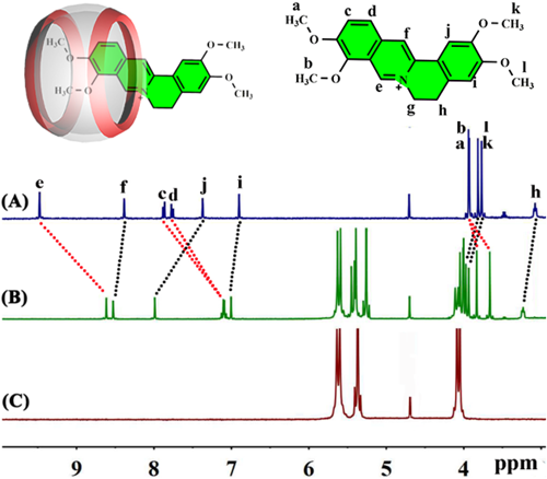
1. Formation of 1 : 1 inclusion complexes in aqueous solutionTo shed some light on the binding characteristics associated with the interaction between Q[7] and PAL in aqueous solution, 1H NMR spectroscopic titration experiments were employed. The 1H NMR spectra of PAL in neutral D2O solutions, both with and without different equivalents of Q[7] present, are depicted in Fig. 2. Significant shifts were evident for the PAL protons Ha, Hb Hc, Hd and He as Q[7] was added, whilst significant downfield shifts were observed for the protons PAL Hf, Hi and Hj. These spectroscopic observations can be rationalized in terms of encapsulation of the methoxyisoquinoline motif, with the functionalized benzene ring remaining in an external position.
2. UV-vis and fluorescence spectroscopyThe binding ability of Q[7] with PAL was next investigated by the use of both UV-vis (Fig. 3) and fluorescence (Fig. 4) spectroscopic titration experiments in aqueous solution. It is evident from Fig. 3a that the characteristic absorption peaks (225, 274 and 343 nm) of PAL are present. The guest absorption peak exhibited a red-shift to 279 nm as Q[7] was added. Concomitantly, a pronounced decrease in the PAL absorbance was observed, suggestive of high binding affinity between Q[7] and PAL. By using the molar ratio method (Fig. 3b), a 1 : 1 binding model produced a good fit, and this 1 : 1 stoichiometry was further confirmed by a continuous variation Job’s plot (Fig. S2).
Moreover, as Q[7] is added, the PAL fluorescence spectrum also exhibits significant changes. It is known that in aqueous solution the guest is not itself fluorescent, however on slow addition of Q[7], the fluorescence emission of PAL was significantly enhanced at λem=491 nm as shown in Fig. 4a. At the same time, with the continuous addition of Q[7], when the stoichiometric ratio of Q[7] : PAL was 1 : 1, the change in the fluorescence intensity of the system tended to be weaker, and the whole system reached equilibrium (Fig. 4b), which is associated with the formation of the 1 : 1 host–guest inclusion complex. In this system, the guest is provided with a micro hydrophobic environment by the Q[7] host.54,55)
3. ITCTo glean more information about the Q[7]/PAL interaction, ambient temperature ITC experiments (Fig. S3) were conducted in a neutral aqueous solution. It is clear also from Fig. S3, where ΔH =−29.14 kJ·mol−1, TΔS=5.40 kJ·mol−1, ΔG =−34.54 kJ·mol−1, that enthalpy and entropy each contribute to host–guest complex formation. This is thought to result partly from an ionic dipole interaction involving the PAL positively charged nitrogen centres and portal oxygens of the host, with a favourable enthalpy resulting from van der Waals interactions of the PAL surface and the Q[7] inner wall. It is also likely that the removal of cavity and portal water molecules together with those associated with the dissolved PAL shell will contribute to the increased entropy. By using the enthalpy and entropy values and the van’t Hoff equation (lnK=−ΔH/RT + ΔS/R), the binding constant for the Q[7]/PAL system was calculated at (1.12±0.2) ×105 M−1.
4. MALDI-TOF mass spectrometryMALDI-TOF mass spectrometry is a technique commonly employed for the observation of host-guest interactions. As depicted in Fig. S4, a strong signal is observed at m/z 1515.52, which compares favorably with the calculated value of m/z 1514.50. This data further supports the formation of a 1 : 1 Q[7]/PAL host-guest inclusion complex.
5. Fluorescence quenching of Q[7]/PAL by PQAs highlighted above, there is no intrinsic fluorescence in aqueous solution associated with PAL, whilst a fairly intense fluorescence occurs when PAL is in the presence of Q[7]. Given this, there is potential for this system to be employed for the identification of common pesticides. From the fluorescence measurements, it was clear that this system exhibited selectivity toward the pesticide PQ. In particular, fluorescence quenching was observed on addition of PQ (1×10⁻⁴ mol·L−1) to the 1 : 1 inclusion complex comprising Q[7] (2×10⁻⁵ mol·L−1) and PAL (2×10⁻⁵ mol·L−1). By contrast, there was no significant fluorescence change when any of the other 12 pesticides (1×10⁻⁴ mol·L−1) shown in Fig. S1 and named in the caption for Fig. 5 below were employed instead of PQ. These observations reveal that in aqueous solution, the selective detection of PQ can be achieved by employing the inclusion complex PAL@Q[7].
6. Effect of PQ concentration on the fluorescence intensity of the Q[7]/PAL systemThe effect of different PQ concentrations on the fluorescence intensity of the complex PAL@Q[7] was next studied. As depicted in Fig. 6a, on increasing the concentration of PQ, the fluorescence intensity of the inclusion complex PAL@Q[7] slowly decreases. The corresponding fluorescence intensity of the PAL@Q[7] inclusion complex for a PQ concentration of 0–2.2×10⁻⁵ mol·L−1 is shown in Fig. 6b.
As is evident from Fig. S5, a near linear relationship is found for the value of the change in fluorescence intensity (ΔI) versus the concentration of PQ over a range of concentrations. The linear range is 0–1.2×10⁻⁵ mol·L−1, and the linear regression equation is ΔI=36.7950C–44.1810 (C represents the concentration (mol·L−1) of PQ) with a correlation coefficient of 0.9903, indicative of good linearity. The detection limit for PQ was calculated at 3.26×10⁻⁷ mol·L−1.
7. The response mechanism of the fluorescent quenchingTo gain an understanding of the reaction mechanism for the fluorescence quenching when PQ is added to Q[7]/PAL, NMR spectroscopic titration experiments were performed. In Fig. 7, the proton peaks associated with both PQ pyridine rings exhibited high field shifts (versus free PQ) when the PQ was added Q[7]@PAL in D2O. These observations are consistent with PQ buried within the Q[7] cavity, i.e., formation of Q[7]@PQ with the PAL having been released (the PAL protons move to lower field).
Based on the reported literature,46) in the structure of PAL, the isoquinoline ring and dimethoxy benzene are unable to form a conjugated system due to their connection via a six-membered ring, which results in PAL itself being non-fluorescent. However, upon encapsulation of PAL by a Q[7] cavity, there is an electrostatic attraction between the positive charge of the heterocyclic nitrogen of the PAL guest and the high-density electron cloud of the carbonyl oxygen on the main body. This effectively forms a conjugation system between the isoquinoline ring and dimethoxy benzene, which results in increased fluorescence intensity. An additional factor favorable for fluorescence enhancement is the reduced freedom of motion of PAL when it is embedded in the hydrophobic cavity of Q[7], which decreases the probability of the radiation-free jump. The enhanced fluorescence will be quenched when the PAL is replaced by other molecules. In this titration study, a significant increase of fluorescence results from the formation of the 1 : 1 host–guest complex when 1.0 eq of PAL was encapsulated by 1.0 eq of Q[7]. By contrast, when PQ was combined with the Q[7]/PAL host–guest system, PQ competitively occupies the Q[7] cavity. As can be seen from the 1H NMR spectra, the peaks of the protons H2, H3 associated with the PQ were shifted to higher field relative to those of free PQ. This indicated that the PQ was located inside the Q[7] cavity (Fig. 7 and Fig. S6), whilst the movement downfield and broadening of the protons associated with PAL was consistent with one included PAL molecule being replaced by the PQ molecule, thereby forming a 1 : 1 inclusion complex. This provided a stable inclusion complex that led to fluorescence quenching, whereas the remaining 12 pesticides screened herein could not replace PAL and thus no significant change in fluorescence was observed. The method proposed in this paper can be compared with other methods reported in the analytical literature, as shown in Table 1.56–58) The results show that the sensitivity of this method is equivalent to that of other methods and has a certain degree of specificity.
Table 1. Comparison of the Different Methods Used for PQ Detection
| Method | Pesticide | Linear range | LOD | Ref |
|---|
| p-DPD@azoCX[4] probe | PQ | 0.35–8.0 uM | 0.35 uM | 54) |
| Pillar[5]arene sensor | PQ | 1 uM–0.1 M | 0.22 uM | 55) |
| Q[8]@dye array | PQ | 0–26.0 uM | 0.80 uM | 56) |
| Q[7]@PAL sensor | PQ | 0–12.0 uM | 0.34 uM | This work |