Abstract
To induce potato variants with enhanced resistance to common scab disease that retain the desirable agronomic traits of the original cultivars, we used a cell culture technique that employs thaxtomin A, the primary phytotoxin that induces scab symptoms. We induced 24 variants from the potato cultivar ‘Saya-akane’, developed in Japan, and selected two with enhanced resistance to the disease by growing them in planters with bacteria-inoculated soil and in a field infested with the disease. We also examined toxin tolerance in micro-tubers of variants that showed a lower degree or percentage of infection in the glasshouse screening, and found no significant difference relative to the original cultivar. To clarify the effect of using thaxtomin A, we examined the efficiency of induction of the potential enhanced resistance by comparing the degree of infection among variants grown in planters with inoculated soil. We observed no significant difference between variants induced on culture medium with and without the toxin. These results suggest that the effect of using the toxin as a positive selection agent is restrictive and that most resistance-enhancing mutations are induced by the cell culture procedure itself.
Introduction
Common scab is one of the most economically important diseases of potato (Solanum tuberosum L.) (Lambert and Loria 1989, Loria et al. 1997). The surfaces of tubers infected with the disease develop scab lesions, marring their appearance, reducing the economic value of table stock, and decreasing yield rates of tubers for food processing. In contrast, tuber yield is scarcely affected by the disease. Common scab is caused by Streptomyces species (Loria et al. 1997), and the scab lesions are thought to be caused mainly by phytotoxins synthesized by the bacteria (King et al. 1989, 1991, Lawrence et al. 1990). Thaxtomin A is the predominant phytotoxin that induces scab lesions (Lawrence et al. 1990). It is thought to inhibit cellulose biosynthesis and to trigger a programmed cell death response (Bischoff et al. 2009, Duval et al. 2005, Fry and Loria 2002, Scheible et al. 2003).
To reduce the damage caused by common scab infection, researchers around the globe have bred new cultivars resistant to the disease (Jansky 2000). A cross-breeding technique is generally employed, using parent cultivars with desirable traits and varieties with resistance to the disease. New cultivars developed by this method often possess many different traits from the parent cultivars, because a rearrangement chance of genetic patterns is restricted one time in potato cross-breeding. Thus, generating new disease-resistant cultivars with the same desirable traits as cultivars with stable market values is difficult and time-consuming.
Plant tissue and cell culture techniques can be used to induce genotypic or phenotypic variation in particular traits (Larkin and Scowcroft 1981). These techniques can offer rapid production of new cultivars without genetic re-assortment, retaining the desirable agronomic traits and market values of the original cultivars (Karp 1991). Successful results have been reported for many crops, including selection of somaclonal variants resistant to several diseases (van den Bulk 1991).
Because the symptoms of common scab disease are caused mainly by the plant’s response to phytotoxins, acquisition of toxin tolerance may reduce scab lesions on tuber surfaces. For selection of potato seedlings with resistance to the disease, treatment using thaxtomin A as a screening agent has been tested in conventional breeding programs (Hiltunen et al. 2006). Wilson et al. (2009, 2010a) used a somatic cell selection approach, with the toxin as a positive selection agent, to induce enhanced resistance to the disease in regenerated potato plants. Using this approach, they obtained numerous somaclonal variants of the potato cultivars ‘Iwa’ (susceptible) and ‘Russet Burbank’ (moderately resistant) with enhanced resistance to the disease. Enhanced resistance was measured by estimating the degree of infection of tubers harvested in glasshouse or field trials. They also showed that the enhanced resistance was retained over a 6-year period, and the selected phenotypes were robust and the genetic changes were stable.
Here, we induced variants derived from potato cultivars developed in Japan through a cell culture technique using thaxtomin A as a positive selection agent, using the method reported by Wilson et al. (2009, 2010a) with slight modifications. We selected variants with potential enhanced resistance to common scab disease by growing them in planters filled with bacteria-inoculated soil and in a field infested with the disease. We measured the toxin tolerance levels of micro-tubers of the induced variants by estimating the degree of necrosis after incubation in medium containing the toxin. We also compared the frequency of induced variants with potential enhanced resistance between samples cultured with and without the toxin. We discuss the efficiency of the cell culture technique using the toxin as a positive selection agent for induction of variants with enhanced resistance.
Materials and Methods
Plants and bacteria
We used five potato cultivars—‘Saya-akane’ (Iketani et al. 2015), ‘Yukitsubura’ (Iketani et al. 2011), ‘Snow March’ (Iketani et al. 2005), ‘Okhotsk Chip’ (Iritani et al. 2009), and ‘Natsufubuki’ (Iketani et al. 2004)—developed by the Hokkaido Prefectural Kitami Agricultural Experiment Station (now the Hokkaido Research Organization Kitami Agricultural Experiment Station). In vitro plantlets of the five cultivars were maintained by subculturing two-node segments every 3 to 4 weeks on potato multiplication (PM) medium (Wilson et al. 2009: MS salts and vitamins [Murashige and Skoog 1962], plus 30 g/L sucrose, 40 mg/L ascorbic acid, 500 mg/L casein hydrolysate, and 0.8% agar, with pH adjusted to 5.8).
We used Streptomyces turgidiscabies isolate 63 for thaxtomin A production and screening in a glasshouse, and isolate OIS9401 for screening in a field. The isolates were provided by the Plant Disease and Pest Section of the Hokkaido Prefectural Central Agricultural Experiment Station (now the Hokkaido Research Organization Central Agricultural Experiment Station), and were maintained on slopes of starch inorganic salts agar medium (Matsumoto 1979: 10 g/L soluble starch, 1 g/L sucrose, 1 g/L yeast extract, 0.1 g/L NaNO3, 0.1 g/L MgSO4·7H2O, 0.1 g/L KH2PO4, 0.1 g/L KCl, and 1.5% agar, with pH adjusted to 8.0).
Thaxtomin A extraction and quantitative analysis
Thaxtomin A was extracted and quantitatively analyzed as follows (Dr. Masahiro Natsume, Tokyo University of Agriculture and Technology, pers. comm.). Isolate 63 was cultured on starch inorganic salts agar medium in Petri dishes at 25°C for a month in the dark. A 7 mm × 7 mm agar block containing the cultured isolate was cut out and inoculated into a 500-mL Erlenmeyer flask containing 300 mL oatmeal broth medium (Babcock et al. 1993: 20 g oatmeal/L distilled water) and incubated at 25°C for 2 weeks on a rotary shaker (120 rpm). After centrifugation at 10,000 × g for 15 min, the aqueous phase was extracted three times with 200 mL CHCl3 in a separatory funnel. The combined extract was washed with distilled water and saturated salt solution. Following addition of 20 g Na2SO4 to a glass bottle containing the extract, the mixture was stirred well and allowed to sit overnight at 20°C. The extract was evaporated to dryness, and the dried pellet was dissolved in 10 mL CHCl3. The concentrated CHCl3 extract was fractionated by 5-g silica gel (Wako Gel C-200, Wako Co., Ltd., Japan) column chromatography with elution by CHCl3: MeOH (9:1 vol/vol). The elutant was collected and evaporated to dryness, and the dried pellet was dissolved in 5 mL CHCl3. The CHCl3 solution was quantitatively analyzed by high-performance liquid chromatography (HPLC: Develosil ODS-UG-5, 30% aq. CH3CN, UV 400 nm; analysis courtesy of Dr. Seiichi Komiyama, Hokkaido Research Organization Central Agricultural Experiment Station) and compared with a thaxtomin A standard (analysis courtesy of Dr. Masahiro Natsume, Tokyo University of Agriculture and Technology).
Thaxtomin A treatment of potato cells and selection of regenerated plants
Potato somaclonal variants were selected using the method reported by Wilson et al. (2009, 2010a) with slight modifications. In vitro plantlets cultured on PM medium for 2 to 3 weeks were used for tissue and cell culture experiments. The stem internodes were cut into 1-cm sections, cut in half lengthways, then placed face down on callus induction (CI) medium (Wilson et al. 2009: MS salts and vitamins, plus 5 g/L sucrose, 40 mg/L ascorbic acid, 500 mg/L casein hydrolysate, 2 mg/L BAP, 0.2 mg/L NAA, 5 mg/L GA3, and 0.8% agar, with pH adjusted to 5.8) for 10 to 14 days at 25°C with a 16 h light photoperiod under fluorescent light (30 to 40 μmol/m2s) to induce somewhat friable calluses. Then the cultured stems with calluses were transferred to CI liquid medium without agar in plastic tubes and incubated for 2 to 3 days on a rotary shaker (120 rpm) under the same conditions. After removal of the stems by sieving through stainless steel mesh, cell clusters were collected in the bottom of the plastic tube and transferred to the liquid medium described above supplemented with 2 mg/L thaxtomin A. The cell suspension was incubated for 2 to 4 days on a rotary shaker (120 rpm) under the same conditions, because Wilson et al. (2009) reported that 1-day culture did not inhibit the background growth of wild-type cells, and was poured into a plastic tube to collect the cell clusters. After removal of the supernatant, sufficient liquid medium without toxin was added to the cell cluster suspension to adjust the cell density to 104–105 plating units/mL by counting cell numbers under a microscope. Then 0.5 mL of the cell cluster suspension was plated on CI medium in a Petri dish and cultured for recovery of surviving potato cells. To assist cell cluster growth, 0.5 mL cell suspension of the other potato cultivars was spread evenly on the CI medium as a nurse culture, a piece of sterile filter paper was placed over the nurse cells, and the toxin-treated potato cells were cultured on top of the filter paper. After 2 to 3 weeks of culture, colonies, which appeared at low rates, were transferred to callus regeneration (CR) medium (Wilson et al. 2009: MS salts and vitamins, plus 5 g/L sucrose, 40 mg/L ascorbic acid, 500 mg/L casein hydrolysate, 1 mg/L zeatin, 0.2 mg/L NAA, 5 mg/L GA3, and 0.8% agar, with pH adjusted to 5.8) and incubated for 2 to 3 months at 25°C with a 16 h photoperiod under fluorescent light (30 to 40 μmol/m2s). Regenerated shoots were transferred to PM medium and grown to plants with roots. The regenerated plants were maintained on PM medium and were used for subsequent screenings.
To examine the frequency at which variants with potential enhanced resistance to common scab disease were generated when thaxtomin A was not used in the culturing procedure, we used the same procedures as above but omitting toxin.
Screening for resistance to common scab disease in a glasshouse
The resistance of the variants to common scab disease was estimated by growing them in soil inoculated with isolate 63 in a glasshouse. After culturing 7 mm × 7 mm agar blocks containing the cultured isolate in 100 mL starch inorganic salts liquid medium at 25°C for 2 weeks on a rotary shaker (120 rpm) as described above, we mixed 2 mL of the liquid culture with 40 mL sterilized distilled water in a plastic tube; added the inoculum to 250 g soil mixed with 25 g wheat bran, which had been autoclaved at 121°C for 1 h, in a 500-mL Erlenmeyer flask; and incubated the soil culture at 25°C for 3 weeks in the dark. After the addition of 9 mL distilled water to 1 g of soil culture, the number of spores in the isolate was counted by hemocytometer, and a suitable amount of soil culture was mixed into the top one-third of sterilized ‘Pot-Ace’ culture soil (Katakura-Chikkarin Co., Ltd., Japan) in a planter (25 cm × 60 cm × 30 cm) to provide a concentration of >1 × 10⁵ spores/g. The top layer was stirred well with a trowel. Three different variants and one original cultivar were planted side by side at 15 cm intervals in the planter. ‘Root preventing sheets’ (Toyobo Co., Ltd., Japan), which prevent other plants’ stolons from crossing the boundary between plots but can transmit water, were inserted at the borders. The temperature of the glasshouse was maintained at between 22 and 25°C. Enough water was supplied to grow the plants until flowering, and then their water supply was minimized to promote infection by the disease. After senescence of the plants, all tubers were harvested. The degree of disease damage of each tuber ≥1 cm in diameter was estimated by scoring of the tuber surface on a 0–4 scale (0, no visible disease on surface; 1, 0%–3%; 2, 4%–13%; 3, 14%–25%; 4, ≥26% tuber surface affected). The degree of infection was calculated as:
|
Degree of infection
=
(
(
1
×
n
1
+
2
×
n
2
+
3
×
n
3
+
4
×
n
4
)
×
100
)
/
(
4
×
number of total tubers checked
) |
where n1 to n4 indicate the number of tubers scoring 1 to 4. The percentage of infected tubers was calculated also.
Screening for resistance to common scab disease in a disease-infested field
We estimated the resistance of the variants to common scab disease by growing them in a field that is infested mainly with S. turgidiscabies at the Kitami Agricultural Experiment Station, northern Japan, during 2012 to 2017. Tubers of the variants and the original cultivar harvested in the glasshouse were planted in the field and allowed to produce tubers for use in the following tests. In the spring of the next year, tubers with no or few scab lesions were planted in the field. Tubers harvested in the field were used as seed potatoes in the following test. Field trials were arranged using a randomized block design without replicates or comprising three replicate plots, each containing four to six plants of each variant and the original cultivar. Herbicide was applied to the field before sprouting, and soil was mounded at the early growth stage. During the growing period, fungicides and insecticides were applied to the plants following the standard cultivation method used at this experiment station. Before the senescence period (early autumn), a withering agent was applied twice to promote senescence of the plants, a necessity of field management. After 2 weeks, all tubers were harvested, and those ≥20 g were counted and weighed. Disease damage was estimated, and the degree of infection and the percentage of infected tubers were calculated as described above. To compare the results of different years, we calculated the ratio of the degree of disease infection as variant/original cultivar in 2016 and 2017.
In 2017, to increase the precision of the test, we spread extra disease spores in the field as follows (Dr. Kenji Asano, Hokkaido Agricultural Research Center, NARO, pers. comm., with slight modifications). Isolate OIS9401 was cultured on starch inorganic salts agar medium in a Petri dish at 25°C for 2 weeks in the dark. Ten pieces of 15 mm × 15 mm agar block containing the cultured isolate were cut out and inoculated into an autoclave bag containing 1 L oatmeal-vermiculite medium (1 L vermiculite, 5.135 g oatmeal, 0.256 g casamino acid, and 350 mL distilled water) and incubated at 25°C for a month in the dark. When the spore concentration was >1 × 10⁸/g (counted by hemocytometer as above), about 80 mL of the culture per plot (six plants) was spread near the roots of sprouting plants.
After we confirmed that the data followed an approximately normal distribution, data for degree of infection and agricultural traits per replicate were averaged and analyzed by one-way analysis of variance using the BellCurve for Excel software (Social Survey Research Information Co., Ltd., Japan). When significant differences were recognized, means were compared by Dunnett’s method for multiple comparisons.
Screening for tolerance to thaxtomin A
Micro-tubers that were induced by adding liquid MS (Murashige and Skoog 1962) medium with 80 g/L sucrose were used for screening of variants with thaxtomin A tolerance. The cut sides of micro-tubers ≥7 mm in diameter were placed on the surface of PM medium containing 0.5 mg/L thaxtomin A in a Petri dish. For each variant and the original cultivar, two replicates comprising four cut micro-tubers were tested. Five variants and the original cultivar were tested in the same Petri dish to compare tolerance levels relative to the original cultivar. After 1 week of incubation at 25°C in the dark, toxin tolerance levels were assessed by checking cut sides for the percentage of necrosis on a 0–4 scale (0, no necrosis; 0.5, 0% to <12.5%; 1, 12.5% to <25%; 2, 25% to <50%; 3, 50% to <75%; 4, 75% to 100% necrosis).
Necrosis index data were normalized for comparison of results among trials by dividing the mean value of each variant by the mean value of the original cultivar. After we confirmed an approximately normal distribution, relative data were further compared by one-way analysis of variance using BellCurve software.
Results
Thaxtomin A extraction and quantitative analysis
HPLC analysis of thaxtomin A crude extract from isolate 63 showed a large single absorbance peak at 400 nm (data not shown). Comparison of the extract with a standard thaxtomin A crystal showed that toxin made up a quarter of the extract. When the crude extract was added to CI medium at 2 mg/L thaxtomin A and leaflets derived from in vitro plantlets were cultured on the medium, no callus was induced, indicating strong toxicity. We therefore concluded that the crude extract was suitable for use as a thaxtomin A solution for subsequent experiments.
Thaxtomin A treatment of potato cells and selection of regenerated plants
After cell culture for 2 to 4 days in CI liquid medium containing 2 mg/L thaxtomin A and for a subsequent 2 to 3 weeks on CI medium, a few colonies appeared in the Petri dishes containing each cultivar (Table 1). No clear trend in colony formation by period of culture with the toxin was observed. After these colonies were transferred to CR medium and cultured for 2 to 3 months, ‘Saya-akane’ formed many regenerated shoots and ‘Natsufubuki’ formed just one (Table 1). The other three cultivars formed none. The growth of variants derived from ‘Saya-akane’ was generally normal, but that of the variant derived from ‘Natsufubuki’ was poor and plants were light green. Thus, we used the variants derived from ‘Saya-akane’, which is susceptible to common scab disease, for the following experiments.
Table 1
Summary of colonies and regenerated plants of each potato cultivar following exposure of somatic cells to thaxtomin A
| Cultivar |
Culture period in CI liquid mediuma containing 2 mg/L thaxtomin A (days) |
Number of plates |
Number of colonies formed on CI mediuma |
Number of plants regenerated on CR mediumb |
| Saya-akane |
2 |
30 |
40 |
26 |
| 3 |
34 |
112 |
15 |
| 4 |
4 |
3 |
0 |
| total |
68 |
155 |
41 |
| Yukitsubura |
2 |
9 |
18 |
0 |
| 3 |
5 |
1 |
0 |
| 4 |
3 |
28 |
0 |
| total |
17 |
47 |
0 |
| Snow March |
2 |
4 |
3 |
0 |
| 3 |
6 |
0 |
0 |
| 4 |
2 |
0 |
0 |
| total |
12 |
3 |
0 |
| Okhotsk Chip |
2 |
18 |
47 |
0 |
| 3 |
27 |
72 |
0 |
| 4 |
4 |
1 |
0 |
| total |
49 |
120 |
0 |
| Natsufubuki |
2 |
11 |
77 |
1 |
| 3 |
14 |
47 |
0 |
| 4 |
2 |
0 |
0 |
| total |
27 |
124 |
1 |
a CI medium: callus induction medium.
b CR medium: callus regeneration medium.
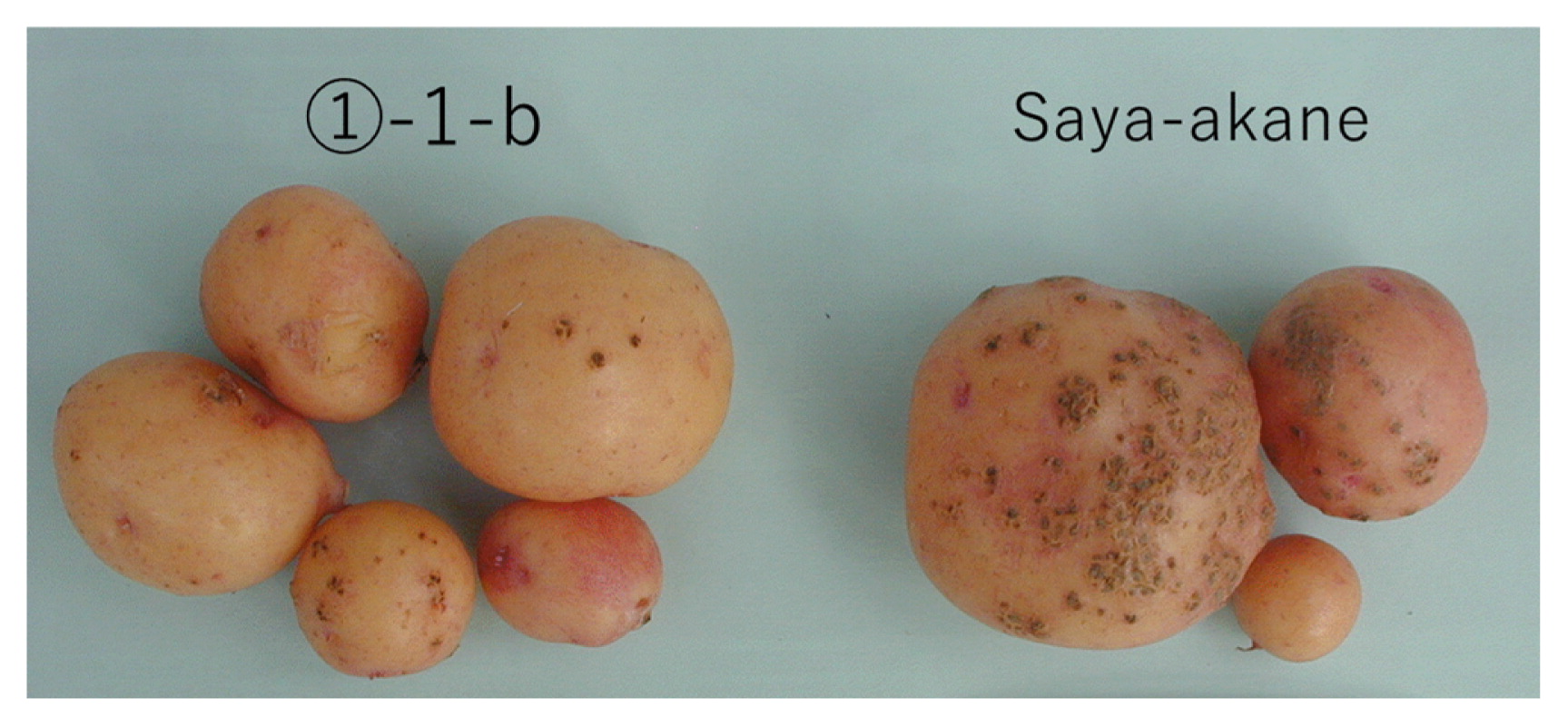
We compared the degree of infection and the percentage of infected tubers of each variant with those of the original cultivar grown in the same planter. Some variants showed approximately equivalent degrees or percentages of infection, or both, to the original cultivar (⑩-2-a, ⑪-1-d, ⑭-1-a) or higher values than the original cultivar (①-2-d, ⑪-1-a, ⑪-1-b, ⑪-1-c, ⑫-1-b, ⑭-1-c), and others showed lower values than the original cultivar (①-1-a, ①-1-b, ①-1-d, ①-1-e, ①-2-a, ①-2-b, ①-2-c, ⑤-4-a, ⑥-1-a, ⑨-1-a, ⑨-2-a, ⑩-1-a, ⑫-1-a, ⑬-1-a, ⑭-1-b) (Table 2, Fig. 1).
Table 2
Comparison of degree of common scab disease infection and percentage of disease-infected tubers between the original cultivar (‘Saya-akane’) and variants in each planter filled with bacteria-inoculated soil
a
| Name of variant |
Degree of disease infectionb |
Percentage of disease-infected tubers (%) |
| Original cultivar |
75.0 |
100 |
| ①-1-a |
25.0 |
100 |
| ①-1-b |
20.0 |
60 |
| ①-1-d |
12.5 |
50 |
| Original cultivar |
50.0 |
100 |
| ①-1-e |
6.3 |
25 |
| ①-2-a |
37.5 |
100 |
| ①-2-b |
16.7 |
67 |
| Original cultivar |
25.0 |
67 |
| ①-2-c |
8.3 |
33 |
| ①-2-d |
66.7 |
83 |
| ⑤-4-a |
18.8 |
50 |
| Original cultivar |
87.5 |
100 |
| ⑥-1-a |
50.0 |
100 |
| ⑨-1-a |
60.0 |
100 |
| ⑨-2-a |
6.3 |
25 |
| Original cultivar |
50.0 |
100 |
| ⑩-1-a |
20.0 |
60 |
| ⑩-2-a |
50.0 |
100 |
| ⑪-1-a |
93.8 |
100 |
| Original cultivar |
41.7 |
67 |
| ⑪-1-b |
41.7 |
100 |
| ⑪-1-c |
50.0 |
100 |
| ⑪-1-d |
37.5 |
75 |
| Original cultivar |
50.0 |
100 |
| ⑫-1-a |
20.8 |
83 |
| ⑫-1-b |
100.0 |
100 |
| ⑬-1-a |
37.5 |
100 |
| Original cultivar |
50.0 |
100 |
| ⑭-1-a |
45.8 |
100 |
| ⑭-1-b |
30.0 |
80 |
| ⑭-1-c |
87.5 |
100 |
a This test was conducted without replicates.
b The degree of common scab disease damage was estimated by scoring of infected tuber surface: 0, no visible disease on tuber surface; 1, 0% to 3%; 2, 4% to 13%; 3, 14% to 25%; 4, ≥26% tuber surface infected. The percentage of infected tubers was calculated. The average degree of disease infection was calculated as:
|
Degree of disease infection
=
(
(
1
×
n
1
+
2
×
n
2
+
3
×
n
3
+
4
×
n
4
)
×
100
)
/
(
4
×
total number of tubers examined
) |
where n1–n4 indicate the number of tubers with a score of 1–4.
Fifteen variants that showed potential enhanced resistance to common scab disease in the glasshouse screening (lower degree or percentage of infection, or both, than the original cultivar) were selected. These variants were screened for resistance to the disease in a field infested mainly by S. turgidiscabies. In 2012, a field test was conducted by planting four tubers per variant and the original cultivar, without replicates. Some variants showed a lower degree of infection (①-1-b, ①-1-e, ⑨-2-a, ⑬-1-a) than the original cultivar (Table 3). In 2013 to 2017, field tests with increased precision were conducted by planting six tubers per variant and the original cultivar in three replicate plots. However, no clear data are available for 2013, as the degree of disease damage was extremely low in general, because the field was too dry (Table 3). In 2014, some variants showed an apparently lower degree of infection (①-1-b, ①-1-e, ⑤-4-a, ⑨-1-a, ⑨-2-a, ⑬-1-a) than the original cultivar (Table 3). However, the differences were not significant (P > 0.05), perhaps because the density of disease spores was uneven across the field.
Table 3
Degree of common scab disease infection of each variant produced in a field infested with the disease (2012–2017)
a
| Name of variant |
Degree of common scab disease infection (ratio of degree of disease infection = variant/original cultivar) |
| 2012b |
2013c |
2014c |
2015c |
2016c |
2017d |
| Original cv. (‘Saya-akane’) |
16.4/4.6 |
0 |
10.9 |
0.5 |
3.6 (1.00) |
11.6 (1.00) |
| ①-1-a |
21.5 |
0 |
15.4 |
– |
– |
– |
| ①-1-b |
0.5 |
0.2 |
6.0 |
0 |
1.8 (0.49) |
5.7* (0.49) |
| ①-1-d |
16.7 |
0.4 |
11.7 |
– |
– |
– |
| ①-1-e |
2.1 |
0.3 |
7.5 |
0.9 |
2.4 (0.65) |
10.8 (0.93) |
| ①-2-a |
15.3 |
0.4 |
11.0 |
– |
– |
– |
| ①-2-b |
12.5 |
0.3 |
14.1 |
– |
– |
– |
| ①-2-c |
10.1 |
0.8 |
10.0 |
– |
– |
– |
| ⑤-4-a |
29.1 |
0.5 |
6.7 |
– |
– |
– |
| ⑥-1-a |
23.1 |
0.1 |
10.5 |
– |
– |
– |
| ⑨-1-a |
9.0 |
0.3 |
6.2 |
– |
– |
– |
| ⑨-2-a |
2.5 |
0.5 |
5.5 |
0.1 |
3.6 (0.99) |
13.2 (1.14) |
| ⑩-1-a |
5.4 |
1.9 |
14.4 |
– |
– |
– |
| ⑫-1-a |
8.3 |
0.8 |
13.9 |
– |
– |
– |
| ⑬-1-a |
0.6 |
0 |
6.1 |
0.4 |
0.1 (0.03) |
4.5* (0.39) |
| ⑭-1-b |
6.7 |
0.9 |
8.7 |
– |
– |
– |
a The degree of common scab disease damage was estimated by scoring of infected tuber surface: 0, no visible disease on tuber surface; 1, 0%–3%; 2, 4%–13%; 3, 14%–25%; 4, ≥26% tuber surface infected. The percentage of infected tubers was calculated. The average degree of disease infection was calculated as:
|
Degree of disease infection
=
(
(
1
×
n
1
+
2
×
n
2
+
3
×
n
3
+
4
×
n
4
)
×
100
)
/
(
4
×
total number of tubers examined
) |
where n1–n4 indicate the number of tubers with a score of 1–4.
b The test in this year was conducted without replicates, and the original cultivar was planted in two plots.
c No significant difference (
P > 0.05).
d *
P = 0.05, relative to original cultivar by one-sided Dunnett’s test.
In 2015, 2016, and 2017, four promising variants (①-1- b, ①-1-e, ⑨-2-a, ⑬-1-a) that showed a lower degree of infection than the original cultivar in 2012 and 2014 were planted in the field, and the degree of disease infection was measured to verify the stability of the potential enhanced resistance. In 2015, disease damage was extremely low, preventing us from obtaining clear data (Table 3). In 2016 and 2017, while three variants (①-1-b, ①-1-e, ⑬-1-a) showed lower ratios of degree of infection than the original cultivar, one variant (⑨-2-a) showed equivalent and higher ratios (Table 3). In 2017, when extra disease spores were spread in the field, the one-sided Dunnett’s test showed a significant difference (P = 0.05) in two of these variants (①-1-b, ⑬-1-a).
Because the variants generally sprouted later than the original cultivar in 2014, the following early growth stages and flowering times were also somewhat later (Table 4). Thus, immediately before the withering agent was applied, the senescence stages of the variants were later than that of the original cultivar. Although no distinct morphological variation was observed (except in ⑬-1-a, which showed flower color change from light pink to pink), variants differed in vigor of growth, as shown by stem length variation. Owing to their delayed growth and the need to apply a withering agent, the total weight of tubers (≥20 g) produced by the variants was significantly lower than that of the original cultivar.
Table 4
Growth and yield of each variant grown in a field infested with common scab disease (2014)
| Name of variant |
Germination time (month/day) |
Flowering time (month/day) |
Timing of senescence on Aug. 13a |
Stem length (cm)c |
Tuber number per plantc |
Tuber weight per plant (g)c |
| Original cv. (‘Saya-akane’) |
6/1 |
7/2 |
3.0 |
62.6 |
16.6 |
581 |
| ①-1-a |
6/5 |
7/5 |
3.3 |
45.9** |
8.6** |
327** |
| ①-1-b |
6/6 |
7/7 |
4.7 |
58.4 |
11.0* |
388** |
| ①-1-d |
6/5 |
7/8 |
3.3 |
47.8** |
11.3* |
364** |
| ①-1-e |
6/9 |
7/12 |
4.0 |
61.7 |
14.8 |
357** |
| ①-2-a |
6/3 |
7/9 |
3.3 |
52.2** |
11.8* |
283** |
| ①-2-b |
6/5 |
7/9 |
4.0 |
63.4 |
11.5* |
374** |
| ①-2-c |
6/5 |
7/6 |
4.7 |
65.1 |
13.0 |
385** |
| ⑤-4-a |
6/5 |
7/9 |
3.7 |
56.7 |
14.0 |
388** |
| ⑥-1-a |
6/3 |
7/10 |
5.0 |
42.4** |
14.1 |
348** |
| ⑨-1-a |
6/6 |
7/7 |
3.7 |
61.3 |
12.6 |
367** |
| ⑨-2-a |
6/5 |
7/4 |
3.0 |
49.0** |
12.1 |
400** |
| ⑩-1-a |
6/3 |
7/3 |
3.7 |
63.8 |
12.8 |
438** |
| ⑫-1-a |
6/3 |
7/4 |
3.7 |
64.7 |
14.4 |
462** |
| ⑬-1-a |
6/3 |
7/4 |
5.0 |
66.3 |
9.9* |
253** |
| ⑭-1-b |
6/6 |
7/8 |
1.0b |
58.5 |
16.1 |
342** |
a Timing of senescence: 1, extremely early; 2, early; 3, middle; 4, late; 5, extremely late.
b ⑭-1-b withered abnormally early.
c **
P = 0.01, *
P = 0.05, relative to original cultivar.
In some variants, the ratios of necrosis index relative to the original cultivar were <1.00 (Table 5). However, there were no significant differences (P > 0.05) among variants and the original cultivar.
Table 5
Necrosis assessment of micro-tubers of variants that showed lower degrees or percentages of infection than the original cultivar in the glasshouse screening
| Name of variant |
Ratio of necrosis indexa b (variant/original cv.) |
| Original cv. (‘Saya-akane’) |
1.00 |
| ①-1-a |
1.04 |
| ①-1-b |
0.72 |
| ①-1-d |
1.07 |
| ①-1-e |
1.06 |
| ①-2-a |
0.80 |
| ①-2-b |
1.35 |
| ①-2-c |
1.14 |
| ⑤-4-a |
0.80 |
| ⑥-1-a |
1.09 |
| ⑨-1-a |
1.14 |
| ⑨-2-a |
1.15 |
| ⑩-1-a |
1.18 |
| ⑫-1-a |
0.99 |
| ⑬-1-a |
1.13 |
| ⑭-1-b |
1.21 |
a Resistance to thaxtomin A was assessed by measuring the necrotic area of the cut side on a 0–4 scale: 0, no necrosis; 0.5, 0% to <12.5%; 1, 12.5% to <25%; 2, 25% to <50%; 3, 50% to <75%; 4, 75% to 100% necrotic area). Ratio of necrosis index indicates variant/original cultivar.
b No significant difference (
P > 0.05).
The average ratios of disease infection (variant/original cultivar) of the treatments for induction of potential enhanced resistance were 0.76 with thaxtomin A and 0.79 without (Table 6). Each treatment was significantly different from the original cultivar (P = 0.05), but treatments did not differ significantly from each other (P > 0.05).
Table 6
Comparison of efficiency of induction of potential enhanced resistance to common scab disease using medium without or with thaxtomin A
a
| Processing category |
Name of variant |
Percentage of disease-infected tubers (%)b |
Degree of disease infectionb |
Ratio of degree of disease infection (variant/original cv.)c |
| Medium without thaxtomin A |
c⑤-2-a |
41.1 |
15.6 |
0.28 |
| c⑥-1-a |
51.8 |
16.1 |
0.29 |
| c⑥-1-b |
100.0 |
51.0 |
0.91 |
| c⑥-2-a |
92.9 |
54.6 |
0.99 |
| c⑥-3-a |
100.0 |
68.1 |
0.88 |
| c⑥-3-b |
100.0 |
85.3 |
1.10 |
| c⑥-3-c |
100.0 |
55.2 |
0.71 |
| c⑬-1-a |
100.0 |
42.7 |
0.78 |
| c⑬-1-b |
71.7 |
43.8 |
0.77 |
| c⑮-1-a |
83.3 |
52.5 |
1.11 |
| c⑮-1-e |
91.7 |
58.3 |
1.19 |
| c⑯-1-a |
54.2 |
30.2 |
0.48 |
| Average |
82.2 |
47.8 |
0.79*d |
| Medium with thaxtomin A |
33-1-a |
100.0 |
53.8 |
1.02 |
| 33-1-b |
100.0 |
48.2 |
0.91 |
| 34-1-a |
80.0 |
30.0 |
0.52 |
| 37-1-a |
83.3 |
50.0 |
0.99 |
| 39-1-a |
58.3 |
29.2 |
0.52 |
| 41-1-a |
71.4 |
32.1 |
0.58 |
| 41-2-a |
63.3 |
38.3 |
0.71 |
| 41-3-a |
91.7 |
51.7 |
0.97 |
| 45-1-a |
100.0 |
43.8 |
0.93 |
| 45-2-a |
71.4 |
25.9 |
0.49 |
| Average |
82.0 |
40.3 |
0.76*d |
| Maintained by sub-culturing |
Ave. of original cv. |
93.4 |
56.9 |
1.00 |
a This test was conducted by planting variants and the original cultivar in a planter filled with bacteria-inoculated soil.
b The value for each variant is the average of two replicates; the value for the original cultivar is the average of 15 plants.
c The ratio of each variant’s degree of disease infection to that of the original cultivar grown in the same planter.
d *
P = 0.05, relative to original cultivar. No significant difference (
P > 0.05) between the average values of the two treatments.
Discussion
We tried to induce variants resistant to common scab disease from potato cultivars developed in Japan using a previously reported cell culture technique that employs thaxtomin A as a positive selection agent. We followed the method reported by Wilson et al. (2009, 2010a) with some modifications, such as degree of thaxtomin A purification. We used five potato cultivars recently developed in Japan for the cell culture and selection experiments. After culturing cells of the five cultivars for 2 to 4 days in medium containing 2 mg/L thaxtomin A and for the subsequent 2 to 3 weeks on medium without the toxin, all cultivars formed a few colonies per Petri dish (Table 1). Because the frequency of colony emergence in toxin-containing medium was not low, it seems that these colonies did not always acquire tolerance to the toxin, and several cells may have occasionally formed colonies without acquiring tolerance. When these colonies were cultured on CR medium, colonies from ‘Saya-akane’, which is known for its high regeneration ability, formed many regenerated shoots (Table 1). Except for one shoot of ‘Natsufubuki’, colonies of the other cultivars produced no shoots. The composition of the medium will need to be adapted if this culture technique is to be applied to other potato cultivars in Japan.
In the disease resistance screening in the glasshouse in inoculated soil, 15 of 24 variants showed lower degrees or percentages of infection than the original cultivar (Table 2). However, because this test was conducted without replicates, it is possible that some of the 15 variants might not have enhanced resistance. Accordingly, in the field, 4 of the 15 variants in 2012 and 6 in 2014 showed relatively lower degrees or percentages of infection than the original cultivar (Table 3). Four of these were selected and grown in the disease-infested field between 2015 and 2017 to test the stability of the potential enhanced disease resistance. Two variants (①-1-b, ⑬-1-a) showed lower ratios of the degree of infection than the original cultivar (P = 0.05) by one-sided Dunnett’s test, but the other two (①-1-e, ⑨-2-a) did not (Table 3). These results indicate that the former two variants might have acquired enhanced resistance through somatic cell selection, and the disease-resistant traits might have been maintained. They also suggest that although the latter two variants had developed enhanced resistance, the disease resistance traits might be gradually lost during successive generations. Previous studies of somaclonal variation in potato show that consistency of phenotypic expression over a 3-year period is sufficient to demonstrate the stability of the selected trait (Kowalski and Cassells 1999). Thus, stability of the enhanced resistance obtained via the cell culture technique should be further investigated.
Experiments on tolerance of micro-tubers to thaxtomin A showed that there were no significant differences among variants and the original cultivar, and that variants with enhanced disease resistance did not always express toxin tolerance (Table 5). These results suggest that the tissue and cell culture procedure itself induced the mutations for enhanced resistance, and in some variants weak toxin tolerance was obtained in addition. As Wilson et al. (2009) reported, potato variants resistant to common scab disease have been obtained through random selection from somaclonal variants in previous studies (Gunn and Wray 1983, Gunn et al. 1985, Saulite 1987, Thompson et al. 1986). These reports show that variants with the trait can be induced relatively easily by somaclonal mutation.
To confirm the supposition that the tissue and cell culture procedure itself induced the enhanced resistance mutations, we compared the ratio of induction of variants with potential enhanced disease resistance between cell cultures with and without the toxin. The average ratio in medium without the toxin was 0.79 and that in medium with the toxin was 0.76 (Table 6). While the difference from the original cultivar was significant, the difference between the two treatments was not significant (P > 0.05). This result suggests that using the toxin as a positive selection agent for induction of enhanced resistance is restrictive. Wilson et al. (2009) reported that the lack of enhanced toxin tolerance observed in most disease-resistant variants suggested that thaxtomin A tolerance was not necessarily the factor driving induction of disease resistance. Wilson et al. (2010a) also reported that although not all disease-resistant variants showed significantly less necrosis than the unselected parent following toxin exposure, the majority of the variants showing the greatest disease resistance expressed enhanced toxin tolerance. The former study used ‘Iwa’, which is relatively toxin tolerant, whereas the latter used ‘Russet Burbank’, which is relatively sensitive. Thus, in cultivars that are relatively sensitive to the toxin, it may be effective to use the toxin to induce variants with enhanced disease resistance. This is supported by a study that found no association between disease resistance and toxin tolerance (Tegg and Wilson 2010), which showed that although the toxin is critical to disease expression, reaction to the toxin is only one component influencing disease resistance, and many other anatomical, physiological, or biochemical factors are critical to defense against the disease. It seems that using the toxin was not effective in ‘Saya-akane’, although the cultivar is sensitive to it.
If the procedure for induction of somaclonal variants can frequently induce enhanced resistance to common scab disease, the mechanism underlying acquisition of resistance to the disease may be similar. The variants induced by Wilson et al. (2010a) were subsequently shown to co-express resistance to powdery scab caused by Spongospora subterranea (Tegg et al. 2013). They were later shown to be resistant to black scurf and tuber soft rot, induced by fungal and bacterial pathogens, by means of pot trials and in vitro assays (Thangavel et al. 2014). The resistance appeared to be tuber-specific, as stolon or stem material did not show enhanced resistance to Rhizoctonia solani, which induces stolon pruning and stem canker. To determine the mechanism of this enhanced disease resistance, Thangavel et al. (2016) examined tuber periderm tissue from disease-resistant clones and their susceptible parent, and examined the relative expression of genes associated with the tuber suberin biosynthesis and innate defense pathways in these tissues. The disease-resistant somaclones reacted to both the pathogen and the toxin by producing additional phellem cell layers in the tuber periderm and accumulating extra suberin polyphenols in these tissues, and the disease-resistant somaclones had greater expression of genes associated with suberin biosynthesis. Thus, they showed that the resistant phenotype was due to induction of increased periderm cell layers and suberization of the tuber periderm, preventing infection. We do not know whether the tubers of the variants with enhanced scab resistance produced here are similarly resistant to other diseases. Further studies should be undertaken to determine the mechanism of the enhanced resistance acquired via the tissue and cell culture technique.
We confirmed that cell culture could be used to induce variants with enhanced resistance to common scab disease in a potato cultivar developed in Japan, with or without thaxtomin A as a positive selection agent. We selected two variants (①-1-b, ⑬-1-a) that showed a lower degree of infection than the original cultivar in a disease-infested field. However, our variants generally had later sprouting times and delayed growth compared with the original cultivar (Table 4). In general, when a withering agent is applied to promote senescence of potatoes for the purpose of field management, delayed growth can lead to low tuber yields, owing to shortening of the ripening period. In addition, inferior growth, with a shorter stem, decreases tuber yield. Thus, the tuber weight produced by each variant was significantly lower than that of the original cultivar (Table 4). Tissue and cell culture can often induce undesirable mutations, reducing yield (Karp 1995). We think that the delayed and inferior growth observed in our variants was caused by somaclonal mutations due to tissue and cell culture. Wilson et al. (2010b) reported a weak trend toward reduced tuber yield (weight) as relative disease severity decreased, but also found highly disease-resistant variants that did not differ significantly in yield from the original cultivar. We think that variants with enhanced resistance to common scab disease that retain the yield of the original cultivar might be obtained if the size of the variant group were large enough. Furthermore, if the genes that underlie the enhanced resistance are identified in the future, methods such as genome editing (Kasai et al. 2016, Umemoto et al. 2016) might be used to efficiently obtain enhanced resistance without the addition of negative traits.
Acknowledgments
We thank Dr. Masahiro Natsume, Professor at Tokyo University of Agriculture and Technology, for useful suggestions on this study and for analysis of a crude extract of thaxtomin A and comparison with standard crystals. This work was mainly supported by a grant from the Stabilization Fund Association of Hokkaido Potato Production.
Literature Cited
- Babcock, M.J., E.C. Eckwall and J.L. Schottel (1993) Production and regulation of potato-scab-inducing phytotoxins by Streptomyces scabies. J. Gen. Microbiol. 139: 1579–1586.
- Bischoff, V., S.J. Cookson, S. Wu and W.R. Scheible (2009) Thaxtomin A affects CESA-complex density, expression of cell wall genes, cell wall composition, and causes ectopic lignifications in Arabidopsis thaliana seedlings. J. Exp. Bot. 60: 955–969.
- Duval, I., V. Brochu, M. Simard, C. Beaulieu and N. Beaudoin (2005) Thaxtomin A induces programmed cell death in Arabidopsis thaliana suspension-cultured cells. Planta 222: 820–831.
- Fry, B.A. and R. Loria (2002) Thaxtomin A: evidence for a plant cell wall target. Physiol. Mol. Plant Pathol. 60: 1–8.
- Gunn, R.E. and P.W. Wray (1983) Protoclone unit. In: Plant Breeding Institute, Annual Report 1983. Plant Breeding Institute, Cambridge, UK, pp. 43–45.
- Gunn, R.E., G.J. Jellis and N.C. Starling (1985) Improved resistance to common scab (Streptomyces scabies) in protoplast-derived potato somaclones previously selected for high yield. Ann. Appl. Biol. 106 (Supplement). Tests of Agrochemicals and Cultivars 6: 162–163.
- Hiltunen, L.H., I. Laakso, V. Chobot, K.S. Hakala, A. Weckman and J.P. Valkonen (2006) Influence of thaxtomins in different combinations and concentrations on growth of micropropagated potato shoot cultures. J. Agric. Food Chem. 54: 3372–3379.
- Iketani, S., M. Iritani, T. Itoh, N. Murakami, H. Matsunaga, K. Senda, K. Sekiguchi, M. Ohnami, T. Yoshida and O. Kanehira (2004) A new potato variety ‘Natsufubuki’. Bull. Hokkaido Res. Org. Agric. Exp. Stn. 87: 9–20.
- Iketani, S., R. Fujita, M. Iritani, T. Itoh, N. Murakami, H. Matsunaga, K. Senda, K. Sekiguchi, M. Ohnami, T. Yoshida et al. (2005) A new potato variety ‘Snow March’. Bull. Hokkaido Res. Org. Agric. Exp. Stn. 89: 13–24.
- Iketani, S., R. Fujita, M. Iritani, T. Itoh, N. Murakami, H. Matsunaga, K. Senda, K. Sekiguchi, M. Ohnami, T. Tsuchiya et al. (2011) A new potato variety ‘Yukitsubura’. Bull. Hokkaido Res. Org. Agric. Exp. Stn. 95: 13–24.
- Iketani, S., K. Senda, M. Iritani, T. Itoh, K. Sekiguchi, M. Ohnami and R. Fujita (2015) Breeding of a new table potato variety ‘Saya-akane’ with high resistance to Phytophthora infestans and high quality. Breed. Res. 17: 25–34.
- Iritani, M., S. Iketani, R. Fujita, K. Senda, T. Itoh, N. Murakami, H. Matunaga, K. Sekiguchi, M. Ohnami, T. Yoshida et al. (2009) A new potato variety ‘Okhotsk Chip’. Bull. Hokkaido Res. Org. Agric. Exp. Stn. 93: 1–12.
- Jansky, S. (2000) Breeding for disease resistance in potato. In: Janick, J. (ed.) Plant Breeding Reviews, 19. John Wiley & Sons Inc., New York, USA, pp. 69–155.
- Karp, A. (1991) On the current understanding of somaclonal variation. Oxford Surveys of Plant Molecular and Cell Biology 7: 1–58.
- Karp, A. (1995) Somaclonal variation as a tool for crop improvement. Euphytica 85: 295–302.
- Kasai, A., S. Bai, H. Hojo and T. Harada (2016) Epigenome editing of potato by grafting using transgenic tobacco as siRNA donor. PLoS ONE 11: e0161729.
- King, R.R., C.H. Lawrence, M.C. Clark and L.A. Calhoun (1989) Isolation and characterisation of phytotoxins associated with Streptomyces scabies. J. Chem. Soc. Chem. Commun. 13: 849–850.
- King, R.R., C.H. Lawrence and M.C. Clark (1991) Correlation of phytotoxin production with pathogenicity of Streptomyces scabies isolates from scab infected potato tubers. Am. Potato J. 68: 675–680.
- Kowalski, B. and A.C. Cassells (1999) Mutation breeding for yield and Phytophthora infestans (Mont.) de Bary foliar resistance in potato (Solanum tuberosum L. cv. Golden Wonder) using computerized image analysis in selection. Potato Res. 42: 121–130.
- Lambert, D.H. and R. Loria (1989) Streptomyces scabies sp. nov. nom. rev. Int. J. Syst. Bacteriol. 39: 387–392.
- Larkin, P.J. and W.P. Scowcroft (1981) Somaclonal variation—a novel source of variability from cell cultures for plant improvement. Theor. Appl. Genet. 60: 197–214.
- Lawrence, C.H., M.C. Clark and R.R. King (1990) Induction of common scab symptoms in aseptically cultured potato tubers by the vivotoxin, thaxtomin. Phytopathology 80: 606–608.
- Loria, R., R.A. Bukhalid, B.A. Fry and R.R. King (1997) Plant pathogenicity in the genus Streptomyces. Plant Dis. 81: 836–846.
- Matsumoto, K. (1979) A spore-formation culture medium and a long-term preservation method for Streptomyces scabies causing potato common scab. Plant Protection 33: 461–463.
- Murashige, R. and F. Skoog (1962) A revised medium for rapid growth and bioassay with tobacco tissue culture. Physiol. Plant. 15: 473–479.
- Saulite, A.P. (1987) Some results of applying experimental mutagenesis to potato breeding. In: Rashal, I.D. (ed.) Genetika i Selektsiya v Latviiskoi SSR. Latviuan, SSR: Zinatne Riga, pp. 68–71.
- Scheible, W.R., B. Fry, A. Kochevenko, D. Schindelasch, L. Zimmerli, S. Somerville, R. Loria and C.R. Somerville (2003) An Arabidopsis mutant resistant to thaxtomin A, a cellulose synthesis inhibitor from Streptomyces species. Plant Cell 15: 1781–1794.
- Tegg, R.S. and C.R. Wilson (2010) Relationship of resistance to common scab disease and tolerance to thaxtomin A toxicity within potato cultivars. Eur. J. Plant Pathol. 128: 143–148.
- Tegg, R.S., T. Thangavel, H. Aminian and C.R. Wilson (2013) Somaclonal selection in potato for resistance to common scab provides concurrent resistance to powdery scab. Plant Pathol. 62: 922–931.
- Thangavel, T., R.S. Tegg and C.R. Wilson (2014) Resistance to multiple tuber diseases expressed in somaclonal variants of the potato cultivar Russet Burbank. The Scientific World Journal 2014: 417697.
- Thangavel, T., R.S. Tegg and C.R. Wilson (2016) Toughing it out—Disease-resistant potato mutants have enhanced tuber skin defenses. Phytopathology 106: 474–483.
- Thompson, A.J., R.E. Gunn, G.J. Jellis, R.E. Boulton and C.N.D. Lacey (1986) The evaluation of potato somaclones. In: Semal, J. (ed.) Somaclonal Variations and Crop Improvement. Martinus Nijhoff Publishers, Dordrecht, the Netherlands, pp. 236–243.
- Umemoto, N., M. Nakayasu, K. Ohyama, M. Yotsu-Yamashita, M. Mizutani, H. Seki, K. Saito and T. Muranaka (2016) Two cytochrome P450 monooxygenases catalyze early hydroxylation steps in the potato steroid glycoalkaloid biosynthetic pathway. Plant Physiol. 171: 2458–2467.
- van den Bulk, R.W. (1991) Application of cell and tissue culture and in vitro selection for disease resistance breeding—a review. Euphytica 56: 269–285.
- Wilson, C.R., G.A. Luckman, R.S. Tegg, Z.Q. Yuan, A.J. Wilson, A. Eyles and A.J. Conner (2009) Enhanced resistance to common scab through somatic cell selection in cv. Iwa with the phytotoxin thaxtomin A. Plant Pathol. 58: 137–144.
- Wilson, C.R., R.S. Tegg, A.J. Wilson, G.A. Luckman, A. Eyles, Z.Q. Yuan, L.H. Hingston and A.J. Conner (2010a) Stable and extreme resistance to common scab of potato obtained through somatic cell selection. Phytopathology 100: 460–467.
- Wilson, C.R., R.S. Tegg and L.H. Hingston (2010b) Yield and cooking qualities of somaclonal variants of cv. Russet Burbank selected for resistance to common scab disease of potato. Ann. Appl. Biol. 157: 283–297.
