Abstract
Matrix-assisted laser desorption/ionization (MALDI) mass spectrometry (MS) has been widely used for analyses of biomolecules and industrial materials. Surface-assisted laser desorption/ionization (SALDI) is studied to complement the ionization ability for the MALDI/MS. In this study, lab-made mist chemical vapor deposition (mist CVD) system was used to produce metal films as ionization assistance materials for SALDI/MS. The system could give Ag film from inexpensive silver trifluoroacetate solution rapidly and simply under atmospheric pressure. Phosphatidylcholines could be detected high sensitively and diacylglycerols (DAGs) could not be detected in MALDI/MS. In the SALDI/MS and the MS imaging with Ag film by mist CVD, both the phosphatidylcholines and the DAGs could be detected and the localized images. In the Ag film-SALDI/MS of lipids, not only Ag-adducted ions but also Na- and K-adducted ions were detected. The Ag film formed by the mist CVD to act as an ionization-assistance material and a cationization agent in SALDI would be useful in MS imaging of biological tissue sections.
INTRODUCTION
Matrix-assisted laser desorption/ionization (MALDI) is a useful method to ionize a substance by irradiating ultraviolet (UV) pulsed laser to mixed crystal of sample and an excess of UV-radiation-absorbing organic compounds. In the MALDI, the UV-radiation-absorbing organic compound is called a matrix. MALDI/mass spectrometry (MS) has been used for analysis of biomolecules and synthetic polymers as industrial materials because of the minimal fragmentation of analytes.1–3) The efficiency of the desorption and ionization for MALDI is dependent on physicochemical properties of samples and matrices.4,5) Influence of amino acid composition on the ion yields of peptides4) and approaches to minimize interference via sample preparation and matrix selection were reported.5) Novel MALDI matrices have been reported to detect lipids such as diacylglycerols (DAGs), ceramides, and sphingomyelins for 3-aminophthalhydrazide, and cholesterol and polyamines for 1,1′-binaphthyl-2,2′-diamine high sensitively.6,7) Almost all steroids and some DAGs could not be detected directly and high sensitively by MALDI/MS although it provides high sensitivity in the analysis of phosphatidylcholines. The derivatization method for the steroids8,9) and application of other ionization method would be considered as the critical steps in the analysis. Surface-assisted laser desorption/ionization (SALDI) is one of the other ionization methods.10–13) SALDI with the use of graphite,11) metal nanoparticles,12,13) and films14–16) as ionization assistance materials instead of UV-radiation-absorbing organic matrix for MALDI has been reported. The ionization ability for the ionization materials of SALDI is dependent on metal species and shapes. Nanoparticles with surface chemical modification have been also developed. Au nanoparticles modified with α-cyano-4-hydroxycinnamic acid for peptides17) and aptamer-modified gold nanoparticles for adenosine triphosphate and glutathione18) have been reported. These analytes could be enriched by the particles and directly analyzed by SALDI/MS. A magnetic iron oxide particle has a potential to concentrate and ionize target peptides and proteins.19) In the use of metal film for SALDI, the sputter-deposited platinum and zirconia films were reported for MS imaging of lipids and drugs.14–16) Generally, the metal target of high quality and purity is used for sputtering performed in vacuum. On the other hand, mist chemical vapor deposition (mist CVD) allows to prepare metal films from metal salt solution under atmospheric pressure.20,21) In the mist CVD, metal film is prepared to expose the metal salt solution misted by ultrasonic waves to high temperature substrate with loss of counter ions of metal salt. The mist CVD method can fabricate several metal oxides for various applications such as semiconductors22) and organic light-emitting diodes.23) This method is used as a novel deposition technique. In this study, the metal film prepared by the lab-made mist CVD system was evaluated as an ionization assistance material for SALDI. Furthermore, localization analysis of lipids in the rat brain tissue section was performed by SALDI/MS imaging with the metal film obtained by the mist CVD.
EXPERIMENTAL
Materials
2,5-Dihydroxybenzoic acid (DHB), oleoyl-stearoyl-phosphatidylcholine (PC36:1), and 1,2-dioleoylglycerol were purchased from Sigma (St. Louis, MO, USA). 1,2-Dipalmitoylglycerol was purchased from Cayman Chemical Company (Ann Arbor, MI, USA). High performance liquid chromatography (HPLC) grade acetonitrile, HPLC grade methanol, and special-grade chloroform were purchased from Wako Pure Chemical Industries (Osaka, Japan). The ultrapure water used in all experiments was purified using a Barnstead Smart2Pure water purification system (Thermo Scientific, Waltham, MA, USA). Silver trifluoroacetate (98%) was purchased from Alpha Aesar (Ward Hill, MA, USA). A DHB solution was dissolved in 50% acetonitrile aqueous at a concentration of 10 mg/mL. Standard lipid samples were dissolved in acetonitrile/chloroform (v/v=1:1) at a concentration of 10 pmol/μL.
Lab-made mist CVD
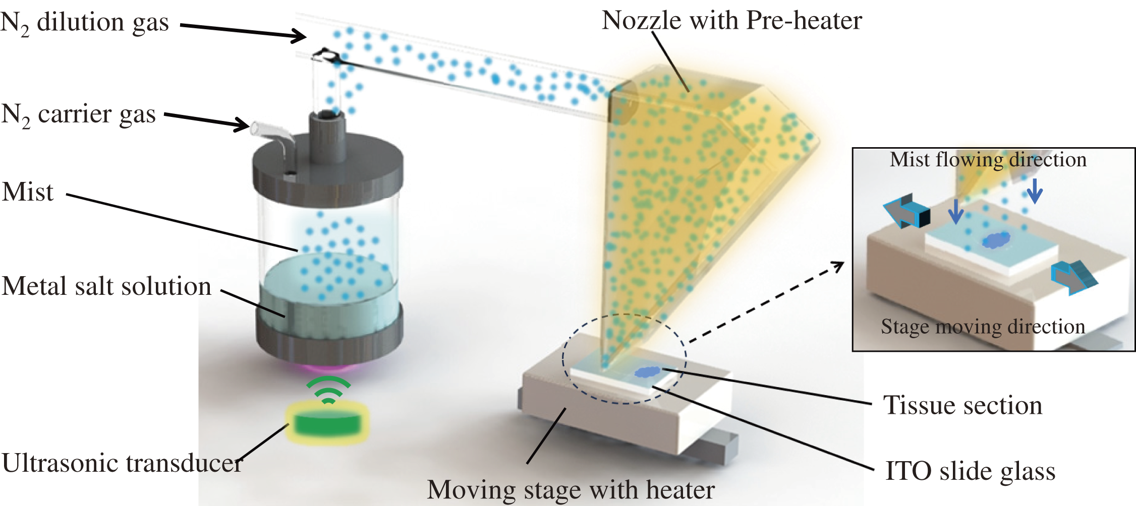
This system was employed to allow uniform deposition. A spray nozzle with a pre-heater at 150°C and moving stage with a heater at 150°C was equipped to ensure uniform mist flow on the area of 20 mm×60 mm. The slide glass or tissue section on the slide glass was set on to the heated moving stage with a speed of 15 mm/s. To prepare film on the slide glass, the moving cycles of 3 times were selected. Precursor mist of metal salt solution was formed by ultrasonic transducers with 2.4 MHz. The total flow rate of N2 carrier and dilution gas was set at 7 L/min. The precursor solution was prepared from 0.02 M silver trifluoroacetate, 50% methanol aqueous solution. The analysis of the film surface was performed using X-ray photoelectron spectroscopy (XPS; Kratos Axis Ultra DLD, Manchester, UK).
SALDI-MS measurements
SALDI/MS experiments were performed using Fourier transform ion cyclotron resonance (FTICR) MS SolariX 9.4 T system (Bruker Daltonics, Billerica, MA, USA) with 400 laser shots and 1 M-word time domain with the smartbeam II laser (Nd:YAG) at a wavelength of 355 nm, and time of flight mass spectrometer (TOFMS) JMS-S3000 SpiralTOF-plus (JEOL Ltd., Tokyo, Japan) with pulsed Nd:YLF laser of 50% intensity, 250 Hz, delay time 100 ns, and laser at a wavelength of 349 nm in the spiral positive mode. The spot diameter was approximately 50 μm. MALDI/ and SALDI/MS were performed by TOFMS. MS imaging was performed by FTICR MS. All spectra were obtained using the positive-ion mode. For the SALDI/MS measurement of standard sample, 1 μL of standard solution was spotted and dried on the prepared Ag film. For the MALDI/MS measurement of standard sample, 1 μL of standard/DHB=1/1(v/v) mixture was spotted and dried. The Ag film was prepared on the tissue section for MS imaging. The selective ion images were acquired at 125 μm lateral for MS imaging.
RESULTS AND DISCUSSION
Ag film prepared by mist CVD
The mist CVD process is shown in Fig. 1. The mist of metal salt solution generated by ultrasonic wave is guided to a nozzle by N2 carrier gas and dilution gas. The mist droplet size is reduced by the preheater equipped in the nozzle. The metal salt in the mist was coated on the indium tin oxide (ITO) slide glass through the nozzle. A metal film was prepared to expose the mist to the heated ITO slide glass. In this study, silver trifluoroacetate was used as the metal salt. The relationship between film growth of silver trifluoroacetate and the temperature of the heater was evaluated. A homogeneous film could not be obtained without the preheater because of large droplets. The temperature of the moving stage heater was set at 150°C, which prevents these lipids from decomposing. The growth of the film formed by the preheater at more than 150°C by the mist CVD was slow to fewer droplets. Dry surface of the sample could not be obtained by the preheater at room temperature. Therefore, the temperature of the preheater was set at 150°C.
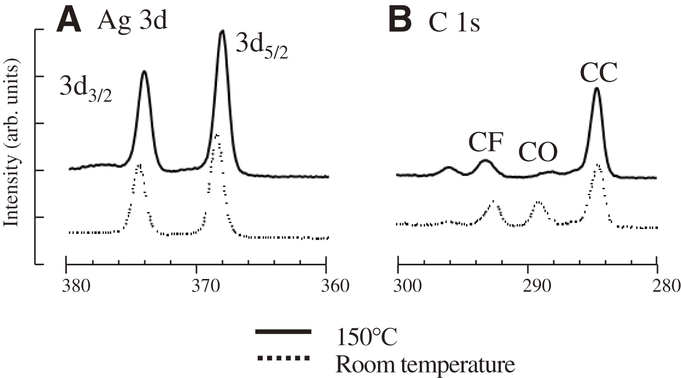
The surfaces of the slide glass coated by silver trifluoroacetate using the lab-made mist CVD system at 150°C or room temperature were analyzed by XPS. Reference binding energies of Ag 3d5/2 for AgOOCCF3 and Ag are 368.7 eV and 368.2 eV. The peak for Ag3d was shifted in the XPS spectrum of heated surface at 150°C as shown in Fig. 2. Additionally, the XPS peak intensities for C1s of CF at 293 eV and CO at 289 eV were also decreased by heating using the mist CVD. It means that trifluoroacetate ions were decreased and Ag ions were grown to the Ag film using the heated mist CVD at 150°C. The growth of the Ag-metal led to the formation of the Ag film. The above results indicate that metal films could be obtained to use both nozzle preheater and stage heater. In previous reports, synthesized nanoparticles12,13) and Pt films prepared by sputtering with expensive metal target in vacuum14,15) were used for SALDI. It takes a lot of time to prepare nanoparticles and films. On the other hand, formation of metal film by the mist CVD with inexpensive metal salt solution could be achieved under atmospheric pressure in a short time.
SALDI/MS with Ag film prepared by mist CVD***
The potential of Ag film obtained by the mist CVD as ionization assistance materials for SALDI/MS was evaluated. Dioleoylglycerol and dipalmitoylglycerol of lipids as standard DAGs were analyzed by SALDI/MS with the Ag film prepared using the heated mist CVD at 150°C. The standard solution was spotted on the Ag film, and dried and measured. The obtained mass spectra are shown in Fig. 3. The ions of [M+Ag]+ and their isotopic ions originated from 109Ag for dioleoylglycerol and dipalmitoylglycerol were observed as strong signals. In the SALDI, the Ag film supplied Ag+ as a cationization agent to molecules. On the other hand, these DAGs could not be detected by MALDI/MS directly.15) The signal to noise (S/N) ratio of these DAG ions by MALDI and SALDI/MS is shown in Fig. 3C. In traditional preparation for MALDI imaging, the handling procedures with a sprayer or an air brush for applying matrix solution to the sample required a manual technique. It was difficult to obtain the homogeneous crystals with high reproducibility. In the mist CVD, the droplet of metal salt solution generated by ultrasonication is small and of uniform size. Therefore, the obtained Ag film has a homogeneous surface.
The results indicate that the Ag film prepared by the mist CVD is useful as an ionization assistance material for SALDI.
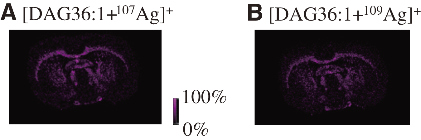
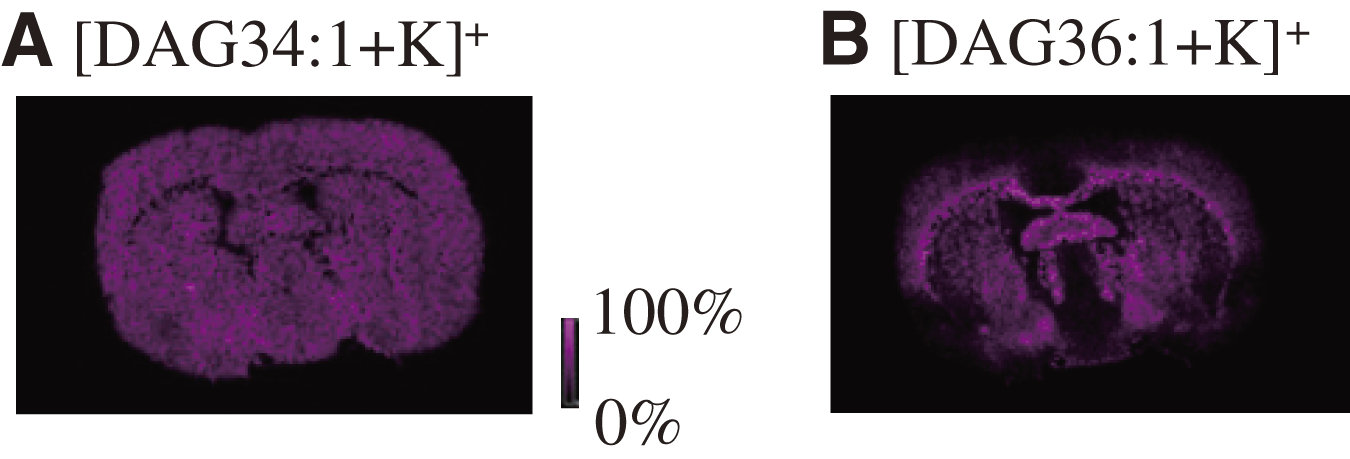
Next, SALDI/MS imaging of rat brain tissue section with the Ag film was performed. In the experiment, it was found that the observed ions at m/z 729.459 and 731.460 were localized in the same region (Fig. 4). These were Ag-adducted ions of [DAG36:1+107Ag]+ and [DAG36:1+109Ag]+. The exact masses are 729.458 and 731.458. Additionally, not only the Ag-adducted ions but also potassium-adducted DAG ions such as DAG34:1 at m/z 633.487 and DAG36:1 at m/z 661.518 were detected (Fig. 5). The exact masses are 633.486 and 661.517. The calculated chemical formulas from the obtained masses of m/z 772.528, 798.542, 826.571, and 820.528 were consistent with those of [PC32:0+K]+, [PC34:1+K]+, [PC36:1+K]+, and [PC36:4+K]+, respectively. The exact masses are 772.525, 798.541, 826.572, and 820.526. These localized images and the average mass spectrum are shown in Fig. 6. Many other peaks could be detected in this SALDI/MS imaging of rat brain tissue section. The results indicate that the Ag film is useful for SALDI/MS imaging of lipids and the film. Ag+ in the Ag film could be attached to the π-bonds (C==C, C==O, and aromatic group) of analytes for Ag–π interactions to ionized analytes in SALDI. In the MS imaging, ion intensities of [M+Ag]+ was low because Ag+ could only attach to the sites of π-bond in the molecule. Furthermore, many potassium-adducted ions were detected due to the large amount of K present in cells. It means that the film could assist the desorption and ionization of other molecules to produce the sodium- and potassium-adducted ions. K- and Na-adducted ions could be formed more than Ag because these ions could attach to the sites of δ− in the molecule.
CONCLUSION
The Ag film prepared from silver trifluoroacetate by the mist CVD was useful as an ionization assistance material for SALDI. The SALDI/MS and the MS imaging with the film allow the detection of lipids. In the localization analysis of DAGs, PCs in the rat brain tissue section was achieved by the SALDI/MS imaging. Metal films for SALDI could be prepared from metal salt solution under atmospheric pressure rapidly, simply, and inexpensively. The method has the potential for various applications of SALDI.
ACKNOWLEDGEMENTS
This work was supported by JSPS KAKENHI (grant numbers 19K05531 and 23K11323) and Matching Planner Program from Japan Science and Technology Agency, JST.
Notes
Mass Spectrom (Tokyo) 2023; 12(1): A0135
REFERENCES
- 1) K. Tanaka, H. Waki, Y. Ido, S. Akita, Y. Yoshida, T. Yoshida, T. Matsuo. Protein and polymer analyses up to m/z 100000 by laser ionization time-of-flight mass spectrometry. Rapid Commun. Mass Spectrom. 2: 151–153, 1988.
- 2) M. Karas, F. Hillenkamp. Laser desorption ionization of proteins with molecular mass exceeding 10,000 Daltons. Anal. Chem. 60: 2299–2301, 1988.
- 3) F. J. Wyzgoski, M. J. Polce, C. Wesdemiotis, M. A. Arnould. Matrix-assisted laser desorption/ionization time-of-flight mass spectrometry investigations of polystyrene and poly(methyl methacrylate) produced by monoacylphosphine oxide photoinitiation. J. Polym. Sci. A Polym. Chem. 45: 2161–2171, 2007.
- 4) D. Asakawa, S. Moriguchi, M. Takayama. Influence of amino acid composition and phosphorylation on the ion yields of peptides in MALDI-MS. J. Am. Soc. Mass Spectrom. 23: 108–115, 2012.
- 5) L. H. Cohen, A. I. Gusev. Small molecule analysis by MALDI mass spectrometry. Anal. Bioanal. Chem. 373: 571–586, 2002.
- 6) Q. Zhou, A. Fülöp, C. Hopf. Recent developments of novel matrices and on-tissue chemical derivatization reagents for MALDI-MSI. Anal. Bioanal. Chem. 413: 2599–2617, 2021.
- 7) C. Sun, W. Liu, Y. Mu, X. Wang. 1,1′-binaphthyl-2,2′-diamine as a novel MALDI matrix to enhance the in situ imaging of metabolic heterogeneity in lung cancer. Talanta 209: 120557, 2020.
- 8) H. Zhang, X. Shi, N. Q. Vu, G. Li, Z. Li, Y. Shi, M. Li, B. Wang, N. V. Welham, M. S. Patankar, P. Weisman, L. Li. On-tissue derivatization with Girard’s reagent P enhances N-glycan signals for formalin-fixed paraffin-embedded tissue sections in MALDI mass spectrometry imaging. Anal. Chem. 92: 13361–13368, 2020.
- 9) E. Takeo, Y. Sugiura, T. Uemura, K. Nishimoto, M. Yasuda, E. Sugiyama, S. Ohtsuki, T. Higashi, T. Nishikawa, M. Suematsu, E. Fukusaki, S. Shimma. Tandem mass spectrometry imaging reveals distinct accumulation patterns of steroid structural isomers in human adrenal glands. Anal. Chem. 91: 8918–8925, 2019.
- 10) W. H. Müller, A. Verdin, E. De Pauw, C. Malherbe, G. Eppe. Surface-assisted laser desorption/ionization mass spectrometry imaging: A review. Mass Spectrom. Rev. 41: 373–420, 2022.
- 11) J. Sunner, E. Dratz, Y. C. Chen. Graphite surface-assisted laser desorption/ionization time-of-flight mass spectrometry of peptides and proteins from liquid solutions. Anal. Chem. 67: 4335–4342, 1995.
- 12) R. Arakawa, H. Kawasaki. Functionalized nanoparticles and nanostructured surfaces for surface-assisted laser desorption/ionization mass spectrometry. Anal. Sci. 26: 1229–1240, 2010.
- 13) T. Yonezawa, T. Asano, M. Matsubara. Surface-assisted laser desorption ionization mass spectrometry (SALDI-MS) of low-molecular-weight medicines and toxic materials using commercial TiO2 nanoparticles. Bull. Chem. Soc. Jpn. 89: 346–353, 2016.
- 14) T. Ozawa, I. Osaka, S. Hamada, T. Murakami, A. Miyazato, H. Kawasaki, R. Arakawa. Direct imaging mass spectrometry of plant leaves using surface-assisted laser desorption/ionization with sputter-deposited platinum film. Anal. Sci. 32: 587–591, 2016.
- 15) T. Ozawa, I. Osaka, T. Ihozaki, S. Hamada, Y. Kuroda, T. Murakami, A. Miyazato, H. Kawasaki, R. Arakawa. Simultaneous detection of phosphatidylcholines and glycerolipids using matrix-enhanced surface-assisted laser desorption/ionization-mass spectrometry with sputter-deposited platinum film. J. Mass Spectrom. 50: 1264–1269, 2015.
- 16) K. Nozaki, Y. Nakabayashi, T. Murakami, A. Miyazato, I. Osaka. Novel approach to enhance sensitivity in surface-assisted laser desorption/ionization mass spectrometry imaging using deposited organic-inorganic hybrid matrices. J. Mass Spectrom. 54: 612–619, 2019.
- 17) J. Duan, M. J. Linman, C.-Y. Chen, Q. J. Cheng. CHCA-modified Au nanoparticles for laser desorption ionization mass spectrometric analysis of peptides. J. Am. Soc. Mass Spectrom. 20: 1530–1539, 2009.
- 18) Y.-F. Hung, H. -T. Chang. Analysis of adenosine triphosphate and glutathione through gold nanoparticles assisted laser desorption/ionization mass spectrometry. Anal. Chem. 79: 4852–4859, 2007.
- 19) W.-Y. Chen, Y.-C. Chen. Affinity-based mass spectrometry using magnetic iron oxide particles as the matrix and concentrating probes for SALDI MS analysis of peptides and proteins. Anal. Bioanal. Chem. 386: 699–704, 2006.
- 20) J. G. Lu, T. Kawaharamura, H. Nishinaka, Y. Kamada, T. Ohshima, S. Fujita. ZnO-based thin films synthesized by atmospheric pressure mist chemical vapor deposition. J. Cryst. Growth 299: 1–10, 2007.
- 21) G. T. Dang, T. Uchida, T. Kawaharamura, M. Furuta, A. R. Hyndman, R. Martinez, S. Fujita, R. J. Reeves, M. W. Allen. Silver oxide Schottky contacts and metal semiconductor field-effect transistors on SnO2 thin films. Appl. Phys. Express 9: 041101, 2016.
- 22) T. Ikenoue, T. Kawai, R. Wakashima, M. Miyake, T. Hirato. Hole mobility improvement in Cu2O thin films prepared by the mist CVD method. Appl. Phys. Express 12: 055509, 2019.
- 23) J. Piao, S. Katori, T. Ikenoue, S. Fujita. Formation of aluminum tris (8-hydroxyquinoline) solution in methanol and fabrication of thin films by ultrasonic spray-assisted vapor deposition. Phys. Status Solidi., A Appl. Mater. Sci. 209: 1298–1301, 2012.